Review
. 2024 Nov;27(4):587-621.
doi: 10.1007/s10456-024-09928-6. Epub 2024 Jun 6.

Breathing new life into tissue engineering: exploring cutting-edge vascularization strategies for skin substitutes

Affiliations
Review

Breathing new life into tissue engineering: exploring cutting-edge vascularization strategies for skin substitutes

M Zohaib Iqbal et al. Angiogenesis. 2024 Nov.

Abstract

Tissue-engineered skin substitutes (TESS) emerged as a new therapeutic option to improve skin transplantation. However, establishing an adequate and rapid vascularization in TESS is a critical factor for their clinical application and successful engraftment in patients. Therefore, several methods have been applied to improve the vascularization of skin substitutes including (i) modifying the structural and physicochemical properties of dermal scaffolds; (ii) activating biological scaffolds with growth factor-releasing systems or gene vectors; and (iii) developing prevascularized skin substitutes by loading scaffolds with capillary-forming cells. This review provides a detailed overview of the most recent and important developments in the vascularization strategies for skin substitutes. On the one hand, we present cell-based approaches using stem cells, microvascular fragments, adipose tissue derived stromal vascular fraction, endothelial cells derived from blood and skin as well as other pro-angiogenic stimulation methods. On the other hand, we discuss how distinct 3D bioprinting techniques and microfluidics, miRNA manipulation, cell sheet engineering and photosynthetic scaffolds like GelMA, can enhance skin vascularization for clinical applications. Finally, we summarize and discuss the challenges and prospects of the currently available vascularization techniques that may serve as a steppingstone to a mainstream application of skin tissue engineering.

Keywords: 3D bioprinting; Angiogenesis; Blood vessels; Dermal substitutes; Endothelial cells; Mesenchymal stem cells; Scaffolds; Skin defect.


Conflict of interest statement

Declarations Competing interests The authors declare no competing interests.

Figures

Fig. 1
Fig. 1
Prevascularization of skin substitutes using HUVECs. A To develop a capillary-like network, ECs are seeded and cultured in a 3D scaffold under optimal culture conditions. B ECs that have been transfected with a caspase-resistant version of Bcl-2 exhibit increased anti-apoptotic activity, survival, and tube formation. C For the formation of long-lasting, completely functional microvessels within the scaffold, the co-cultivation of ECs with mural cells are required [modified after 23]
Fig. 2
Fig. 2
A scheme showing the preparation of prevascularized dermal hydrogel. HDMECs containing both BEC (red) and LEC (green) were combined with dermal fibroblasts in a 3D collagen type I based scaffold promotes capillary formation after 3 weeks in vitro culture (prepared with BioRender)
Fig. 3
Fig. 3
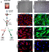
Analysis of endothelial cells (watECs) and adipose stromal cells (watASCs) of SVF. A Isolation and FACS sorting of watECs (red) and watASCs (green). Phase contrast microscopy showing cobblestone pattern of (B) watECs and spindle-shaped pattern of (F) watASCs. C–E Immunofluorescence staining of watECs showing positive expression of CD31, VEGFR2 and Dil-Ac-LDL. G–I Immunofluorescent evaluation of watASCs showing positive staining of CD90 and vimentin and negative for Dil-Ac-LDL. Scale bars: B, F: 200 µm; C–E, G–I: 50 µm [modified after 5]
Fig. 4
Fig. 4
In vitro analysis of bioengineered capillaries. A–C Light microscopy of SVF-derived watECs cultured in 3D hydrogels for 3–7 days to visualize the presence of vacuolar structures (3 days) and their fusion into branched capillaries (7 days) D CD31 staining (red) of hydrogels after 21 days of culturing revealed a network of well-connected capillaries. E Electron microscopy confirmed the location of lumen (L) surrounded by several ECs and pericytes (Pc) and presence of basement membrane (blue arrows). Scale bars: A–C 50 µm; D 100 µm; E 1 µm [modified after 5]
Fig. 5
Fig. 5
In vivo analysis of SVF seeded DESS A Immunofluorescence staining of DESS showing a connection between human capillaries (red) and rat capillaries (green) 4 days after transplantation. B Blood perfusion of capillaries of transplanted capillaries is confirmed by visibility of rat erythrocytes (red) inside their lumina. C Co-staining of CD31 and CD90 confirming the location of the capillaries in dermal compartment 7 days after DESS transplantation. D Presence of pericytes around the transplanted capillaries is confirmed through human/rat αSMA (pericyte marker) staining. Scale: A 40 µm; B–D 50 µm [modified after 5]
Fig. 6
Fig. 6
Vascularization in tissue engineering by MVF. A Epididymal fat pads isolated from mouse tissue. B–C Phase-contrast of MVF freshly isolated from fat pads. D–E Immunohistochemistry of MVFs showing endothelial cells (green), α-smooth muscle actin (red) and cell nuclei (blue) [modified after 47]
Fig. 7
Fig. 7
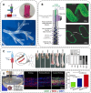
Applications of coaxial printing in vasculature engineering: A Vessels were fabricated by extruding sodium alginate onto a rotating rod (upper), followed by assembly into multiscale vasculature (lower). B Coiled-rope structures (upper right) were formed within a glass tube by crosslinking and adjusting the viscosity of the core and sheath fluids (left), providing space for endothelial cell lumen formation (lower right). C Artificial bio-blood vessels (BBV) were designed using a sacrificial core fluid (left), showcasing enhanced limb salvage in a mouse model when loaded with cells (EBBV) and a statin drug (EABBV) (center right). D Coaxially printed vessels (left) demonstrated cell differentiation in vitro (center) compared to single fluid printing (right) in a rat model. Scale bars: A 5 mm; B 200 μm; D 200 μm [Modified after 72]
Fig. 8
Fig. 8
Vascularization strategies using ECs in vitro: A Microneedle-based Removable Method: (i) A needle-molding technique to form fluidic channels within hydrogels. (ii) Endothelialized microchannels under the influence of fluid flow. B Micropatterned Planar Hydrogel Slab Bonding Method: (i) Fluidic hydrogels created via micromolding from PDMS or silicon molds, followed by bonding. (ii) Micrographs exhibit the shape of microvessel networks within collagen gel constructed from a micropatterned silicon stamp, along with confocal sections of endothelialized microfluidic vessels immunostained with CD31. Scale bar, 100 μm. C Dissolvable Materials-Based Sacrificial Micromolding Method: (i) A schematic showcases a 3D interconnected microvessel network formed by casting a carbohydrate glass lattice as the sacrificial element with a 3D printer. (ii) Micrographs reveal HUVECs expressing mCherry attached to the hydrogel wall for generating the microvessel network and an endothelial monolayer-lined vascular lumen surrounded by 10T1/2 cells after 9 days in culture. Scale bars, 1 mm and 200 μm. D EC Lining Inside a PDMS-Based Microfluidic Channel: (i) A schematic depicts a PDMS-based microfluidic channel, alongside a confocal image of endothelial cells within the channel. (ii) Confocal reconstruction images show the complete lumen formed by HUVECs inside the PDMS tube, along with fluorescence micrographs of cross-sectional views of an endothelialized PDMS tube stained with CD31/nuclei. Scale bars, 100 μm (top) or 200 μm (bottom) [modified after 73]
Fig. 9
Fig. 9
Development of MAP gel using microfluidics. A Application of MAP gel to produce any kind of 3D shape through 25-gauge syringe. B–C Detection of vasculature by staining for endothelial cell marker PECAM-1 in MAP scaffolds, 5 days after transplantation onto mice injured skin [modified after 76]
Fig. 10
Fig. 10
Schematic diagram for preparation and application of dECM (prepared with BioRender)
Fig. 11
Fig. 11
Schematic illustration of 3D printing of vascular network template using sacrificial materials (prepared with BioRender)
Fig. 12
Fig. 12
In vitro development of prevascularized human DESS by SkinFactory. a–b Immunofluorescence whole-mount staining revealed the blood capillaries (CD31) formation in hydrogels after 3 and 4 weeks of HDMECs cultivation via SkinFactory. c Detection of mural cells (CD90) around blood capillaries (CD31) after 3 weeks. d–f immunofluorescence staining of HDMECs cultured in hydrogels under uncompressed (d), compressed on HDMECs seeding (e) and compressed after 3 weeks of HDMECs seeding (f). Scale bar are 100 µm [modified after [145]
Fig. 13
Fig. 13
Transplantation of bioprinted prevascularized DESS (pigmented) developed by SkinFactory. a Dermal fibroblasts (CD90), blood capillaries (CD31-upper panel) and lymphatic capillaries (Lyve1-lower panel) are visible in neodermis after 1 and 2 weeks of transplantation. b Hematoxylin and eosin staining (upper panel) reveals the following structures: the stratum corneum (indicated by a white star), the stratum spinosum (indicated by a black star), pigmented clusters of melanocytes (indicated by black arrowheads), unpigmented clusters of basal keratinocytes (indicated by white arrowheads), melanocyte dendrites (indicated by blue arrows), and melanosome supranuclear caps in keratinocytes (indicated by black arrows). In a four-color immunofluorescence staining, the presence and localization of four distinct cell types are demonstrated: fibroblasts (labeled with CD90 and shown in blue), endothelial cells (labeled with CD31 and shown in white), keratinocytes (labeled with CK1 and shown in red), and melanocytes (labeled with HMB45 and shown in green). The dermo-epidermal border is indicated by the white dotted line. Scale: 200 µm [modified after 145]

References

    1. Graham GP, Helmer SD, Haan JM, Khandelwal A (2013) The use of Integra® dermal regeneration template in the reconstruction of traumatic degloving injuries. J Burn Care Res 34(2):261–266. 10.1097/BCR.0b013e3182853eaf - PubMed
    1. Shevchenko RV, James SL, James SE (2010) A review of tissue-engineered skin bioconstructs available for skin reconstruction. J Royal Soc Interface 7(43):229–258 - PMC - PubMed
    1. Hosper NA, Eggink AJ, Roelofs LA, Wijnen RM, van Luyn MJ, Bank RA, Feitz WF (2010) Intra-uterine tissue engineering of full-thickness skin defects in a fetal sheep model. Biomaterials 31(14):3910–3919. 10.1016/j.biomaterials.2010.01.129 - PubMed
    1. Klar AS, Güven S, Biedermann T, Luginbühl J, Böttcher-Haberzeth S, Meuli-Simmen C, Meuli M, Martin I, Scherberich A, Reichmann E (2014) Tissue-engineered dermo-epidermal skin grafts prevascularized with adipose-derived cells. Biomaterials 35(19):5065–5078. 10.1016/j.biomaterials.2014.02.049. (Epub 2014 Mar 27 PMID: 24680190) - PubMed
    1. Klar AS, Güven S, Zimoch J, Zapiórkowska NA, Biedermann T, Böttcher-Haberzeth S, Meuli M (2016) Characterization of vasculogenic potential of human adipose-derived endothelial cells in a three-dimensional vascularized skin substitute. Pediatr Surg Int 32:17–27 - PubMed

LinkOut - more resources