. 2024 Jun 11;121(24):e2404383121.
doi: 10.1073/pnas.2404383121. Epub 2024 Jun 6.

The ARK2N-CK2 complex initiates transcription-coupled repair through enhancing the interaction of CSB with lesion-stalled RNAPII

Affiliations

The ARK2N-CK2 complex initiates transcription-coupled repair through enhancing the interaction of CSB with lesion-stalled RNAPII

Yefei Luo et al. Proc Natl Acad Sci U S A. .

Abstract

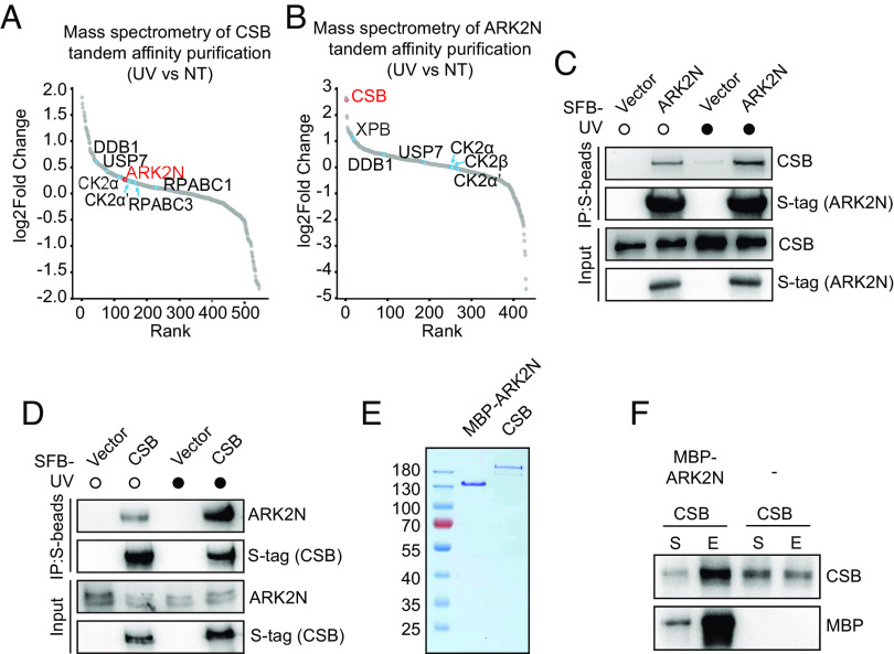
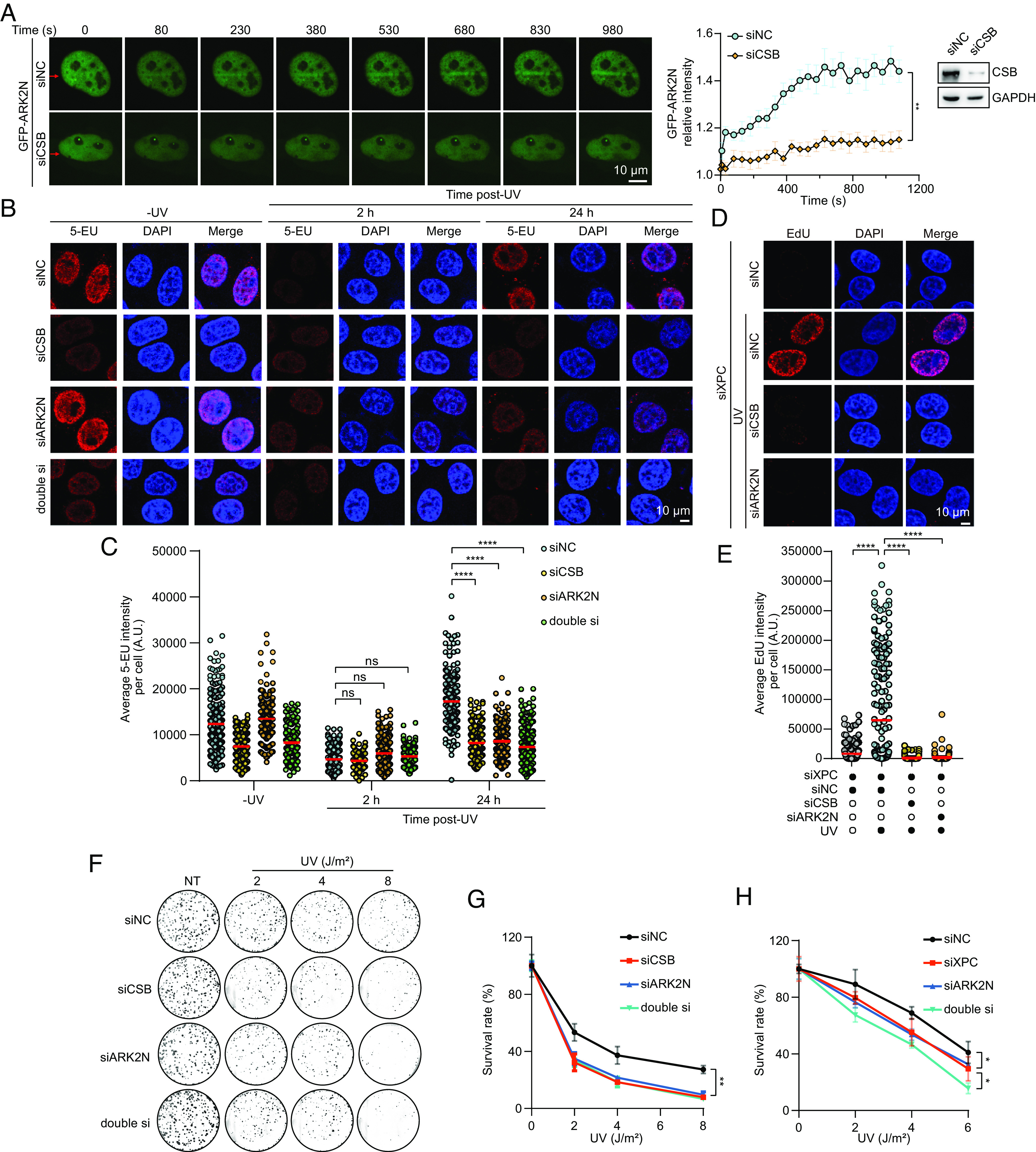
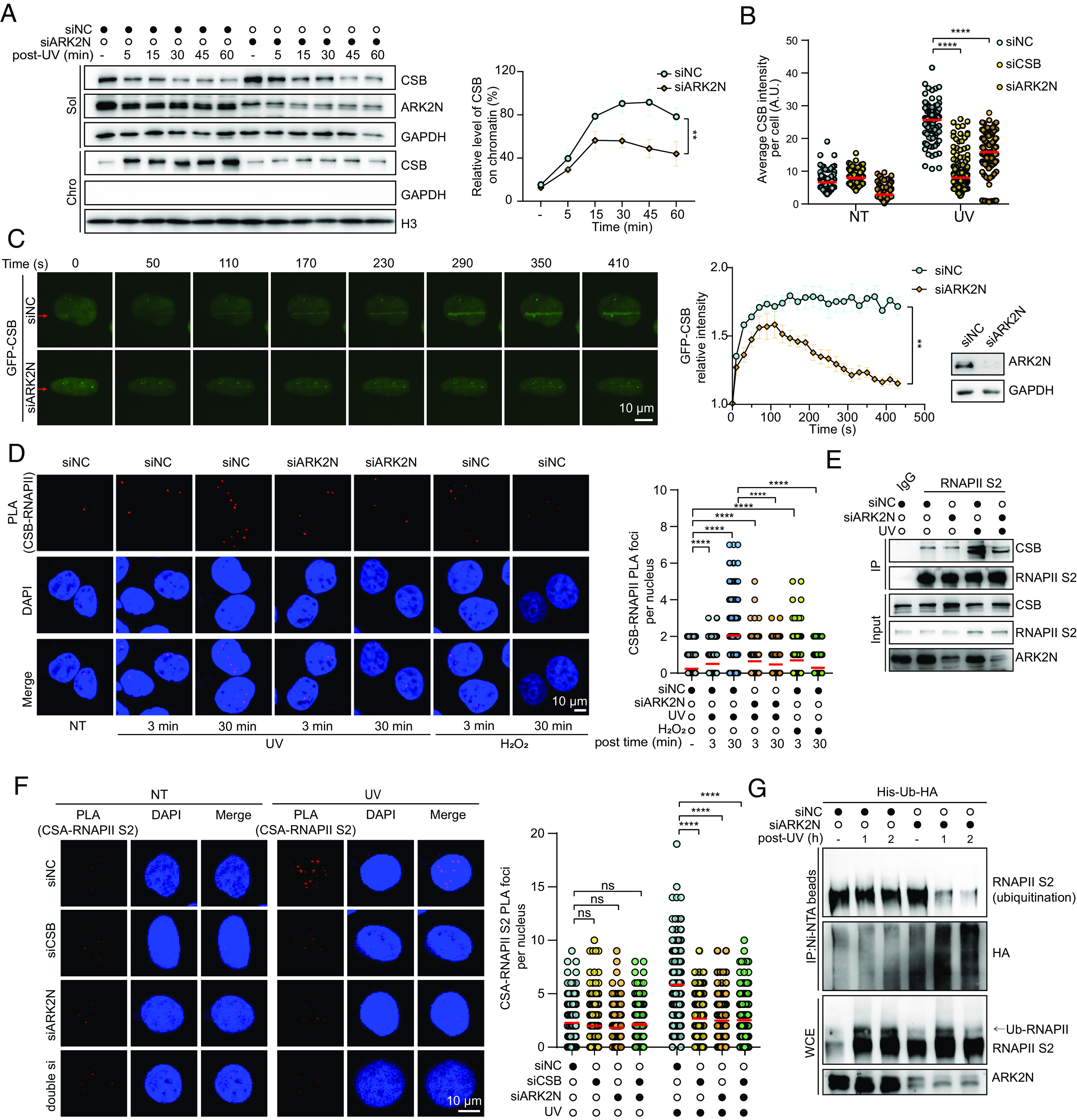
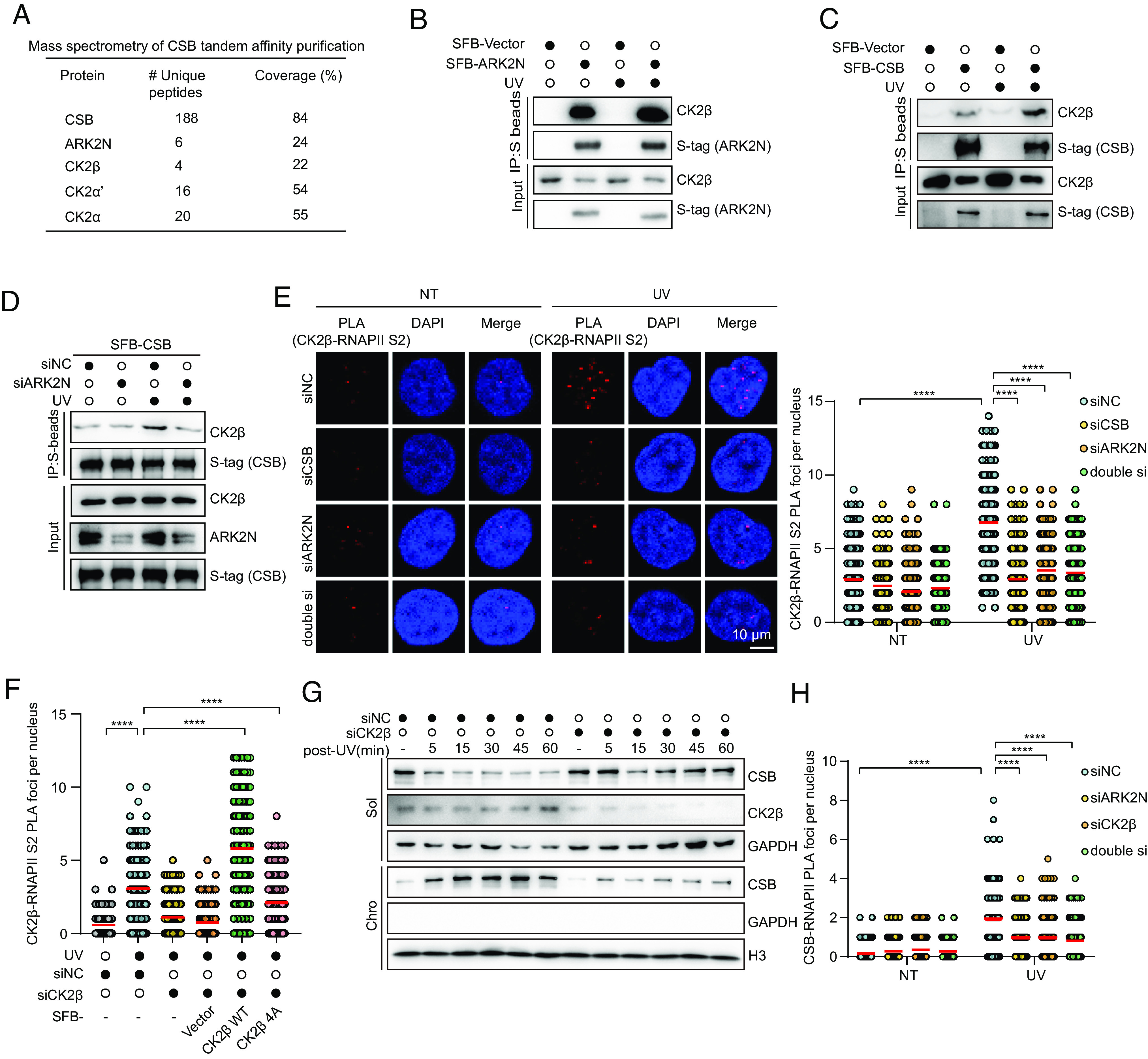
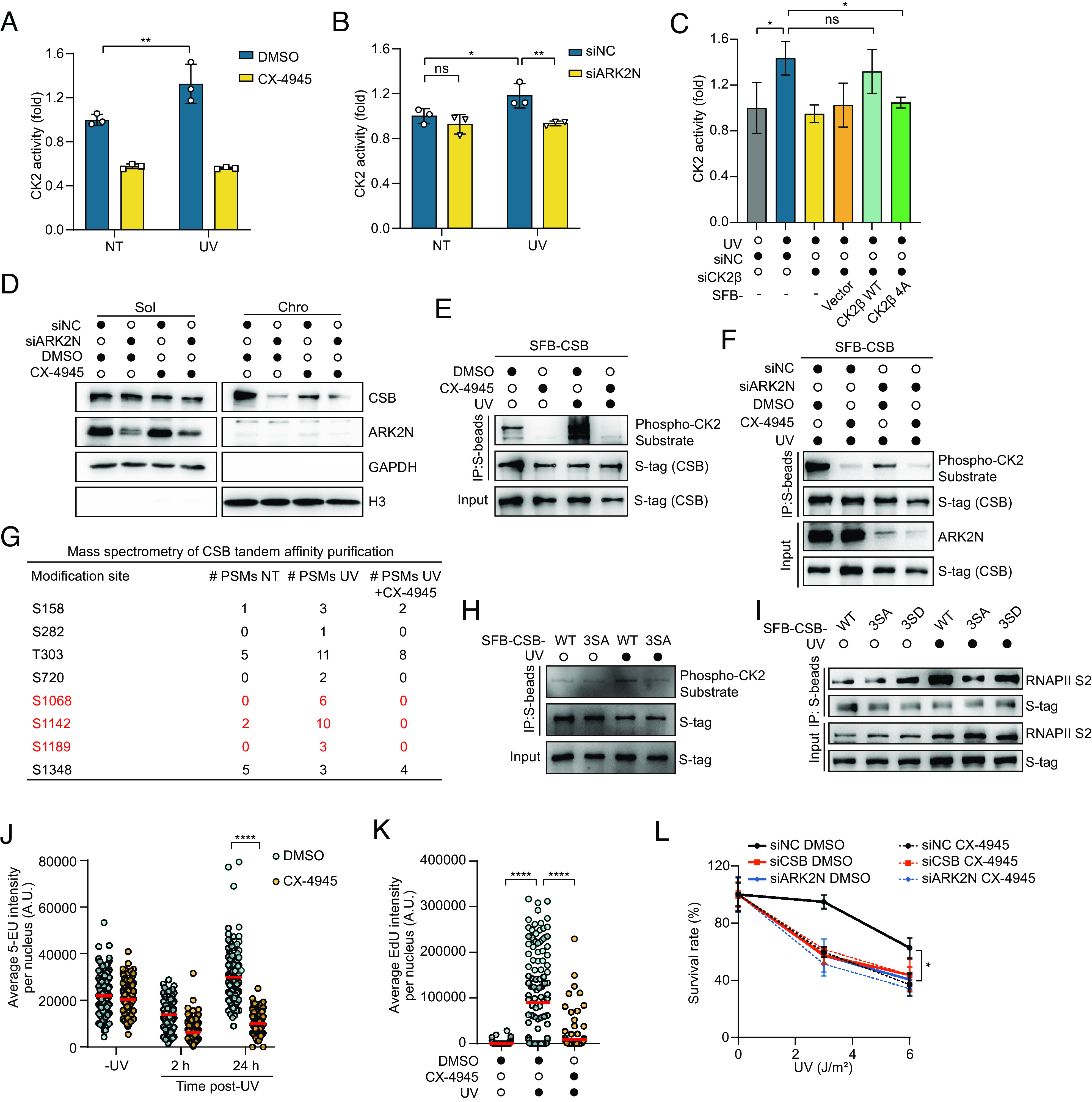
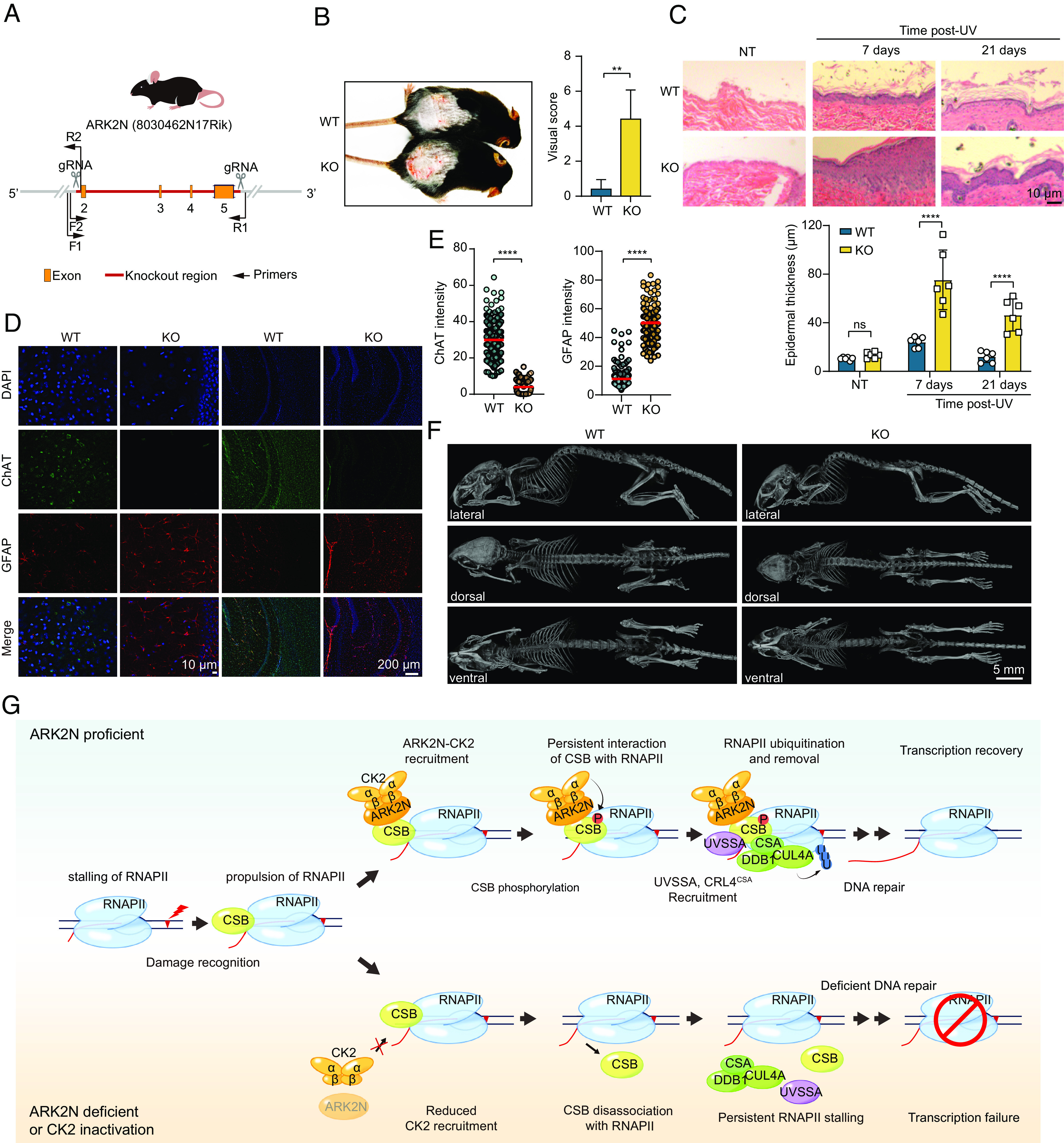
Transcription is extremely important for cellular processes but can be hindered by RNA polymerase II (RNAPII) pausing and stalling. Cockayne syndrome protein B (CSB) promotes the progression of paused RNAPII or initiates transcription-coupled nucleotide excision repair (TC-NER) to remove stalled RNAPII. However, the specific mechanism by which CSB initiates TC-NER upon damage remains unclear. In this study, we identified the indispensable role of the ARK2N-CK2 complex in the CSB-mediated initiation of TC-NER. The ARK2N-CK2 complex is recruited to damage sites through CSB and then phosphorylates CSB. Phosphorylation of CSB enhances its binding to stalled RNAPII, prolonging the association of CSB with chromatin and promoting CSA-mediated ubiquitination of stalled RNAPII. Consistent with this finding, Ark2n-/- mice exhibit a phenotype resembling Cockayne syndrome. These findings shed light on the pivotal role of the ARK2N-CK2 complex in governing the fate of RNAPII through CSB, bridging a critical gap necessary for initiating TC-NER.

Keywords: ARK2N; CK2; CSB; Cockayne syndrome; transcription-coupled nucleotide excision repair.


Conflict of interest statement

Competing interests statement:The authors declare no competing interest.

Figures

Fig. 1.
Fig. 1.
ARK2N is identified as a partner of CSB. (A and B) The TAP-MS coupled rank plot analysis showed the differential abundance of protein peptide segments associated with CSB (A) and ARK2N (B) in cells treated with UV (20 J/m2, 30 min release) compared to untreated cells. (C and D) The interaction between exogenously expressed ARK2N (D) or CSB (E) and endogenous CSB or ARK2N was investigated in HEK293T cells under untreated conditions or conditions involving UV irradiation (20 J/m2) followed by a 30-min release period. (E) CSB protein bought in Origene and MBP-ARK2N protein purified in yeast were stained by Coomassie blue. (F) CSB and MBP-ARK2N proteins were incubated in vitro, followed by the pull-down experiment with MBP beads. The supernatant (S) containing unbound proteins and the eluate (E) containing proteins interacting with MBP-ARK2N were analyzed by western blotting using the indicated antibodies.
Fig. 2.
Fig. 2.
ARK2N is an indispensable factor in the TC-NER pathway. (A) Real-time fluorescence imaging detected the recruitment of ARK2N to UV microirradiation-induced damage sites in live HeLa cells transfected with the indicated siRNAs. The data are presented as the means ± SEMs (n = 8, **P < 0.01). (B and C) HeLa cells transfected with the indicated siRNAs were subjected to RNA synthesis recovery (RRS) assay after being either untreated or exposed to UV (9 J/m2) and allowed to recover for the indicated time. Representative images and quantification of the intensity of 5-EU per nucleus are shown. (n > 170, ****P < 0.0001, ns: not significant). (D and E) The TCR-UDS assay was performed in XPC-deficient HeLa cells transfected with the indicated siRNAs, following exposure to UV (8 J/m2). The data are presented as the means (n > 130, ****P < 0.0001). (F and G) The colony formation assay was conducted in HeLa cells transfected with the indicated siRNAs and treated with the indicated doses of UV irradiation. The survival rates were calculated by determining the colony numbers. The data are presented as the means ± SEMs (n = 3, **P < 0.01). (H) The colony formation assay was conducted in HeLa cells transfected with the indicated siRNAs and treated with the indicated doses of UV irradiation. The survival rates were calculated by determining the colony numbers. The data are presented as the means ± SEMs (n = 3, *P < 0.05).
Fig. 3.
Fig. 3.
ARK2N prolongs the occupancy of CSB at damaged chromatin sites by enhancing its interaction with RNAPII. (A) The soluble and chromatin fractions were extracted from HeLa cells transfected with the indicated siRNAs, either exposed to UV irradiation (20 J/m2, 30 min release) or subjected to control treatment. Representative immunoblot images are shown (Left). The percentage of CSB in the chromatin-enriched fraction was quantified by calculating the proportion of the gray value between CSB and H3. Histone H3 was used as the marker for the chromatin fraction. The data are presented as the means ± SEMs (n = 3) (Right). (B) The immunofluorescence intensity of CSB associated with damaged chromatin was evaluated in HeLa cells transfected with the indicated siRNAs following UV irradiation (20 J/m2, 30 min release). The data are presented as the means (****P < 0.0001). (C) Real-time fluorescence imaging detected the recruitment of CSB to UV microirradiation-induced damage sites in live HeLa cells transfected with the indicated siRNAs. The data are presented as the means ± SEMs (n = 8). (D) Colocalization of CSB and RNAPII in HeLa cells was assessed by PLA analysis under NT (no treatment) condition or after a 30-min release following UV irradiation (20 J/m2) or a 30-min treatment with H2O2 (2 mM). Representative images and quantification of PLA foci per nucleus are shown (n > 160, ****P < 0.0001). (E) HeLa cells transfected with the indicated siRNAs, with or without UV treatment, were lysed and subjected to immunoprecipitation using control IgG or RNAPII p-Ser2 antibody, followed by immunoblotting with the indicated antibodies. (F) Colocalization of CSA and RNAPII p-Ser2 in HeLa cells was assessed by PLA analysis under NT condition or after a 30-min release following UV irradiation (20 J/m2). Representative images and quantification of PLA foci per nucleus are shown (n > 150, ****P < 0.0001, ns: not significant). (G) HeLa cells expressing His-Ub-HA and transfected with the indicated siRNAs were treated with UV irradiation (20 J/m2, 30 min release). The cell lysates were incubated with Ni-NTA beads in guanidine denaturing buffer and examined by immunoblotting.
Fig. 4.
Fig. 4.
ARK2N–CK2 is essential for initiating TC-NER. (A) TAP-MS analysis demonstrated a strong association between CSB and both ARK2N and CK2 subunits. (B and C) The interaction between exogenously expressed ARK2N (B) or CSB (C) and endogenous CK2β was investigated in HEK293T cells under untreated conditions or conditions involving UV irradiation (20 J/m2, 30 min release). (D) The interaction between exogenously expressed CSB and endogenous CK2β was examined in HEK293T cells transfected with the specified siRNAs under untreated conditions or conditions involving UV irradiation (20 J/m2, 30 min release). (E) Colocalization of CK2β and RNAPII p-Ser2 in HeLa cells was assessed by PLA analysis under NT condition or following UV irradiation (20 J/m2, 15 min release). Representative images and quantification of PLA foci per nucleus are shown (n > 105, ****P < 0.0001). (F) Colocalization of CK2β and RNAPII p-Ser2 in HeLa cells expressing SFB-CK2-WT or 4A by PLA analysis following UV irradiation (20 J/m2, 15 min release). Quantification of PLA foci per nucleus is shown (n > 105, ****P < 0.0001). (G) The soluble and chromatin fractions were extracted from HeLa cells transfected with the indicated siRNA, either exposed to UV irradiation (20 J/m2, 30 min release) or subjected to control treatment. Immunoblot analysis was performed using indicated antibodies. (H) Colocalization of CSB and RNAPII in HeLa cells transfected with the indicated siRNAs was assessed by PLA analysis under NT condition or following UV irradiation (20 J/m2, 30 min release). Quantification of PLA foci per nucleus is shown (n > 120, ****P < 0.0001).
Fig. 5.
Fig. 5.
ARK2N facilitates CK2-mediated phosphorylation of CSB and enhances the interaction between CSB and RNAPII. (A) HeLa cells were treated with DMSO or CX-4945 (8 μM, 8 h), followed by either untreated or UV irradiation (20 J/m2, 30 min release). Cells were then lysed and incubated with the kinase reaction buffer to evaluate CK2 activity. (n = 3, **P < 0.01). (B) The activity of CK2 in HeLa cells transfected with the indicated siRNAs following UV irradiation (20 J/m2, 30 min release) (n = 3, *P < 0.05, **P < 0.01, ns: not significant). (C) The activity of CK2 in HeLa cells transfected with the indicated siRNAs and expressing SFB-Vector, SFB-CK2-WT, or SFB-CK2-4A following UV irradiation (20 J/m2, 30 min release) (n = 3, *P < 0.05, ns: not significant). (D) The soluble and chromatin fractions were extracted from HeLa cells transfected with the indicated siRNAs and treated with DMSO or CX-4945 (8 μM, 8 h), then exposed to UV irradiation (20 J/m2, 30 min release). Immunoblot analysis was performed using indicated antibodies. (E) HEK293T cells expressing SFB-CSB were treated as indicated and then subjected to immunoprecipitation using S-beads, followed by immunoblotting analysis. (F) HeLa cells expressing SFB-CSB were treated as indicated and then subjected to immunoprecipitation using S-beads, followed by immunoblotting analysis. (G) HEK293T cells stably expressing CSB were treated with control, UV irradiation (20 J/m2), and UV irradiation combined with CX-4945 (8 μM, 8 h) treatment. Following treatment, the cells were lysed, and CSB protein was immunoprecipitated using S-beads. The phosphorylation sites of the CSB protein were then analyzed using mass spectrometry. (H) Hela cells expressing SFB-CSB-WT or SFB-CSB-3SA were treated as indicated and then subjected to immunoprecipitation using S-beads, followed by immunoblotting analysis. (I) Hela cells expressing SFB-CSB-WT, SFB-CSB-3SA, or SFB-CSB-3SD were treated as indicated and then subjected to immunoprecipitation using S-beads, followed by immunoblotting analysis. (J) HeLa cells transfected with the indicated siRNAs were subjected to the RRS assay after being treated with DMSO or CX-4945 (8 μM, 8 h); then, the cells were untreated or exposed to UV (9 J/m2) and allowed to recover for the indicated time. Quantification of the intensity of 5-EU per nucleus is shown. (n > 130, ****P < 0.0001). (K) The TCR-UDS assay was performed in XPC-deficient HeLa cells treated with DMSO or CX-4945 (8 μM, 8 h), following exposure to UV (8 J/m2). The data are presented as the means (n > 130, ****P < 0.0001). (L) The colony formation assay was conducted in HeLa cells transfected with the indicated siRNAs and treated with the indicated doses of UV irradiation. The cells were then treated with DMSO or CX-4945 (8 μM, 24 h). The survival rates were calculated by determining the colony numbers. The data are presented as the means ± SEMs (n = 3, *P < 0.05).
Fig. 6.
Fig. 6.
ARK2N deficiency in mice results in Cockayne syndrome–like phenotype. (A) Schematic representation of the ARK2N Knockout mice. (B) UV-induced erythema in the skin of ARK2N-WT and ARK2N-KO mice. Shaven animals were exposed to UV-C irradiation (1,000 J/m2/d) for 4 consecutive days (photographs were taken 1 wk after the first exposure). Photoaging of the mice was graded according to the scoring standard as described. The data are presented as the means ± SDs (n = 6, **P < 0.01). (C) H&E staining of skin sections from the shaven mice at the indicated time after being either untreated or exposed to UV irradiation as described in (B). The epidermal thickness was determined via ImageJ. The data are presented as the means ± SDs (n = 6, ****P < 0.0001). (D and E) Immunofluorescence staining of the hippocampus was performed in ARK2N-WT and ARK2N-KO mice (120 d). The data are presented as the means (n > 130, ****P < 0.0001). (F) Computed tomography images in ARK2N-WT and ARK2N-KO mice (female, 120 d). (G) Model for the involvement of ARK2N–CK2 in TC-NER.

Similar articles

Cited by

References

    1. Chen F. X., Smith E. R., Shilatifard A., Born to run: Control of transcription elongation by RNA polymerase II. Nat. Rev. Mol. Cell Biol. 19, 464–478 (2018). - PubMed
    1. Hirose Y., Manley J. L., RNA polymerase II is an essential mRNA polyadenylation factor. Nature 395, 93–96 (1998). - PubMed
    1. Lans H., Hoeijmakers J. H. J., Vermeulen W., Marteijn J. A., The DNA damage response to transcription stress. Nat. Rev. Mol. Cell Biol. 20, 766–784 (2019). - PubMed
    1. Belotserkovskii B. P., Mirkin S. M., Hanawalt P. C., DNA sequences that interfere with transcription: Implications for genome function and stability. Chem. Rev. 113, 8620–8637 (2013). - PubMed
    1. Milano L., Gautam A., Caldecott K. W., DNA damage and transcription stress. Mol. Cell 84, 70–79 (2024). - PubMed

MeSH terms

LinkOut - more resources