Nocturnal Hypoxemia Is Associated with Sarcopenia in Patients with Chronic Obstructive Pulmonary Disease
- PMID: 38843487
- PMCID: PMC11376365
- DOI: 10.1513/AnnalsATS.202312-1062OC
Nocturnal Hypoxemia Is Associated with Sarcopenia in Patients with Chronic Obstructive Pulmonary Disease
Abstract
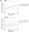
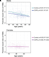
Rationale: Chronic obstructive pulmonary disease (COPD) is the third leading cause of death worldwide. Our previous studies have identified that nocturnal hypoxemia causes skeletal muscle loss (i.e., sarcopenia) in in vitro models of COPD. Objectives: We aimed to extend our preclinical mechanistic findings by analyzing a large sleep registry to determine whether nocturnal hypoxemia is associated with sarcopenia in patients with COPD. Methods: Sleep studies from patients with COPD (n = 479) and control subjects without COPD (n = 275) were analyzed. Patients with obstructive sleep apnea, as defined by apnea-hypopnea index ⩾ 5, were excluded. Pectoralis muscle cross-sectional area (PMcsa) was quantified using computed tomography scans performed within 1 year of the sleep study. We defined sarcopenia as less than the lowest 20% residuals for PMcsa of control subjects, which was adjusted for age and body mass index (BMI) and stratified by sex. Youden's optimal cut-point criteria were used to predict sarcopenia based on mean oxygen saturation during sleep. Additional measures of nocturnal hypoxemia were analyzed. The pectoralis muscle index (PMI) was defined as PMcsa normalized to BMI. Results: On average, males with COPD had a 16.6% lower PMI than control males (1.41 ± 0.44 vs. 1.69 ± 0.56 cm2/BMI; P < 0.001), whereas females with COPD had a 9.4% lower PMI than control females (0.96 ± 0.27 vs. 1.06 ± 0.33 cm2/BMI; P < 0.001). Males with COPD with nocturnal hypoxemia had a 9.5% decrease in PMI versus COPD with normal O2 (1.33 ± 0.39 vs. 1.47 ± 0.46 cm2/BMI; P < 0.05) and a 23.6% decrease compared with control subjects (1.33 ± 0.39 vs. 1.74 ± 0.56 cm2/BMI; P < 0.001). Females with COPD with nocturnal hypoxemia had an 11.2% decrease versus COPD with normal O2 (0.87 ± 0.26 vs. 0.98 ± 0.28 cm2/BMI; P < 0.05) and a 17.9% decrease compared with control subjects (0.87 ± 0.26 vs. 1.06 ± 0.33 cm2/BMI; P < 0.001). These findings were largely replicated using multiple measures of nocturnal hypoxemia. Conclusions: We defined sarcopenia in the pectoralis muscle using residuals that take into account age, BMI, and sex. We found that patients with COPD have a lower PMI than patients without COPD and that nocturnal hypoxemia was associated with an additional decrease in the PMI of patients with COPD. Additional prospective analyses are needed to determine a protective threshold of oxygen saturation to prevent or reverse sarcopenia due to nocturnal hypoxemia in COPD.
Keywords: COPD; cachexia; nocturnal hypoxemia; sarcopenia; skeletal muscle wasting.
Figures


References
-
- Yang IA, Jenkins CR, Salvi SS. Chronic obstructive pulmonary disease in never-smokers: risk factors, pathogenesis, and implications for prevention and treatment. Lancet Respir Med . 2022;10:497–511. - PubMed
-
- Remels AH, Gosker HR, Langen RC, Schols AM. The mechanisms of cachexia underlying muscle dysfunction in COPD. J Appl Physiol . 2013;114:1253–1262. - PubMed
MeSH terms
Grants and funding
- R56 HL141744/HL/NHLBI NIH HHS/United States
- U01 DK062470/DK/NIDDK NIH HHS/United States
- U01 DK061732/DK/NIDDK NIH HHS/United States
- T32 HL155005/HL/NHLBI NIH HHS/United States
- K12 HL141952/HL/NHLBI NIH HHS/United States
- U01 AA021890/AA/NIAAA NIH HHS/United States
- K12HL141952/NH/NIH HHS/United States
- U01 AA026976/AA/NIAAA NIH HHS/United States
- R01 HL158746/HL/NHLBI NIH HHS/United States
- R01 HL119792/HL/NHLBI NIH HHS/United States
- P50 AA024333/AA/NIAAA NIH HHS/United States
- R01 HL155064/HL/NHLBI NIH HHS/United States
- R01 GM119174/GM/NIGMS NIH HHS/United States
- R01 HL161674/HL/NHLBI NIH HHS/United States
- R21 AR071046/AR/NIAMS NIH HHS/United States
- R21 HL170206/HL/NHLBI NIH HHS/United States
- R01 DK113196/DK/NIDDK NIH HHS/United States
LinkOut - more resources
Full Text Sources
Medical

