Review
. 2025 Feb;14(5):e2400965.
doi: 10.1002/adhm.202400965. Epub 2024 Jul 3.

Unlocking Transplant Tolerance with Biomaterials

Affiliations
Review

Unlocking Transplant Tolerance with Biomaterials

John-Paul A Pham et al. Adv Healthc Mater. 2025 Feb.

Abstract

For patients suffering from organ failure due to injury or autoimmune disease, allogeneic organ transplantation with chronic immunosuppression is considered the god standard in terms of clinical treatment. However, the true "holy grail" of transplant immunology is operational tolerance, in which the recipient exhibits a sustained lack of alloreactivity toward unencountered antigen presented by the donor graft. This outcome is resultant from critical changes to the phenotype and genotype of the immune repertoire predicated by the activation of specific signaling pathways responsive to soluble and mechanosensitive cues. Biomaterials have emerged as a medium for interfacing with and reprogramming these endogenous pathways toward tolerance in precise, minimally invasive, and spatiotemporally defined manners. By viewing seminal and contemporary breakthroughs in transplant tolerance induction through the lens of biomaterials-mediated immunomodulation strategies-which include intrinsic material immunogenicity, the depot effect, graft coatings, induction and delivery of tolerogenic immune cells, biomimicry of tolerogenic immune cells, and in situ reprogramming-this review emphasizes the stunning diversity of approaches in the field and spotlights exciting future directions for research to come.

Keywords: biomaterials; immunoengineering; immunomodulation; transplantation.


Conflict of interest statement

The authors declare no conflict of interest.

Figures

Figure 1
Figure 1
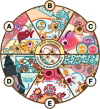
Engineering strategies for promoting transplant tolerance with biomaterials. Biomaterials can be engineered to interface with the immune system in a transplant setting in a variety of ways. A) The physicochemical properties of biomaterials can endow immunogenicity, such as size allowing smaller particles to readily traffic to organs of interest like the lymph node or pancreas while larger particles are uptaken by peripheral phagocytes. B) Immunoisolation is a strategy by which transplant alloantigen is concealed from the recipient immune system by a selectively permeable polymeric barrier shell, or coating the graft vasculature such that recipient immune cells are not activated. C) Biomaterials can be used as depots for in situ delivery of soluble immunosuppressive cytokines or for the delivery of seeded, ex vivo educated regulatory T cells or tolerogenic dendritic cells. D) Biomaterials can be surface‐functionalized with bioactive signaling components, such as Fas ligand (FasL) and PD‐L1, which make them biomimetics of tolerance‐inducing cells. E) Biomaterials for in situ gene reprogramming are at the cutting edge of the field, using ionizable lipid nanoparticles and charge‐altering releasable transporters to enhance the in situ delivery of mRNA and siRNA to alloreactive T cells.
Figure 2
Figure 2
Mechanisms of action and adverse effects of contemporary clinical immunosuppressants. Corticosteroids act by binding and activating the cytoplasmic glucocorticoid receptor (GR), which leads to upregulation of anti‐inflammatory genes like GILZ, IL‐10, and IκB‐α. They can also bind to and inactivate transcription factors like NF‐κB via transrepression, leading to downregulation of inflammatory genes that are typically transcriptionally activated by NF‐κB. Mycophenolate mofetil (MMF) inhibits enzymes essential for DNA synthesis. With DNA synthesis inhibited, affected immune cells cannot proliferate. Calcineurin inhibitors prevent the dephosphorylation and activation of NFAT1c, a transcription factor responsible for upregulating cytokines responsible for T cell proliferation like IL‐2. Upon binding with calcium and immunophilins, calcineurin inhibitors can complex with immunophilins and sterically block the active site. Mammalian target of rapamycin (mTOR) inhibitors operate by blocking the assembly of the mTORc1 complex, which prevents affected lymphocytes from progressing through the cell cycle. Adapted with permission.[ 4 , 5 , 9 , 12 , 13 , 14 , 15 , 17 , 19 , 20 , 22 , 23 , 38 , 39 , 49 , 50 , 51 , 52 , 53 , 54 , 55 ]
Figure 3
Figure 3
Advantages and disadvantages of biomaterial geometries and physicochemical properties in transplant immunomodulation. Information for nanoparticles and microparticles. Adapted with permission.[ 81 , 90 , 91 , 92 , 93 , 245 , 246 , 250 ] Information for nonspherical geometries. Adapted with permission.[ 94 , 95 , 96 , 128 , 137 , 292 ] Information for porosity and mechanical response. Adapted with permission.[ 142 , 143 , 144 , 184 , 185 , 186 , 187 , 188 , 189 , 190 , 191 , 192 , 193 , 194 , 195 , 196 , 197 , 198 , 261 ] Information for membrane geometries. Adapted with permission.[ 184 , 185 , 186 , 187 , 188 , 189 , 190 , 191 , 192 , 201 , 202 , 203 , 204 , 210 ] Information for synthetic polymers. Adapted with permission.[ 99 ] Information for natural polymers. Adapted with permission.[ 152 ] Information for lipid nanoparticles. Adapted with permission.[ 75 , 77 , 173 , 315 , 316 , 317 , 318 , 319 , 320 , 321 , 322 , 323 , 324 , 325 , 326 ] Information for decellularization. Adapted with permission.[ 145 , 146 , 147 , 148 , 149 , 150 , 151 , 152 ] Information for inorganic nanoparticles. Adapted with permission.[ 116 , 117 , 118 , 119 , 120 , 121 , 122 , 123 , 124 , 125 , 126 , 127 , 128 , 129 , 130 , 131 ] Information for extracellular vesicles. Adapted with permission.[ 153 , 156 , 157 , 158 , 159 , 160 , 161 , 162 , 163 , 164 ] Information for anionic Nanocarriers. Adapted with permission.[ 92 , 93 ] Information for zwitterionic hydrogels. Adapted with permission.[ 140 , 141 ]
Figure 4
Figure 4
Differentiation of peripherally induced regulatory T cells and mechanisms of Treg‐mediated immune suppression. Regulatory T cells can be induced from soluble cytokine signaling from the immune microenvironment, or synthetically from infusible or cotransplanted biomaterial carriers encapsulating immunosuppressive cytokines and prodrugs. IL‐2 and TGF‐B are two cytokines that are known to be critical for mediating functional Treg homeostasis upon binding to their cognate receptors on T cells, while rapamycin can bias differentiation toward a Treg phenotype by metabolically reprogramming the T cell by suppressing the mTOR pathway, inhibiting glycolysis, and rediverting internal metabolite processing through mitochondrial respiration and fatty acid oxidation pathways. Supporting this soluble cytokine or prodrug signaling is concomitant exposure of T cells to suboptimal antigen stimulation and costimulation via CD28. Downstream cytokine signaling and low costimulatory signaling promote the expression of FoxP3 through the translocation of transcription factors to the promoter and conserved noncoding sequence (CNS) enhancer regions of the FoxP3 locus. Treg induction can also be supported by coinhibitory ligand–receptor signaling, with PD‐L1 presentation to PD‐1 on T cells attenuating the activation signaling cascade associated with antigen ligation through the T cell receptor (TCR). These surface‐bound coinhibitory ligands are endogenously presented on tolerogenic dendritic cells to promote antigen‐specific tolerance, but can also be synthetically delivered using biomaterial carriers. Following differentiation into a Treg phenotype, this T cell subpopulation can maintain local peripheral tolerance through various mechanisms targeting Teffs and dendritic cells as independent secretion of immunosuppressive cytokines or cytolytic molecules, sequestering IL‐2 to prevent Teff proliferation, and upregulation of coinhibitory ligands as PD‐1 or CTLA‐4. Adapted with permission.[ 15 , 19 , 20 , 22 , 23 , 24 , 84 , 86 , 227 , 228 , 245 , 254 , 255 , 256 , 257 , 258 ]
Figure 5
Figure 5
Biomaterial‐mediated strategies for enhancing Treg induction or recruitment in promoting tolerance. A) Infusion of anionic carboxylated particles carrying antigen results in uptake by proinflammatory APCs and promotes their diversion to the liver and spleen. There, these proinflammatory APCs can undergo apoptosis or conversion into a tolerogenic phenotype where they can then promote Treg activation. Adapted with permission.[ 268 ] B) Intralymph node injection of degradable microparticles encapsulating alloantigen and rapamycin resulted in locally increased frequencies of antigen‐specific Tregs underlying enhanced protection against allogeneic transplant rejection and autoimmune disease incidence rates in mouse models. Adapted with permission.[ 245 ] C) Cotransplantation of microparticles combinatorically delivering Treg‐inducing cytokines and prodrugs results in a synergistic enhancement of in situ Treg conversion in an allogeneic transplant site. Adapted with permission.[ 247 , 248 ] D) Scaffolds can be used to spatially direct antigen‐specific protection in a transplant recipient. In this example, allogeneic islets and ex vivo expanded Tregs specific against islet antigen resulted in longer‐lasting graft survival and the spontaneous development of systemic tolerance from the spread of antigen‐specific protectivity (known as infectious tolerance) between cotransplanted Tregs and recipient Tregs. Adapted with permission.[ 232 , 233 ] E) The phenotype stability and proliferative capacity of ex vivo expanded Tregs was improved by “backpacking” IL‐2 protein nanogels to the cell surface before therapeutic administration. Adapted with permission.[ 239 ] F) Transplantation of degradable hydrogels engineered from an innately immunomodulatory polymer and further delivering antigen‐primed tolerogenic DCs enhanced localized Treg induction and recruitment. Adapted with permission.[ 259 ]
Figure 6
Figure 6
Cell‐based and synthetic biomaterial carrier therapies for targeted immunosuppression in transplantation. A) In ex vivo CAR‐T cell engineering, autologous T cells are first isolated from a patient via leukapheresis, which separates out white blood cells from plasma. Immunomagnetic separation is then typically used to extract only T cells. Adapted with permission.[ 314 ] B) DNA vectors encoding the chimeric antigen receptor (CAR) or surface protein of interest can be directly introduced to isolated T cells using electroporation‐based transfection methods, or by using lentiviral carriers generated by introducing plasmids to a packaging cell line, and then transducing isolated T cells with lentiviruses. Following this, a lengthy in vitro expansion period using a variety of stimulatory cytokines is necessary to obtain a sufficient therapeutic dose of CAR‐T cells. Adapted with permission.[ 314 ] C) Overview of the components comprising a CAR or surface ligand, which relies on endogenous or synthetic intracellular signaling pathways to induce a desired function or phenotype upon ligation of the presented receptor. Adapted with permission.[ 314 ] D) In comparison to CAR‐T cells, biomaterials‐mediated immunomodulatory ligand presentation can be achieved by conjugating ligands of interest like TGF‐β1 to carriers like glass beads using Staudinger ligation to azide groups, which boosts Treg generation. Adapted with permission.[ 273 ] E) Biotinylated PEG microgels can bind chimeric PD‐L1 or FasL with a streptavidin group, similarly promoting Treg conversion, alloreactive cell apoptosis, and extending cotransplanted islet survival. Adapted with permission.[ 114 , 118 , 285 , 290 , 291 ] F) Charge‐altering lipid nanoparticles (NPs) exhibit unique properties that allow efficient loading of gene‐based cargo like mRNA. Furthermore, these cationic NPs are easily uptaken through the negatively charged cell membrane before decomposing into neutral components to facilitate endosomal escape while releasing their cargo. Optimizing the composition and charge of the lipid formulations used for the NPs can precisely direct NP targeting to certain parenchymal and immune cell subpopulations, opening the door for in situ genetic reprogramming of immune cells toward tolerogenic phenotypes. Adapted with permission.[ 77 , 173 , 319 , 320 , 321 , 322 , 323 , 324 , 325 , 326 ]

References

    1. Meloche R. M., World J. Gastroenterol. 2007, 13, 6347. - PMC - PubMed
    1. Fishman J. A., Rubin R. H., N. Engl. J. Med. 1998, 338, 1741. - PubMed
    1. Kato T., Gaynor J. J., Yoshida H., Montalvano M., Takahashi H., Pyrsopoulos N., Nishida S., Moon J., Selvaggi G., Levi D., Ruiz P., Schiff E., Tzakis A., Transplantation 2007, 84, 829. - PubMed
    1. Shapiro A. M. J., Lakey J. R. T., Ryan E. A., Korbutt G. S., Toth E., Warnock G. L., Kneteman N. M., Rajotte R. V., N. Engl. J. Med. 2000, 343, 230. - PubMed
    1. Shapiro A. M. J., Ricordi C., Hering B. J., Auchincloss H., Lindblad R., Robertson R. P., Secchi A., Brendel M. D., Berney T., Brennan D. C., Cagliero E., Alejandro R., Ryan E. A., DiMercurio B., Morel P., Polonsky K. S., Reems J.‐A., Bretzel R. G., Bertuzzi F., Froud T., Kandaswamy R., Sutherland D. E. R., Eisenbarth G., Segal M., Preiksaitis J., Korbutt G. S., Barton F. B., Viviano L., Seyfert‐Margolis V., Bluestone J., et al., N. Engl. J. Med. 2006, 355, 1318. - PubMed

MeSH terms

Substances

LinkOut - more resources