

Development and organization of the retinal orientation selectivity map
- PMID: 38844438
- PMCID: PMC11156980
- DOI: 10.1038/s41467-024-49206-z
Development and organization of the retinal orientation selectivity map
Abstract
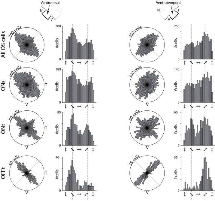
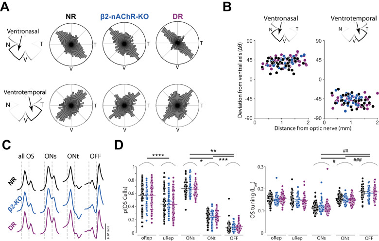
Orientation or axial selectivity, the property of neurons in the visual system to respond preferentially to certain angles of visual stimuli, plays a pivotal role in our understanding of visual perception and information processing. This computation is performed as early as the retina, and although much work has established the cellular mechanisms of retinal orientation selectivity, how this computation is organized across the retina is unknown. Using a large dataset collected across the mouse retina, we demonstrate functional organization rules of retinal orientation selectivity. First, we identify three major functional classes of retinal cells that are orientation selective and match previous descriptions. Second, we show that one orientation is predominantly represented in the retina and that this predominant orientation changes as a function of retinal location. Third, we demonstrate that neural activity plays little role on the organization of retinal orientation selectivity. Lastly, we use in silico modeling followed by validation experiments to demonstrate that the overrepresented orientation aligns along concentric axes. These results demonstrate that, similar to direction selectivity, orientation selectivity is organized in a functional map as early as the retina.
© 2024. The Author(s).
Conflict of interest statement
The authors declare no competing interests.
Figures


Update of
-
Development and Organization of the Retinal Orientation Selectivity Map.bioRxiv [Preprint]. 2024 Mar 28:2024.03.27.585774. doi: 10.1101/2024.03.27.585774. bioRxiv. 2024. Update in: Nat Commun. 2024 Jun 6;15(1):4829. doi: 10.1038/s41467-024-49206-z. PMID: 38585937 Free PMC article. Updated. Preprint.
References
MeSH terms
Grants and funding
LinkOut - more resources
Full Text Sources
