. 2024 Jun 6;15(1):4809.
doi: 10.1038/s41467-024-48462-3.

Tau propagation in the brain olfactory circuits is associated with smell perception changes in aging

Affiliations

Tau propagation in the brain olfactory circuits is associated with smell perception changes in aging

Ibai Diez et al. Nat Commun. .

Abstract

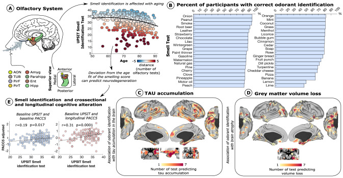
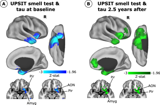
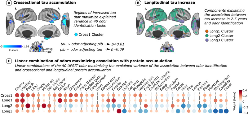
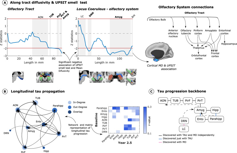
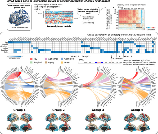
The direct access of olfactory afferents to memory-related cortical systems has inspired theories about the role of the olfactory pathways in the development of cortical neurodegeneration in Alzheimer's disease (AD). In this study, we used baseline olfactory identification measures with longitudinal flortaucipir and PiB PET, diffusion MRI of 89 cognitively normal older adults (73.82 ± 8.44 years; 56% females), and a transcriptomic data atlas to investigate the spatiotemporal spreading and genetic vulnerabilities of AD-related pathology aggregates in the olfactory system. We find that odor identification deficits are predominantly associated with tau accumulation in key areas of the olfactory pathway, with a particularly strong predictive power for longitudinal tau progression. We observe that tau spreads from the medial temporal lobe structures toward the olfactory system, not the reverse. Moreover, we observed a genetic background of odor perception-related genes that might confer vulnerability to tau accumulation along the olfactory system.


Conflict of interest statement

M.W.A. is a co-founder and owns shares in Aromha, Inc. The other authors declared no potential conflicts of interest with respect to the research, authorship and/or publication of this article.

Figures

Fig. 1
Fig. 1. Olfactory system dysfunction in healthy aging and its association with neurodegeneration.
A Primary olfactory regions of the brain and smell identification ability in healthy aging are displayed. The colors in the scatterplot represent the deviation of each participant’s estimated smell identification test based on their age (N = 418). B Sorted bar plot of the percentage of participants correctly identifying the 40 odorants composing the UPSIT test. C, D Amount of odorants able to distinguish high accumulation of tau and atrophy at voxel-level (N = 155). E Association between baseline smell identification and cognitive performance at baseline and after ~ 2.5 years using a linear regression model adjusting for age, gender, APOEε4 status, smoking history, and years of education (N = 155). AON anterior olfactory nucleus, TUR olfactory tubercle, PirF frontal piriform cortex, PirT temporal piriform cortex, Ent entorhinal, Amyg amygdala, and Hipp hippocampus. Source data are provided as a Source Data file.
Fig. 2
Fig. 2. Smell identification test association with tau accumulation and spreading (N = 89).
A Z-stat of the association between UPSIT smell identification test with baseline tau deposition. B Z-stat of the association between baseline UPSIT smell identification test and tau accumulation ~2.5 years after. A voxel-wise general linear model was used to compute these associations. Only results surviving cluster-wise multiple comparisons are displayed with a p-value < 0.05. AON anterior olfactory nucleus, Pir piriform cortex, Amyg amygdala.
Fig. 3
Fig. 3. Olfactory biomarkers of cross-sectional and longitudinal tau accumulation.
We performed a dimensionality reduction approach (principal component analysis; PCA) on a bipartite network of the association between odor item identification and tau and amyloid accumulation. A We obtained a linear combination of odorants that maximized cross-sectional tau accumulation prediction. Olfactory and medial temporal regions are strongly associated with odor identification information (Cross 1 Cluster; N = 155). B The same approach was repeated with the longitudinal tau data ( ~ 2.5 years), and three different clusters with different odor combinations were found representing tau accumulation at different brain regions - disease stages (Long1, Long2, Long3 clusters; N = 89). A voxel-wise general linear model was used to compute the association of tau accumulation with the linear combination of odorants A and B. Only regions surviving multiple comparisons with a p-value < 0.05 are shown. All the previous associations were adjusted for age, sex, APOEε4 status, and smoking history. Results survived when adjusting for amyloid. See Supplementary Fig. 3 for association with amyloid. Tau accumulation was associated with odor identification when controlling for amyloid but not vice versa. C Displays the linear combination of 40 odor items (olfactory biomarkers) predicting cross-sectional and longitudinal tau accumulation in the (A, B) clusters. Both the size of the circle and the color represent the coefficients of PCA, the contribution of each variable to the component (the importance of the odorant to predict tau accumulation). Source data are provided as a Source Data file.
Fig. 4
Fig. 4. Spreading of tau to olfactory regions.
A Along-tract statistics of the association of microstructural damage and UPSIT smell test are shown on both the olfactory tract and fibers connecting the locus coeruleus to the olfactory system (N = 82). A general linear model was used to compute the statistics across the tracts, adjusting for age, sex, APOEε4 status, and smoking history. Grey regions represent tract regions surrounding or passing anatomical structures of interest. Brain slices showing the evaluated tract and a diagram of known olfactory system connections are also displayed. B Bipartite graph between baseline and longitudinal tau accumulation showing a progression of tau from medial temporal regions towards the olfactory tract. The mean diffusivity of the dorsal raphe nucleus was also included. The node size represents the weighted degree and amount of out and in tau propagation. C Tau progression backbone between olfactory and medial temporal regions computed with conditional independent testing. Arrows were drawn based on directionality results obtained from a bipartite graph. AON anterior olfactory nucleus, TUR olfactory tubercle, PirF frontal piriform cortex, PirT temporal piriform cortex, Ent entorhinal, Amyg amygdala, Hipp hippocampus, Parahipp parahippocampus, LC Locus Coeruleus, DRN Dorsal Raphe Nucleus. Source data are provided as a Source Data file.
Fig. 5
Fig. 5. Co-expression of genes related to olfactory perception and associated neurodegenerative traits.
Brain transcriptome data from AHBA was projected to the Desikan brain atlas and the spatial brain co-expression of 390 genes annotated as sensory perception of smell were computed. Four groups of genes with similar co-expression were identified. Using the GWAS Catalog, phenotypes associated with olfactory genes in Genecard Suite were used to calculate bipartite networks associating its genes with traits related to neurodegeneration (see Supplementary Fig. 6). For each group of co-expressed olfactory genes, a circular diagram is shown with the associations and their mean brain expression. These associations were classified into five domains: tau, amyloid, Alzheimer’s disease, aging, cognition, and brain. For visualization purposes, the brain maps of the mean expression of each cluster were z-scored separately for each hemisphere. A matrix representing the association of each gene with each domain is also displayed to identify the association of each gene with the different domains. Source data are provided as a Source Data file.

References

    1. Price. “Olfactory System” in The Human Nervous System. (Academic Press, 979-998, 1990).
    1. Sherman & Guillery. Exploring the Thalamus and Its Role in Cortical Function, Second Edition. (MIT Press, 2006).
    1. Shepherd & Greer. “Olfactory Bulb” in The Synaptic Organization of the Brain, 4th Edn, Ed. G. M. Shepherd. (New York: Oxford University Press, 1998).
    1. Lane G, Zhou G, Noto T, Zelano C. Assessment of direct knowledge of the human olfactory system. Exp. Neurol. 2020;329:113304. doi: 10.1016/j.expneurol.2020.113304. - DOI - PMC - PubMed
    1. Doty RL. The olfactory vector hypothesis of neurodegenerative disease: Is it viable? Ann. Neurol. 2008;63:7–15. doi: 10.1002/ana.21327. - DOI - PubMed

MeSH terms