. 2024 Jun 6;14(1):12995.
doi: 10.1038/s41598-024-63562-2.

Neuroinflammation is dependent on sex and ovarian hormone presence following acute woodsmoke exposure

Affiliations

Neuroinflammation is dependent on sex and ovarian hormone presence following acute woodsmoke exposure

Kartika Wardhani et al. Sci Rep. .

Abstract

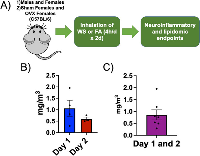
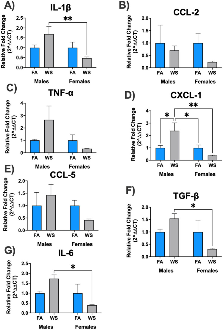
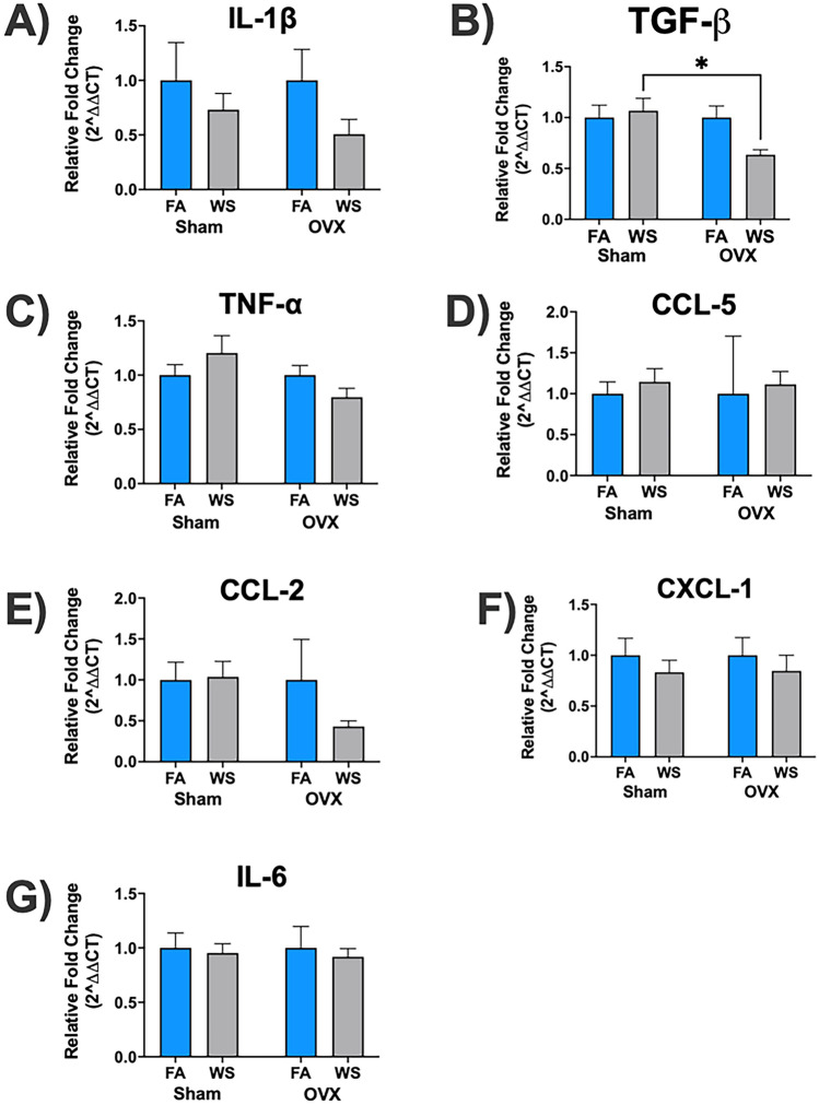
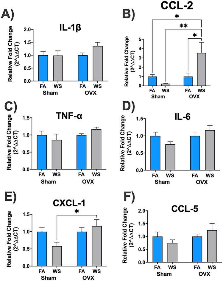
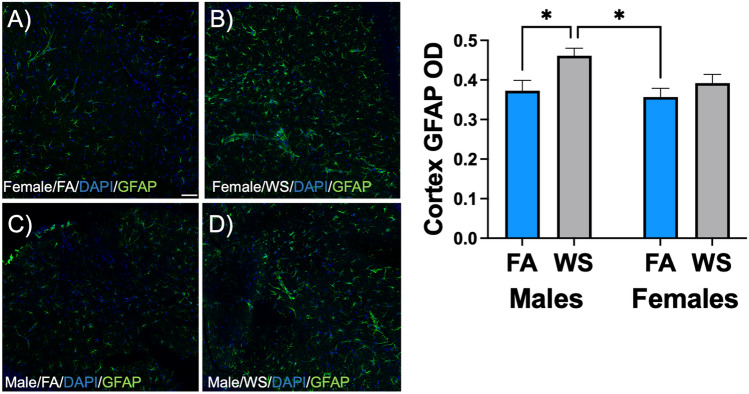
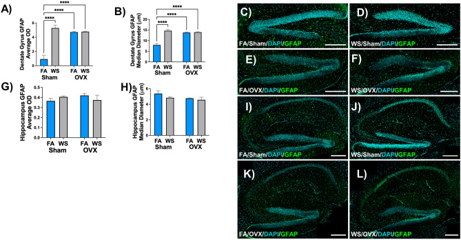
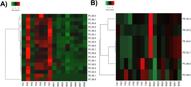
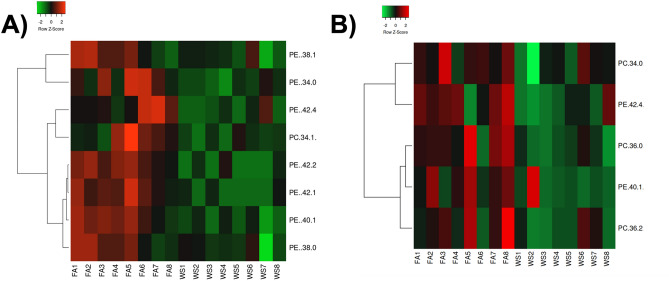
Woodsmoke (WS) exposure is associated with significant health-related sequelae. Different populations can potentially exhibit varying susceptibility, based on endocrine phenotypes, to WS and investigating neurological impacts following inhaled WS is a growing area of research. In this study, a whole-body inhalation chamber was used to expose both male and female C57BL/6 mice (n = 8 per group) to either control filtered air (FA) or acute WS (0.861 ± 0.210 mg/m3) for 4 h/d for 2 days. Neuroinflammatory and lipid-based biological markers were then assessed. In a second set of studies, female mice were divided into two groups: one group was ovariectomized (OVX) to simulate an ovarian hormone-deficient state (surgical menopause), and the other underwent Sham surgery as controls, to mechanistically assess the impact of ovarian hormone presence on neuroinflammation following FA and acute WS exposure to simulate an acute wildfire episode. There was a statistically significant impact of sex (P ≤ 0.05) and statistically significant interactions between sex and treatment in IL-1β, CXCL-1, TGF-β, and IL-6 brain relative gene expression. Hippocampal and cortex genes also exhibited significant changes in acute WS-exposed Sham and OVX mice, particularly in TGF-β (hippocampus) and CCL-2 and CXCL-1 (cortex). Cortex GFAP optical density (OD) showed a notable elevation in male mice exposed to acute WS, compared to the control FA. Sham and OVX females demonstrated differential GFAP expression, depending on brain region. Overall, targeted lipidomics in phosphatidylcholine (PC) and phosphatidylethanolamine (PE) serum and brain lipids demonstrated more significant changes between control FA and acute WS exposure in female mice, compared to males. In summary, male and female mice show distinct neuroinflammatory markers in response to acute WS exposure. Furthermore, ovarian hormone deficiency may impact the neuroinflammatory response following an acute WS event.

Keywords: Inflammation; Inhalation; Menopause; Sex-differences; Toxicology; Wildfire.


Conflict of interest statement

The authors declare no competing interests.

Figures

Figure 1
Figure 1
Graphical abstract. (A) Male and female C57BL/6 mice were exposed to either acute woodsmoke (WS) or control HEPA-filtered air (FA) for a 2-day exposure protocol with 4-h periods, daily (n = 8 per group). In a second set of experiments, Sham-operated and OVX female mice (6–8 weeks old) were then exposed to the same experimental conditions. (B) Typical average WS conditions (concentration ± standard error) for Day 1 (1.059 ± 0.349 mg/m3) and Day 2 (0.597 ± 0.080 mg/m3) during the acute WS exposures. (C) Combined aggregated concentrations for Day 1 and Day 2 overall (0.861 ± 0.210 mg/m3).
Figure 2
Figure 2
Sex-dependent brain mRNA gene expression levels in response to acute WS exposure (A-G). Brain mRNA relative gene expression levels demonstrated a response influenced by sex when exposed to acute WS. The data were analyzed using a two-way ANOVA, and the P-values for each variable (sex, treatment, and interactions between sex and treatment) on brain mRNA genes, where P < 0.05 signified statistical significance, were presented in the Supplemental Table S1(a). Asterisks denoted statistically significant mean differences between groups, as identified by Tukey’s post-hoc test (*P < 0.05; **P < 0.01). Data were presented as mean normalized values ± SEM. Each treatment group consisted of n = 8 mice.
Figure 3
Figure 3
Ovariectomy (OVX)-dependent effects on mRNA gene expression levels in the hippocampus following acute WS exposure (A-G). Analysis was conducted using a two-way ANOVA from control FA and acute WS exposures in Sham-operated and OVX female mice. The P-values for each variable (surgery, treatment, and interactions between surgery and treatment) in relation to hippocampal mRNA gene expression levels, where P < 0.05 signified statistical significance, were presented in the Supplemental Table S1(b). Asterisks denoted statistically significant mean differences between groups, as identified by Tukey’s post-hoc test (*P < 0.05). Data were expressed as mean normalized values ± SEM. Each treatment group had n = 8 mice.
Figure 4
Figure 4
Ovarian hormone-dependent responses of cortical mRNA gene expression to acute WS exposure (A-F). Analysis was performed using a two-way ANOVA comparing control FA and acute WS exposures in Sham-operated and OVX female mice. The P-values for each variable (surgery, treatment, and interactions between surgery and treatment) concerning mRNA gene expression in the cortex, where P < 0.05 indicated statistical significance, were presented in the Supplemental Table S1(c). Asterisks denoted statistically significant mean differences between groups, as identified by Tukey’s post-hoc test (*P < 0.05; **P < 0.01). Data were reported as mean normalized values ± SEM. Each treatment group had n = 8 mice.
Figure 5
Figure 5
Sex-specific modulation of glial fibrillary acidic protein (GFAP) staining in the cortex of (A, B) female and (C, D) male C57BL/6 mice following acute WS exposure. The morphology of astrocytes and cell nuclei in the cortical brain regions were visualized by GFAP (green) and DAPI (blue), respectively, and were captured at excitation peaks of 490 nm and 359 nm, and emission peaks at 535 nm and 457 nm, with a scale bar of 50 µm (bottom right corner of 5A). Normalized GFAP optical density (OD) in the cortex was analyzed using a two-way ANOVA. The P-values for each variable (sex, treatment, and interactions between sex and treatment) concerning the cortical brain regions and GFAP marker, where P < 0.05 signified statistical significance, were presented in the Supplemental Table S1(d). Asterisks denoted statistically significant mean differences between groups, as identified by Tukey’s post-hoc test (*P < 0.05). Data were presented as mean normalized values ± SEM. Each treatment group consisted of n = 8 mice.
Figure 6
Figure 6
Ovarian hormone-dependent alterations in glial fibrillary acidic protein (GFAP) staining within the dentate gyrus and hippocampus of female C57BL/6 mice following acute WS exposure. (A, B) Dentate gyrus GFAP quantification with associated representative images (C-F), and (G, H) hippocampus GFAP quantification with corresponding representative images (I–L) in Sham-operated and OVX female mice exposed to control FA and acute WS. Normalized GFAP optical density (OD) in the dentate gyrus and hippocampus were analyzed using a two-way ANOVA. The P-values for each variable (surgery, treatment, and interactions between surgery and treatment) regarding the dentate gyrus and hippocampal brain regions and GFAP marker, where P < 0.05 signified statistical significance, were presented in the Supplemental Table S1(e). Asterisks denoted statistically significant mean differences between groups, as identified by Tukey’s post-hoc test (****P < 0.0001). Data were presented as mean normalized values ± SEM. The morphology of astrocytes and cell nuclei in the dentate gyrus and hippocampal brain regions were visualized by GFAP (green) and DAPI (blue), respectively, and were captured at excitation peaks of 490 nm and 359 nm, and emission peaks at 535 nm and 457 nm, with a scale bar of 250 µm. Each treatment group consisted of n = 8 mice.
Figure 7
Figure 7
Relative expression of phosphatidylethanolamine (PE) and phosphatidylcholine (PC) lipids in plasma from female and male C57BL/6 mice. (A) heatmap of significant plasma lipid changes in female mice exposed to control FA (n = 7) and acute WS (n = 8), (B) significant plasma lipid changes in male mice. Statistics were calculated with a Mann–Whitney test and *P ≤ 0.05 was considered statistically significant.
Figure 8
Figure 8
Relative expression of phosphatidylcholine (PC) and phosphatidylethanolamine (PE) in acute WS- and control FA-exposed C57BL/6 mouse brains. Compared to control FA-exposed mice, acute WS exposure significantly downregulated (A) PC and PE brain lipids in female mice, (B) PC and PE brain lipids in males. Statistics were calculated with a Mann–Whitney test and P ≤ 0.05 was considered statistically significant. Heatmaps display only significantly differentially expressed lipids. Each treatment group consisted of n = 8 mice.

Similar articles

Cited by

References

    1. Elser H, Rowland ST, Marek MS, Kiang MV, Shea B, Do V, Benmarhnia T, Schneider AL, Casey JA. Wildfire smoke exposure and emergency department visits for headache: A case-crossover analysis in California, 2006–2020. Headache. 2023;63:94–103. doi: 10.1111/head.14442. - DOI - PMC - PubMed
    1. Casey JA, Kioumourtzoglou M-A, Elser H, Walker D, Taylor S, Adams S, Aguilera R, Benmarhnia T, Catalano R. Wildfire particulate matter in Shasta County, California and respiratory and circulatory disease-related emergency department visits and mortality, 2013–2018. Environ. Epidemiol. 2021;5:e124–e127. doi: 10.1097/EE9.0000000000000124. - DOI - PMC - PubMed
    1. Rooney MS, Arku RE, Dionisio KL, Paciorek C, Friedman AB, Carmichael H, Zhou Z, Hughes AF, Vallarino J, Agyei-Mensah S. Spatial and temporal patterns of particulate matter sources and pollution in four communities in Accra, Ghana. Sci. Total Environ. 2012;435:107–114. doi: 10.1016/j.scitotenv.2012.06.077. - DOI - PubMed
    1. Bede-Ojimadu O, Orisakwe OE. Exposure to wood smoke and associated health effects in Sub-Saharan Africa: A systematic review. Ann. Global Health. 2020;86:32–58. doi: 10.5334/aogh.2725. - DOI - PMC - PubMed
    1. O’Donnell M, Behie A. Effects of wildfire disaster exposure on male birth weight in an Australian population. Evol. Med. Public Health. 2015;2015:344–354. doi: 10.1093/emph/eov027. - DOI - PMC - PubMed