

DNA damage and repair in patients undergoing myocardial perfusion single-photon emission computed tomography
- PMID: 38844507
- PMCID: PMC11156974
- DOI: 10.1038/s41598-024-63537-3
DNA damage and repair in patients undergoing myocardial perfusion single-photon emission computed tomography
Abstract
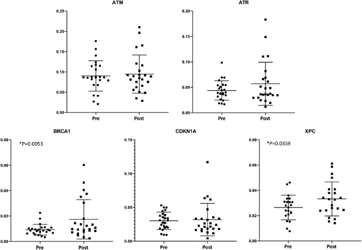
As patient exposure to ionizing radiation from medical imaging and its risks are continuing issues, this study aimed to evaluate DNA damage and repair markers after myocardial perfusion single-photon emission computed tomography (MPS). Thirty-two patients undergoing Tc-99m sestamibi MPS were studied. Peripheral blood was collected before radiotracer injection at rest and 60-90 min after injection. The comet assay (single-cell gel electrophoresis) was performed with peripheral blood cells to detect DNA strand breaks. Three descriptors were evaluated: the percentage of DNA in the comet tail, tail length, and tail moment (the product of DNA tail percentage and tail length). Quantitative PCR (qPCR) was performed to evaluate the expression of five genes related to signaling pathways in response to DNA damage and repair (ATM, ATR, BRCA1, CDKN1A, and XPC). Mann-Whitney's test was employed for statistical analysis; p < 0.05 was considered significant. Mean Tc-99m sestamibi dose was 15.1 mCi. After radiotracer injection, comparing post-exposure to pre-exposure samples of each of the 32 patients, no statistically significant differences of the DNA percentage in the tail, tail length or tail moment were found. qPCR revealed increased expression of BRCA1 and XPC, without any significant difference regarding the other genes. No significant increase in DNA strand breaks was detected after a single radiotracer injection for MPS. There was activation of only two repair genes, which may indicate that, in the current patient sample, the effects of ionizing radiation on the DNA were not large enough to trigger intense repair responses, suggesting the absence of significant DNA damage.
Keywords: Comet assay; DNA; Myocardial perfusion imaging; Radiation.
© 2024. The Author(s).
Conflict of interest statement
The authors declare no competing interests.
Figures

Similar articles
-
Evaluation of DNA damage induced by ionizing radiation from myocardial perfusion imaging: a pilot study.BMC Cardiovasc Disord. 2022 Sep 3;22(1):394. doi: 10.1186/s12872-022-02839-8. BMC Cardiovasc Disord. 2022. PMID: 36057570 Free PMC article.
-
Can carbonated lime drink intake prior to myocardial perfusion imaging with Tc-99m MIBI reduce the extracardiac activity that degrades the image quality and leads to fallacies in interpretation?Clin Nucl Med. 2010 Mar;35(3):160-4. doi: 10.1097/RLU.0b013e3181cc63a1. Clin Nucl Med. 2010. PMID: 20173445 Clinical Trial.
-
Eleven-year prognostic value of dobutamine stress (99m)Tc-sestamibi myocardial perfusion imaging in patients with limited exercise capacity.Am J Cardiol. 2015 Apr 1;115(7):884-9. doi: 10.1016/j.amjcard.2015.01.011. Epub 2015 Jan 14. Am J Cardiol. 2015. PMID: 25661571
-
Effects of adenosine and a selective A2A adenosine receptor agonist on hemodynamic and thallium-201 and technetium-99m-sestaMIBI biodistribution and kinetics.JACC Cardiovasc Imaging. 2009 Oct;2(10):1198-208. doi: 10.1016/j.jcmg.2009.06.013. JACC Cardiovasc Imaging. 2009. PMID: 19833310
-
Slightly increased level of DNA migration in the comet assay: does statistical significance equal biological significance?Mutagenesis. 2025 Apr 24;40(2):99-110. doi: 10.1093/mutage/geaf004. Mutagenesis. 2025. PMID: 39963750 Free PMC article. Review.
Cited by
-
C1QBP Modulates DNA Damage Response and Radiosensitivity in Hepatocellular Carcinoma by Regulating NF-κB Activity.Int J Mol Sci. 2025 May 9;26(10):4513. doi: 10.3390/ijms26104513. Int J Mol Sci. 2025. PMID: 40429658 Free PMC article.
References
-
- National Council on Radiation Protection and Measurements. Report No. 184—Medical Radiation Exposure of Patients in The United States. Bethesda, MD (2019).
-
- Chen J, Einstein AJ, Fazel R, Krumholz HM, Wang Y, Ross JS, Ting HH, Shah ND, Nasir K, Nallamothu BK. Cumulative exposure to ionizing radiation from diagnostic and therapeutic cardiac imaging procedures: a population-based analysis. J. Am. Coll. Cardiol. 2010;56:702–711. doi: 10.1016/j.jacc.2010.05.014. - DOI - PMC - PubMed
MeSH terms
Substances
Grants and funding
LinkOut - more resources
Full Text Sources
Research Materials
Miscellaneous
