. 2024 Jun 7;8(1):68.
doi: 10.1186/s41747-024-00463-z.

Time-of-flight MRA of intracranial vessels at 7 T

Affiliations

Time-of-flight MRA of intracranial vessels at 7 T

Mirco Cosottini et al. Eur Radiol Exp. .

Abstract

Background: Three-dimensional time-of-flight magnetic resonance angiography (TOF-MRA) is a largely adopted non-invasive technique for assessing cerebrovascular diseases. We aimed to optimize the 7-T TOF-MRA acquisition protocol, confirm that it outperforms conventional 3-T TOF-MRA, and compare 7-T TOF-MRA with digital subtraction angiography (DSA) in patients with different vascular pathologies.

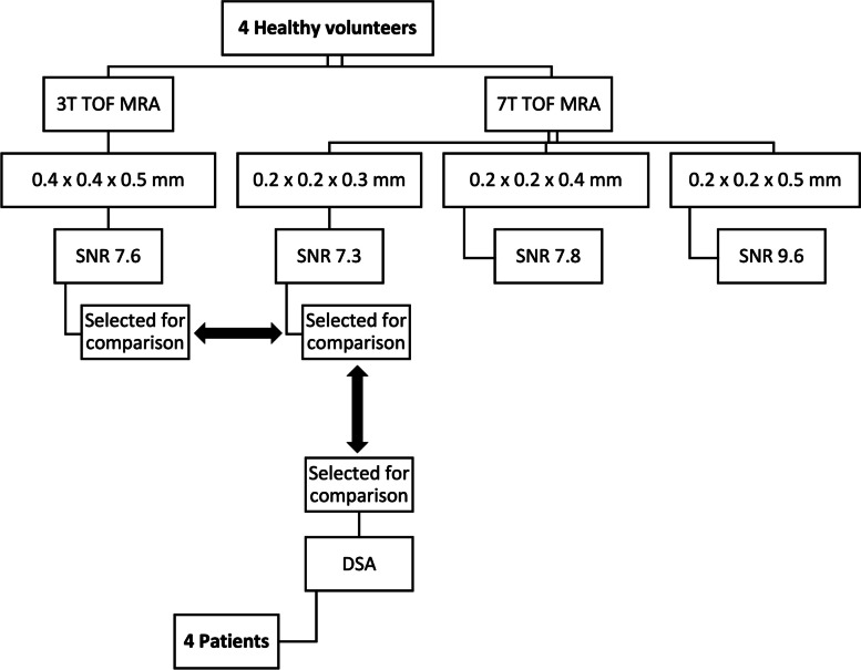
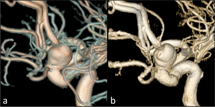
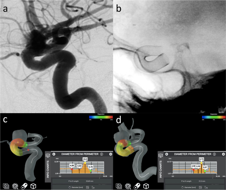
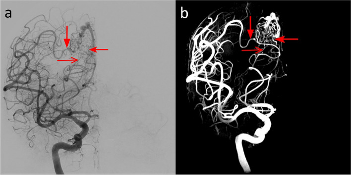
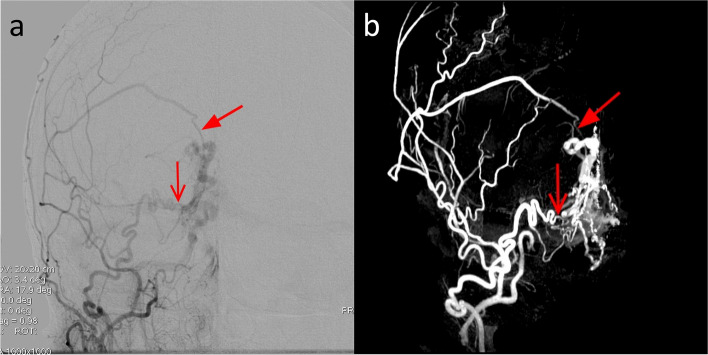
Methods: Seven-tesla TOF-MRA sequences with different spatial resolutions acquired in four healthy subjects were compared with 3-T TOF-MRA for signal-to-noise and contrast-to-noise ratios as well as using a qualitative scale for vessel visibility and the quantitative Canny algorithm. Four patients with cerebrovascular disease (primary arteritis of the central nervous system, saccular aneurism, arteriovenous malformation, and dural arteriovenous fistula) underwent optimized 7-T TOF-MRA and DSA as reference. Images were compared visually and using the complex-wavelet structural similarity index.

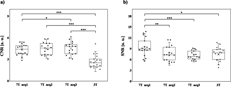
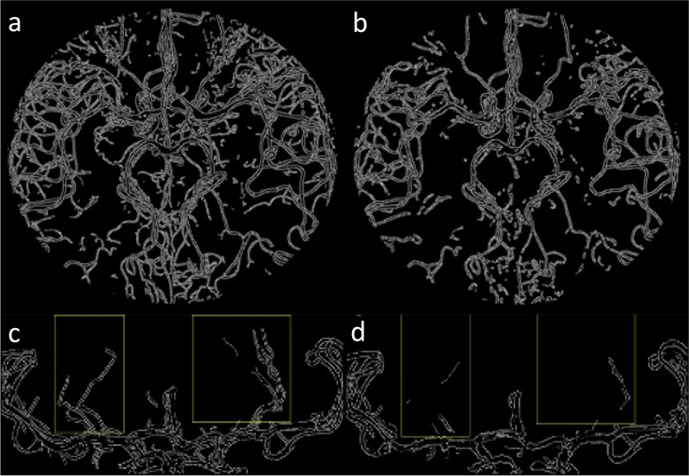
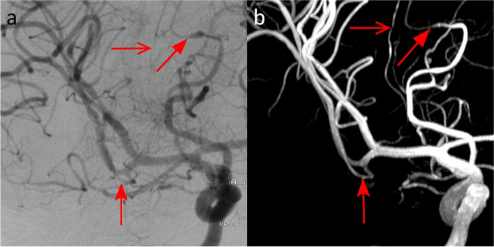
Results: Contrast-to-noise ratio was higher at 7 T (4.5 ± 0.8 (mean ± standard deviation)) than at 3 T (2.7 ± 0.9). The mean quality score for all intracranial vessels was higher at 7 T (2.89) than at 3 T (2.28). Angiogram quality demonstrated a better vessel border detection at 7 T than at 3 T (44,166 versus 28,720 pixels). Of 32 parameters used for diagnosing cerebrovascular diseases on DSA, 27 (84%) were detected on 7-T TOF-MRA; the similarity index ranged from 0.52 (dural arteriovenous fistula) to 0.90 (saccular aneurysm).

Conclusions: Seven-tesla TOF-MRA outperformed conventional 3-T TOF-MRA in evaluating intracranial vessels and exhibited an excellent image quality when compared to DSA. Seven-tesla TOF-MRA might improve the non-invasive diagnostic approach to several cerebrovascular diseases.

Relevance statement: An optimized TOF-MRA sequence at 7 T outperforms 3-T TOF-MRA, opening perspectives to its clinical use for noninvasive diagnosis of paradigmatic pathologies of intracranial vessels.

Key points: • An optimized 7-T TOF-MRA protocol was selected for comparison with clinical 3-T TOF-MRA for assessing intracranial vessels. • Seven-tesla TOF-MRA outperformed 3-T TOF-MRA in both quantitative and qualitative evaluation. • Seven-tesla TOF-MRA is comparable to DSA for the diagnosis and characterization of intracranial vascular pathologies.

Keywords: Angiography (digital subtraction); Central nervous system vascular malformations; Magnetic fields; Magnetic resonance angiography; Primary angiitis of the central nervous system.


Conflict of interest statement

The authors declare that they have no competing interests.

Figures

Fig. 1
Fig. 1
Flowchart of the study. SNR Signal-to-noise ratio, TOF-MRA Time-of-flight magnetic resonance angiography
Fig. 2
Fig. 2
CNR (a) and SNR (b) calculated for the three 7-T TOF-MRA sequences at different spatial resolutions (black color) and for the clinical 3-T TOF-MRA images (dark gray color). Each dot represents the mean among subjects of CNR (a) or SNR (b) for a specific ROI. Data distribution is shown through box plots, where the box is determined by the 25th and 75th percentiles and the median and the mean are represented by the horizontal and the cross line inside the box, respectively. The whiskers represent the 5th and 95th percentiles. To compare results for CNR (a) or SNR (b) obtained by different acquisitions, nonparametric paired tests (Wilcoxon signed-rank test) were performed between each couple of datasets. Statistically significant differences of CNR and SNR between different acquisition schemes are reported in the graph according to the following legend: one asterisk (*) indicates p < 0.050, two asterisks (**) p < 0.010, and three asterisks (***) p < 0.001. CNR Contrast-to-noise ratio, SNR Signal-to-noise ratio, TOF-MRA Time-of-flight magnetic resonance angiography
Fig. 3
Fig. 3
Graphical representation of the pixels pertaining to the vessels’ borders obtained with the Canny algorithm on the axial maximum intensity projection images at 7 T (a) and 3 T (b), and on the lenticulostriate arteries (within the boxes) imaged at 7 T (c) and 3 T (d)
Fig. 4
Fig. 4
Digital subtraction angiography (a) and 7-T TOF-MRA (b) images in the patient with primary arteritis of the central nervous system. The selective contrast injection of the right internal carotid artery revealed multiple segments of vessels narrowing on both anterior and middle cerebral arteries: most of these stenoses (arrows) were confirmed in the multiplanar volume reconstruction of 7-T TOF-MRA (b). TOF-MRA Time-of-flight magnetic resonance angiography
Fig. 5
Fig. 5
DSA (a) and 7-T TOF-MRA (b) in the patient with carotid artery aneurysm. Three-dimensional reconstruction of a rotational DSA with images acquired during the selective injection of the left internal carotid artery (a) showed the presence of a saccular aneurysm of the ophthalmic segment of the internal carotid artery. The volume rendering of the 7-T TOF-MRA allowed an optimal depiction of the aneurysmal geometric characteristics (b). DSA Digital subtraction angiography, TOF-MRA Time-of-flight magnetic resonance angiography
Fig. 6
Fig. 6
Intraprocedural DSA angiograms of the stent placement (a, b) and the stent placement simulations obtained with an automatic software (Ankyras, Galgo Medical, Barcelona, Spain) from the DSA (c) and 7-T TOF-MRA dataset (d). The red, brown, yellow, and green spherical markers indicate the spatial location of the parameter value shown in the side graph. DSA Digital subtraction angiography, TOF-MRA Time-of-flight magnetic resonance angiography
Fig. 7
Fig. 7
DSA (a) and 7-T TOF-MRA (b) images of the patient with plessiform AVM of the occipital lobe. The AVM had a superficial nidus fed by arterial afferents arising from carotid and vertebral circulations. The DSA angiogram obtained by the selective injection of the right internal carotid artery (a) showed multiple feeders from the temporo-occipital artery arising from the middle cerebral arteries (filled arrow). In this patient, being the posterior cerebral artery supplied by the carotid circulation via the posterior communicating artery, some afferents from the parieto-occipital artery and calcarine artery are also opacified (open arrow and dented arrows)
Fig. 8
Fig. 8
DSA (a) and 7-T TOF-MRA (b) in the patient with DAVF type III of Cognard. The selective injection of the right external carotid arteries (a) depicted meningeal feeders arising from the right middle meningeal artery (light blue arrows) and from the right occipital artery (red arrow)

References

    1. Benjamin EJ, Muntner P, Alonso A, et al. Heart disease and stroke statistics-2019 update: a report from the American Heart Association. Circulation. 2019;139:e56–e528. doi: 10.1161/CIR.0000000000000659. - DOI - PubMed
    1. Greenberg SM, Ziai WC, Cordonnier C, et al. 2022 guideline for the management of patients with spontaneous intracerebral hemorrhage: a guideline from the American Heart Association/American Stroke Association. Stroke. 2022;53:e282–e361. doi: 10.1161/STR.0000000000000407. - DOI - PubMed
    1. Hoh BL, Ko NU, Amin-Hanjani S, et al. 2023 guideline for the management of patients with aneurysmal subarachnoid hemorrhage: a guideline from the American Heart Association/American Stroke Association. Stroke. 2023;54:e314–e370. doi: 10.1161/STR.0000000000000436. - DOI - PubMed
    1. Powers WJ, Rabinstein AA, Ackerson T, et al. 2018 guidelines for the early management of patients with acute ischemic stroke: a guideline for healthcare professionals from the American Heart Association/American Stroke Association. Stroke. 2018;49:e46–e110. doi: 10.1161/STR.0000000000000158. - DOI - PubMed
    1. Feldmann E, Wilterdink JL, Kosinski A, et al. The Stroke Outcomes and Neuroimaging of Intracranial Atherosclerosis (SONIA) trial. Neurology. 2007;68:2099–2106. doi: 10.1212/01.wnl.0000261488.05906.c1. - DOI - PubMed

LinkOut - more resources