

MedYOLO: A Medical Image Object Detection Framework
- PMID: 38844717
- PMCID: PMC11612059
- DOI: 10.1007/s10278-024-01138-2
MedYOLO: A Medical Image Object Detection Framework
Abstract
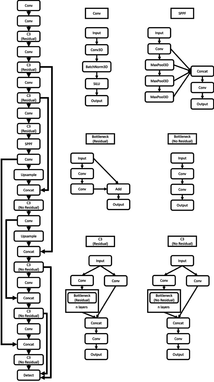
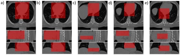
Artificial intelligence-enhanced identification of organs, lesions, and other structures in medical imaging is typically done using convolutional neural networks (CNNs) designed to make voxel-accurate segmentations of the region of interest. However, the labels required to train these CNNs are time-consuming to generate and require attention from subject matter experts to ensure quality. For tasks where voxel-level precision is not required, object detection models offer a viable alternative that can reduce annotation effort. Despite this potential application, there are few options for general-purpose object detection frameworks available for 3-D medical imaging. We report on MedYOLO, a 3-D object detection framework using the one-shot detection method of the YOLO family of models and designed for use with medical imaging. We tested this model on four different datasets: BRaTS, LIDC, an abdominal organ Computed tomography (CT) dataset, and an ECG-gated heart CT dataset. We found our models achieve high performance on a diverse range of structures even without hyperparameter tuning, reaching mean average precision (mAP) at intersection over union (IoU) 0.5 of 0.861 on BRaTS, 0.715 on the abdominal CT dataset, and 0.995 on the heart CT dataset. However, the models struggle with some structures, failing to converge on LIDC resulting in a mAP@0.5 of 0.0.
Keywords: Computed tomography; Convolutional neural network; Deep learning; Magnetic resonance; Medical imaging; Object detection.
© 2024. The Author(s) under exclusive licence to Society for Imaging Informatics in Medicine.
Conflict of interest statement
Declarations. Ethics Approval: This retrospective study was reviewed and deemed exempt by Mayo Clinic’s Institutional Review Board. Consent to Participate: The informed consent was waived by ethics groups. Consent for Publication: The informed consent was waived by ethics groups. Competing Interests: The authors declare no competing interests.
Figures


References
-
- Baumgartner M., Jäger P.F., Isensee F., Maier-Hein K.H.: nnDetection: A Self-configuring Method for Medical Object Detection. Medical Image Computing and Computer Assisted Intervention – MICCAI 2021. 10.1007/978-3-030-87240-3_51, 2021
-
- Redmon J, Farhadi A: YOLO9000: Better, Faster, Stronger. 2017 IEEE Conference on Computer Vision and Pattern Recognition. 10.48550/arXiv.1612.08242, 2016
-
- Jocher G, et al.: ultralytics/yolov5: v7.0 - YOLOv5 SOTA Realtime Instance Segmentation. 10.5281/zenodo.3908559, 2022
-
- U. Baid, et al.: The RSNA-ASNR-MICCAI BraTS 2021 Benchmark on Brain Tumor Segmentation and Radiogenomic Classification. 10.48550/arXiv.2107.02314, 2021
MeSH terms
LinkOut - more resources
Full Text Sources
Medical