. 2024 Jun 6;7(1):693.
doi: 10.1038/s42003-024-06375-3.

C-type natriuretic peptide/cGMP/FoxO3 signaling attenuates hyperproliferation of pericytes from patients with pulmonary arterial hypertension

Affiliations

C-type natriuretic peptide/cGMP/FoxO3 signaling attenuates hyperproliferation of pericytes from patients with pulmonary arterial hypertension

Swati Dabral et al. Commun Biol. .

Abstract

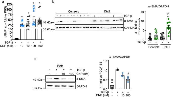
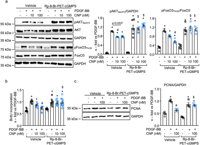
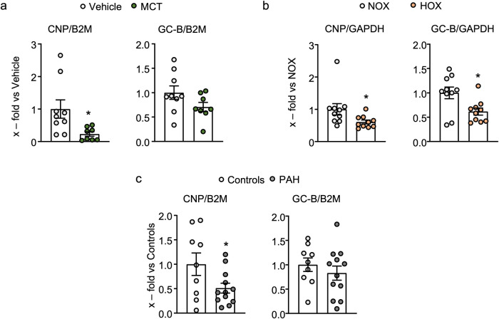
Pericyte dysfunction, with excessive migration, hyperproliferation, and differentiation into smooth muscle-like cells contributes to vascular remodeling in Pulmonary Arterial Hypertension (PAH). Augmented expression and action of growth factors trigger these pathological changes. Endogenous factors opposing such alterations are barely known. Here, we examine whether and how the endothelial hormone C-type natriuretic peptide (CNP), signaling through the cyclic guanosine monophosphate (cGMP) -producing guanylyl cyclase B (GC-B) receptor, attenuates the pericyte dysfunction observed in PAH. The results demonstrate that CNP/GC-B/cGMP signaling is preserved in lung pericytes from patients with PAH and prevents their growth factor-induced proliferation, migration, and transdifferentiation. The anti-proliferative effect of CNP is mediated by cGMP-dependent protein kinase I and inhibition of the Phosphoinositide 3-kinase (PI3K)/AKT pathway, ultimately leading to the nuclear stabilization and activation of the Forkhead Box O 3 (FoxO3) transcription factor. Augmentation of the CNP/GC-B/cGMP/FoxO3 signaling pathway might be a target for novel therapeutics in the field of PAH.


Conflict of interest statement

The authors declare no competing interests.

Figures

Fig. 1
Fig. 1. Unaltered CNP/GC-B/cGMP signaling in pericytes from patients with pulmonary arterial hypertension (PAH).
a Effects of CNP and ANP on cGMP levels of human lung microvascular endothelial cells (n = 3 wells from one biological sample), vascular smooth muscle cells (n = 6 wells from three biological replicates), and pericytes (n = 8 wells from four biological replicates) (1-way ANOVA). b and c Lung pericytes isolated from control individuals and patients with PAH exhibit similar cGMP responses to CNP and similar expression levels of the GC-B receptor and cGKI (b: n = 8 wells from four biological replicates (1-way ANOVA). c n = 12 from 6 controls and n = 10 from 5 PAH patients (unpaired 2-tailed Student’s t-test). d CNP similarly stimulates the phosphorylation of VASP at Ser239, the cGKI-specific site, in control and PAH pericytes (n = 4 biological replicates per group (2-way ANOVA)). *P < 0.05 vs. PBS (−).
Fig. 2
Fig. 2. CNP attenuates the stimulatory effects of PDGF-BB on pericyte proliferation and migration.
a and b In comparison to control pericytes, PAH pericytes had higher proliferation (a) and migration rates (b) at baseline and in response to PDGF-BB (30 ng/ml, 24 h) (a BrdU incorporation, n = 10 wells from three biological replicates for control and n = 16 wells from four biological replicates for PAH; non-parametric Kruskal–Wallis analyses. b Scratch assays with 8–14 wells from three biological replicates from each group; 2-way ANOVA). c PDGF-BB (30 ng/ml, 15 min pretreatment) did not significantly alter the cGMP responses of control pericytes to CNP (n = 12 wells from four biological replicates per condition; non-parametric Kruskal–Wallis analyses). d–f CNP (pretreatment for 30 min) attenuated PDGF-BB (30 ng/ml) induced proliferation (d), cyclin D1 (e) and PCNA protein expression (f) of control and PAH pericytes (d: n = 10–18 wells from three biological replicates per group; non-parametric Kruskal–Wallis analyses; e and f: n = 4 biological replicates from control and PAH pericytes; 1-way ANOVA). g CNP (100 nM, 24 h) attenuated PDGF-BB-induced migration of control and PAH pericytes. Top panels: representative pictographs of control (left) and PAH pericytes (right) at 0, 16, and 24 h of the scratch assay (scale bar: 500 mm); Bottom panels, evaluation of the wounding areas in comparison to the initial wound (n = 8–13 wells from three biological replicates per group; 2-way ANOVA). For a: *p < 0.05 vs. Controls, #p < 0.05 vs. Basal. For b: *p < 0.05 vs. PBS-Control, #p < 0.05 vs. PDGF-BB-Control. For c: *p < 0.05 vs. PBS (–). For d–g: *p < 0.05 vs. PBS (–), #p < 0.05 vs. PDGF-BB.
Fig. 3
Fig. 3. CNP attenuates the stimulatory effects of TGF-β on α-SMA expression.
a TGF-β (10 ng/ml, 15 min) did not significantly alter the cGMP responses of control pericytes to CNP (n = 12 from four biological replicates; non-parametric Kruskal–Wallis analyses). b Immunoblotting: TGF-β (10 ng/ml, 24 h) enhanced α-smooth muscle actin (α-SMA) expression more in PAH pericytes than in controls (n = 10 from five biological replicates in each group; non-parametric Kruskal–Wallis analyses). c Immunoblotting: Pretreatment with CNP (10 and 100 nM, 15 min) significantly attenuated TGF-β (10 ng/ml, 24 h)-induced α-SMA expression in PAH pericytes (n = 3 biological replicates; 1-way ANOVA). For a: *p < 0.05 vs. PBS (–). For b: *p < 0.05 vs. PBS (–), #p < 0.05 vs. TGF-β in controls. For c: *p < 0.05 vs. PBS (–), #p < 0.05 vs. TGF-β.
Fig. 4
Fig. 4. CNP attenuates PDGF-BB-induced pro-proliferative signaling pathways in human control and PAH lung pericytes.
a CNP (10 and 100 nM, 15 min) increased VASP (Ser239) phosphorylation in PDGF-BB-pretreated control ad PAH pericytes. b Pretreatment with CNP prevented the PDGF-BB (30 ng/ml, 30 min)-induced phosphorylation of AKT (Ser473) and ERK1/2 (Thr202/Tyr204) in control and PAH pericytes (n = 4 from control and PAH pericytes; 1-way ANOVA). *p < 0.05 vs. PBS (–), #p < 0.05 vs. PDGF-BB.
Fig. 5
Fig. 5. FoxO3 mediates the counter-regulation of PDGF-BB-induced pericyte proliferation by CNP.
a CNP (10 and 100 nM, 15 min) prevented the PDGF-BB (30 ng/ml, 30 min)-induced phosphorylation of FoxO3 (Thr32) in control and PAH pericytes (n = 4 control pericyte samples, n = 5 from four PAH pericytes; 1-way ANOVA). b CNP (100 nM) pretreatment prevented the PDGF-BB-induced FoxO3 nuclear exclusion as assessed by immunocytochemical staining of FoxO3, followed by nuclear fluorescence intensity measurement by Image J (n = 5 from three biological replicates; each value is the mean of three images; 1-way ANOVA). Scale bar: 100 mm. c and d Transfection of control pericytes with siFoxO3 reduced FoxO3 protein expression (c: n = 6 independent experiments from three biological replicates; 1-way ANOVA), and this prevented the inhibitory effects of CNP on PDGF-BB induced pericyte proliferation (d: n = 9 wells from three biological replicates; 2-way ANOVA). For a and b: *p < 0.05 vs. PBS (–), #p < 0.05 vs. PDGF-BB. For c: *P < 0.05 vs. untransfected control (–), #p < 0.05 vs. si-Control. For d: *p < 0.05 vs. PBS (–), #p < 0.05 vs. PDGF-BB, $p < 0.05 vs. corresponding vehicle-treated group (−).
Fig. 6
Fig. 6. Inhibition of cGKI prevents the effects of CNP on PDGF-BB-induced proliferative signaling.
a The cGKI inhibitor Rp-8-Br-PET-cGMPS (100 µM) prevented the effect of CNP on PDGF-BB (30 ng/ml, 30 min)-induced phosphorylation of AKT (Ser473) and FoxO3 (Thr32) (n = 5–6 from four biological replicates; 2-way ANOVA). b and c Rp-8-Br-PET-cGMPS (10 µM) attenuated the inhibitory effects of CNP on PDGF-BB-induced proliferation of control pericytes as analysed by b BrdU incorporation assay (n = 9–10 wells from three biological replicates; non-parametric Kruskal–Wallis analyses) and c immunoblotting for PCNA (n = 4 biological replicates; 2-way ANOVA). *p < 0.05 vs PBS (–), #p < 0.05 vs. PDGF-BB, $p < 0.05 vs. corresponding vehicle-treated group. Please note that in the samples derived from cells treated with Rp-8-Br-PET-cGMPS the immunoreactive signal obtained for total AKT was markedly increased (second lane in the original westerns depicted in (a)). Presently, we do not have an explanation for this reproducible observation. Due to the brief incubation time (<1 h), we believe that this enhanced immunoreactivity is not derived from increased AKT protein expression but related to changes in AKT conformation and enhanced binding of the anti-AKT antibody to its epitope. Due to these changes, the signal of pAKTSer473 was normalized to GAPDH and not to total AKT (middle panel in Fig. 6a).
Fig. 7
Fig. 7. CNP/cGKI signaling activates the phosphatase PTEN, thereby inhibiting PDGF-BB-mediated AKT phosphorylation. Fig. 7.
a Scheme of the postulated and investigated signaling pathway. PDGF-BB, via its PDGFR- β, triggers the activation of the PI3K, which phosphorylates PIP2 to PIP3. PIP3 then activates PDK1-AKT signaling. Subsequently, AKT phosphorylates and inactivates FoxO3, which enhances lung pericyte proliferation. CNP, via GC-B/cGMP signaling, activates cGKI. It has been shown that cGKI elicits inactivating phosphorylations of RhoA at Ser188 and of GSK3b at Ser9, thereby preventing their inhibitory phosphorylations of PTEN,. Activated PTEN dephosphorylates PIP3 and prevents AKT activation, resulting in an increase of nuclear FoxO3 and a concomitant reduction in pericyte proliferation. b PDGF-BB stimulated the phosphorylation of PTEN at Ser380/Thr382/383 and CNP prevented this effect in the absence (vehicle) but not in the presence of the cGKI inhibitor Rp-8-Br-PET-cGMPS (100 µM). c The cGKI activator, 8-Bromo-cGMP (0.01–10 µM), prevented PDGF-BB (30 ng/ml)-induced phosphorylation of PTEN in control pericytes (b and c: n = 4 biological replicates; b: 2-way ANOVA; c: 1-way ANOVA). *p < 0.05 vs. PBS (–), #p < 0.05 vs. PDGF-BB, $p < 0.05 vs. corresponding vehicle-treated group. PDGF-BB platelet-derived growth factor-BB, PDGFR-β platelet-derived growth factor beta, PI3K phosphoinositide 3-kinase, PIP2 phosphatidylinositol 4,5-bisphosphate, PIP3 phosphatidylinositol 3,4,5-trisphosphate, PDK1 phosphoinositide-dependent kinase 1, AKT protein kinase B, FoxO3 forkhead box O3, CNP C-type natriuretic peptide, GC-B guanylyl cyclase-B, cGMP cyclic guanosine monophosphate, cGKI cGMP-dependent kinase I, RhoA Ras homolog family member A, PTEN phosphatase and tensin homolog.
Fig. 8
Fig. 8. Pulmonary CNP mRNA expression is reduced in clinical PAH and experimental PH.
a and b Lung CNP and GC-B mRNA expression in a monocrotaline (MCT) vs. vehicle-treated rats, b normoxia (NOX) vs. Hypoxia (HOX: 21 days) exposed mice and c PAH patients vs. controls. Values are the ratios of CNP or GC-B mRNA level relative to GAPDH (b) or b2 microglobulin (a and c), determined by qRT-PCR and expressed as x-fold versus vehicle-treated rats (a), NOX mice (b), or human control lungs (c) (a: n = 9 samples from the vehicle and n = 8 from MCT-treated rats; b: n = 10 samples each from NOX vs. HOX exposed mice; a and b: Mann–Whitney test for CNP and unpaired 2-tailed Student’s t-test for GC-B; c: n = 9 samples from controls and n = 12 samples from PAH patients, unpaired 2-tailed Student’s t-test). *p < 0.05.

References

    1. Humbert M, et al. Pathology and pathobiology of pulmonary hypertension: state of the art and research perspectives. Eur. Respir. J. 2019;53:1801887. doi: 10.1183/13993003.01887-2018. - DOI - PMC - PubMed
    1. Humbert M, et al. Cellular and molecular pathobiology of pulmonary arterial hypertension. J. Am. Coll. Cardiol. 2004;43:13S–24S. doi: 10.1016/j.jacc.2004.02.029. - DOI - PubMed
    1. Yuan K, et al. Lung pericytes in pulmonary vascular physiology and pathophysiology. Compr. Physiol. 2021;11:2227–2247. doi: 10.1002/cphy.c200027. - DOI - PMC - PubMed
    1. Schermuly RT, Ghofrani HA, Wilkins MR, Grimminger F. Mechanisms of disease: pulmonary arterial hypertension. Nat. Rev. Cardiol. 2011;8:443–455. doi: 10.1038/nrcardio.2011.87. - DOI - PMC - PubMed
    1. He S, Zhu T, Fang Z. The role and regulation of pulmonary artery smooth muscle cells in pulmonary hypertension. Int. J. Hypertens. 2020;2020:1478291. doi: 10.1155/2020/1478291. - DOI - PMC - PubMed

Publication types

MeSH terms