. 2024 Jun 6;24(1):508.
doi: 10.1186/s12870-024-05205-2.

An improved method to study Phytophthora cinnamomi Rands zoospores interactions with host

Affiliations

An improved method to study Phytophthora cinnamomi Rands zoospores interactions with host

Lucía Del Castillo-González et al. BMC Plant Biol. .

Abstract

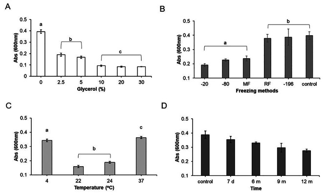
Phytophthora cinnamomi Rands is a highly prevalent phytopathogen worldwide, ranking among the top ten in terms of distribution. It inflicts crown rot, canker, and root rot on numerous plant species, significantly impacting the biodiversity of both flora and fauna within affected environments. With a host range spanning over 5,000 species, including important plants like Quercus suber, Quercus ilex, Castanea sativa, and commercially significant crops such as avocado (Persea americana), maize (Zea mays), and tomato (Solanum lycopersicum), Phytophthora cinnamomi poses a substantial threat to agriculture and ecosystems. The efficient dissemination of the oomycete relies on its short-lived asexually motile zoospores, which depend on water currents to infect host roots. However, managing these zoospores in the laboratory has long been challenging due to the complexity of the life cycle. Current protocols involve intricate procedures, including alternating cycles of growth, drought, and flooding. Unfortunately, these artificial conditions often result in a rapid decline in virulence, necessitating additional steps to maintain infectivity during cultivation. In our research, we sought to address this challenge by investigating zoospore survival under various conditions. Our goal was to develop a stable stock of zoospores that is both easily deployable and highly infective. Through direct freezing in liquid nitrogen, we have successfully preserved their virulence. This breakthrough eliminates the need for repeated culture transfers, simplifying the process of plant inoculation. Moreover, it enables more comprehensive studies of Phytophthora cinnamomi and its interactions with host plants.

Keywords: Phytophthora cinnamomi Rands; Quercus Sp; Solanum lycopersicum; Zoospores; qRT-PCR.


Conflict of interest statement

The authors declare no competing interests.

Figures

Fig. 1
Fig. 1
Obtaining Phytophthora cinnamomi zoospores. A.1.P. cinnamomi growth in Potato Dextrose Agar medium (PDA) in darkness, at 24 °C for 3 weeks. A.2. Inducing mycelium growth by growing P. cinnamomi on V8 agar medium in darkness, at 24 °C for 7 days on a miracloth disc. A.3. Inducing sporangia formation in V8 clarified liquid medium under fluorescence light and shaking, at 24 °C for 48 h. A.4. Second sporangia induction in mineral salt solution, enriched with chelated iron solution, under fluorescent light and shaking at 24 °C for 24 h. A.5. Inducing zoospore formation in sterile water under shaking, at 4 °C for 90 min. A.6. Filtration of zoospores. A.7. Freezing of zoospores in liquid nitrogen. A.8. Storage at -80 °C until use. B. Bright field of mature sporangium. C. P. cinnamomi culture stained with trypan blue, correponding to chlamydospore and hyphal swelling after the step of sporangia induction with mineral salt solution. Scale bar = 50 μm. D. Bright field snapshot image taken of obtained fresh zoospores. Scale bar = 20 μm. Images were taken at 40x magnification. E.P. cinnamomi culture stained with trypan blue, correponding to chlamydospore and hyphal swelling after the step of sporangia induction with mineral salt solution where some zoospores are observed. Scale bar = 90 μm
Fig. 2
Fig. 2
Viability of Phytophthora cinnamomi zoospores measured via MTT and Neubauer chamber. (A) The scatter plot shows the concentration of zoospores (×108/mL), at 50, 100 and 500 dilutions, counted using a Neubauer chamber plotted against the absorbance signal (measured at 600 nm using MTT; see Sect. 5). Significant differences were based on one-way analysis of variance (ANOVA) with a variance check (p < 0.05). Lowercase letters (a, b, c and d) indicate significant differences (R = 0.907). Error bars indicate standard deviation (SD) (n = 20). (B) Changes in zoospore survival at 23 °C measured via MTT. Significant differences were based on a non-parametric Krustal-Wallis´s test with a variance check (p < 0.05). Error bars indicate standard deviation (SD) (n = 14). The data were analyzed using the Stat-graphics Centurion 19 program
Fig. 3
Fig. 3
Viability of Phytophthora cinnamomi cells determined by fluorescence. (A) Bright image of chlamydospore and hypha. (B) fluorescent image of A. Scale bars = 50 μm. (C) Snapshot of zoospores (white arrowed), and Duroc sperm cells at time 0 by SQS® system. (D) Snapshot of zoospores (white arrowed), and Duroc sperm cells after 1 h by SQS® system. Scale bars = 30 μm. E and F. Bright (left), and fluorescence (right), images of zoospores by SQS® system. F inset. Detail that alive cells are arrowed in green while dead cells are arrowed in red. Scale bars = 20 μm. Images with 40x magnification. G. Percentage of zoospore survival after 0, 0.5, 1, 2, 3, 4, and 24 h, determined by taking at least five snapshots per time, measured with SQS® machine and counted with ImageJ® Software (see Sect. 5). Error bars indicate standard deviation (SD) (n = 35). The data were analyzed with Stat-graphics Centurion 19 program, with a variance check (p < 0.05) indicated by one asterisk
Fig. 4
Fig. 4
Survival of Phythophthora cinnamomi zoospores to freezing and defrosting. (A) Survival of zoospores after freezing in glycerol (0, 2.5, 5, 10, 20, and 30%). Lowercase letters (a, b, and c) indicate significant differences. Error bars indicate standard deviation (SD) (n = 115). (B) Survival of zoospores using next freezing methods: −20 °C (− 20), − 80 °C (− 80), Mister Frosty (MF), ramp freezing (for 22–18 − 4 to − 20 °C) (RF), liquid nitrogen (− 196 °C). For A and B significant differences were based on one-way analysis of variance (ANOVA) with a variance check (p < 0.05). Lowercase letters (a and b) indicate significant differences. Error bars indicate standard deviation (SD) (n = 16). (C) Survival of zoospores after defrosting at 4, 22, 24, and 37 °C. Lowercase letters (a, b, and c) indicate significant differences. Error bars indicate standard deviation (SD) (n = 30). (D) Survival of zoospores stocks at seven days (d) and six, nine and twelve months (m). The zoospore stock analyzed was 2 × 108 /mL. At least four independent biological assays were performed with similar results. Error bars indicate standard deviation (SD) (n = 14). For C and D significant differences were based on Krustal-Wallis´s test with a variance check (p < 0.05). All data were analyzed using the Stat-graphics Centurion 19 program
Fig. 5
Fig. 5
Symptoms of Solanum lycopersicum seedlings inoculated with Phytophthora cinnamomi zoospore stocks. Fourteen-day-old seedlings were grown on soil and inoculated with a final concentration of fresh zoospores (F), or non-fresh zoospore stocks (2 × 10 7 Zs/ml, see Sect. 5). A and C. Control plants (water). B and D. Symptoms of inoculated plants after four days of inoculation (4 dpi). Scale bar = 1 cm. E. Necrotic lesions detected by trypan blue staining, control (left image), and inoculated plant (right) at 4dpi. Scale bars = 1 cm. F. Disease ratio at 4 dpi, on seedlings performed with F, and NF. Symptom severity was assessed on a scale where “0” is absence of symptoms, “1” plant growth inhibition, “2” light chlorosis on leaves, “3” significant chlorosis on leaves and root necrosis, “4” high necrosis on leaves and roots and decayed seedlings. (n = 240). G. Oomycete biomass was quantified into vegetal tissue at 4dpi, inoculated with F (fresh) and NF (non-fresh) zoospores, measured by qRT-PCR using specific primers for P. cinnamomi β-Tubulin and normalized to the Solanum lycopersicum β-actin (see Sect. 5). The data were analyzed using the Stat-graphics Centurion 19 program, with a variance check (p < 0.05) indicated by asterisk. Error bars indicate standard deviation (SD) (n = 3)

Similar articles

Cited by

References

    1. Burgess TI, Scott JK, Mcdougall KL, et al. Current and projected global distribution of Phytophthora cinnamomi, one of the world’s worst plant pathogens. Global Change Biol. 2017;23(4):1661–74. doi: 10.1111/gcb.13492. - DOI - PubMed
    1. Kamoun S, Furzer O, Jones JDG, et al. The top 10 oomycete pathogens in molecular plant pathology. Mol Plant Pathol. 2015;16(4). 10.1111/mpp.12190 - PMC - PubMed
    1. Shakya SK, Grünwald NJ, Fieland VJ, et al. Phylogeography of the wide-host range panglobal plant pathogen Phytophthora cinnamomi. Mol Ecol. 2021;30(20). 10.1111/mec.16109 - PubMed
    1. Hardham AR, Blackman LM. Phytophthora cinnamomi. Mol Plant Pathol. 2018;19(2):260–85. doi: 10.1111/MPP.12568. - DOI - PMC - PubMed
    1. Corcobado T, Cubera E, Pérez-Sierra A, Jung T, Solla A. First report of Phytophthora gonapodyides involved in the decline of Quercus ilex in xeric conditions in Spain. New Dis Rep. 2010;22(1). 10.5197/j.2044-0588.2010.022.033

MeSH terms

LinkOut - more resources