. 2024 Jun 6;30(1):78.
doi: 10.1186/s10020-024-00845-4.

Wogonin upregulates SOCS3 to alleviate the injury in Diabetic Nephropathy by inhibiting TLR4-mediated JAK/STAT/AIM2 signaling pathway

Affiliations

Wogonin upregulates SOCS3 to alleviate the injury in Diabetic Nephropathy by inhibiting TLR4-mediated JAK/STAT/AIM2 signaling pathway

Yufeng Liu et al. Mol Med. .

Abstract

Background: Diabetic nephropathy (DN) is a life-threatening renal disease and needs urgent therapies. Wogonin is renoprotective in DN. This study aimed to explore the mechanism of how wogonin regulated high glucose (HG)-induced renal cell injury.

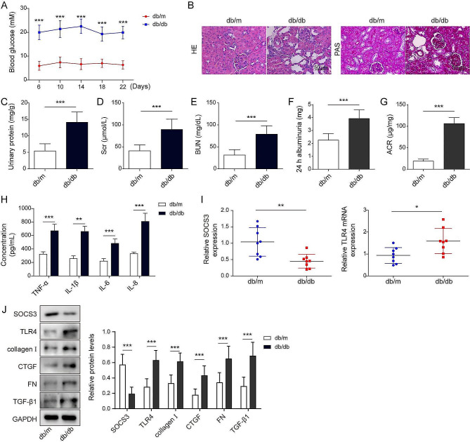
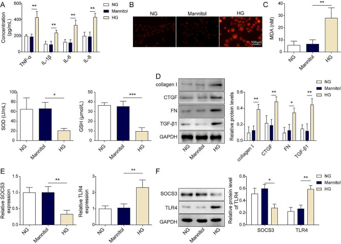
Methods: Diabetic mice (db/db), control db/m mice, and normal glucose (NG)- or HG-treated human tubule epithelial cells (HK-2) were used to evaluate the levels of suppressor of cytokine signaling 3 (SOCS3), Toll-like receptor 4 (TLR4), inflammation and fibrosis. Lentivirus was used to regulate SOCS3 and TLR4 expressions. After oral gavage of wogonin (10 mg/kg) or vehicle in db/db mice, histological morphologies, blood glucose, urinary protein, serum creatinine values (Scr), blood urea nitrogen (BUN), superoxide dismutase (SOD), glutathione (GSH), and reactive oxygen species (ROS) were assessed. RT-qPCR and Western blot evaluated inflammation and fibrosis-related molecules.

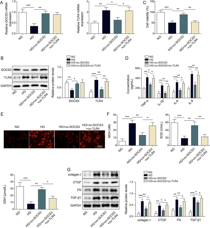
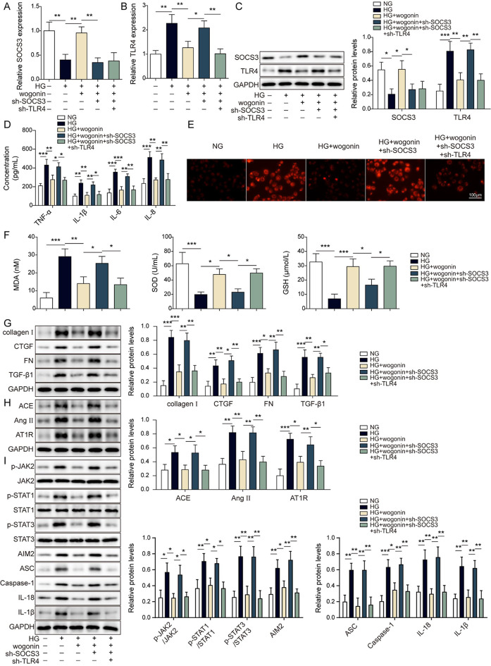
Results: HG exposure induced high blood glucose, severe renal injuries, high serumal Src and BUN, low SOD and GSH, and increased ROS. HG downregulated SOCS3 but upregulated TLR4 and JAK/STAT, fibrosis, and inflammasome-related proteins. Wogonin alleviated HG-induced renal injuries by decreasing cytokines, ROS, Src, and MDA and increasing SOD and GSH. Meanwhile, wogonin upregulated SOCS3 and downregulated TLR4 under HG conditions. Wogonin-induced SOCS3 overexpression directly decreased TLR4 levels and attenuated JAK/STAT signaling pathway-related inflammation and fibrosis, but SOCS3 knockdown significantly antagonized the protective effects of wogonin. However, TLR4 knockdown diminished SOCS3 knockdown-induced renal injuries.

Conclusion: Wogonin attenuates renal inflammation and fibrosis by upregulating SOCS3 to inhibit TLR4 and JAK/STAT pathway.

Keywords: Diabetic nephropathy; Inflammasome; JAK/STAT signaling pathway; SOCS3; TLR4; Wogonin.


Conflict of interest statement

The authors declare that they have no competing interests to disclose.

Figures

Fig. 1
Fig. 1
SOCS3 and TLR4 expression in the diabetic and control renal tissues. (A). Blood glucose levels were detected in tail vein blood. (B). Histopathologically examine the renal tissues using hematoxylin, eosin (HE), and periodic acid-Schiff base (PAS) staining. Scale bar = 50 μm. (C-E). An automatic biochemical analyzer measured the content of urinary protein, serum creatinine values (Scr), and blood urea nitrogen (BUN), respectively. (F-G) 24-hour albumin and urinary albumin-to-creatinine ratio (ACR) were detected by ELISA assay. (H). ELISA assay detected the contents of cytokines in mice sera. (I). SOCS3 and TLR4 expressions were detected by RT-qPCR in renal tissue. (J). Western blot measured the protein levels of SOCS3, TLR4, collagen I, CTGF, FN, and TGF-β1 in renal tissues. Data are representative of three independent experiments. *P < 0.05, **P < 0.01, ***P < 0.001 versus the corresponding control
Fig. 2
Fig. 2
Effects of high glucose on the expressions of SOCS3 and TLR4 in HK-2 cells. (A). The secretions of cytokines were measured by ELISA assay. (B). ROS levels were detected by DCFH-DA fluorescence imaging at different times after mannitol or HG incubation. Scale bar = 100 μm. (C). The corresponding assay kit detected MDA, SOD, and GSH levels after mannitol or HG treatment. (D). The protein levels of collagen I, CTGF, FN, and TGF-β1 were detected by western blot assay after mannitol or HG treatment. (E-F). The mRNA and protein levels of SOCS3 and TLR4 were evaluated in HK-2 cells with mannitol or HG treatment by RT-qPCR and western blot, respectively. *P < 0.05, **P < 0.01, ***P < 0.001 versus the corresponding control. Data are representative of three independent experiments
Fig. 3
Fig. 3
Wogonin attenuated high glucose-induced injuries in HK-2 cells. HK-2 cells were treated with vehicle, wogonin (1–64 µM) single or by treated with vehicle control, HG, or HG + wogonin (8 µM) for 24 h. Then, (A-B) cell survival was assessed by CCK-8 assay. (C) The cytokines levels were measured by ELISA. (D) ROS levels were detected by DCFH-DA. Scale bar = 100 μm. (E) MDA, SOD, and GSH were detected by the corresponding assay kit. (F) The protein levels of collagen I, CTGF, FN, and TGF-β1 were detected by western blot. (G) The mRNA levels of SOCS3 and TLR4 were detected by RT-qPCR. (H) The protein levels of SOCS3 and TLR4 were detected by western blot. *P < 0.05, **P < 0.01, ***P < 0.001 versus the corresponding control. Data are representative of three independent experiments
Fig. 4
Fig. 4
SOCS3 overexpression alleviates high glucose-induced renal cell injury by inhibiting TLR4 HK-2 cells were transfected with ov-SOCS3 single or together ov-TLR4 for 24 h, followed by 24 h treatment by vehicle or high glucose. (A-B) The mRNA and protein levels of SOCS3 and TLR4 were detected by RT-qPCR and western blot, respectively. (C) The cell viability was evaluated by CCK-8 assay. (D) The contents of cytokines were determined by ELISA assay. (E) A DCFH-DA Assay Kit detected the ROS levels. Scale bar = 100 μm. (F) The individual commercial kits measured the contents of MDA, SOD, and GSH. (G) The protein levels of collagen I, CTGF, FN, and TGF-β1 were measured by western blot. *P < 0.05, **P < 0.01, ***P < 0.001 versus the corresponding control. Data are representative of three independent experiments
Fig. 5
Fig. 5
The knockdown of TLR4 inhibited AIM2 inflammasome activation via the JAK/STAT signaling pathway to alleviate high glucose exposure-induced HK-2 cell injury. HK-2 cells were transfected with shRNA empty vector or sh-TLR4, followed by exposure to high glucose. (A-B) The mRNA and protein levels of TLR4 were detected by RT-qPCR (A) and western blot assay (B). (C) The contents of cytokines were measured by ELISA assay. (D) The ROS levels were detected by DCFH-DA fluorescence imaging. Scale bar = 100 μm. (E) The individual commercial kits evaluated MDA, SOD, and GSH. (F) The protein levels of collagen I, CTGF, FN, and TGF-β1 were detected by western blot assay. (G) The protein levels of ACE, Ang II, and AT1R were detected by western blot assay. (H) The protein levels of p-JAK2, JAK2, p-STAT1, STAT1, p-STAT3, STAT3, AIM2, ASC, caspase-1, IL-18 and IL-1β were detected by western blot assay. *P < 0.05, **P < 0.01, ***P < 0.001 versus the corresponding control. Data are representative of three independent experiments
Fig. 6
Fig. 6
Wogonin alleviated high glucose-induced injury via inhibitionTLR4-regulatedated JAK/STAT pathway and AIM2 inflammasome activation through upregulation of SOCS3 HK-2 cells were individually transfected with shRNA empty vector, sh-SOCS3 single with sh-TLR4 for 24 h, followed by HG or HG + wogonin stimulation. (A-B) The mRNA levels of SOCS3 (A) and TLR4 (B) were detected by RT-qPCR. (C) The protein levels of SOCS3 and TLR4 were measured by western blot assay. (D) ELISA evaluated the contents of cytokines. (E) The ROS levels were assessed by DCFH-DA fluorescence imaging. Scale bar = 100 μm. (F) The respective commercial kits determined the contents of MDA, SOD, and GSH. (G) The protein levels of collagen I, CTGF, FN, and TGF-β1 were detected by western blot assay. (H) The protein levels of ACE, Ang II, and AT1R were detected by western blot assay. (I) The protein levels of p-JAK2, JAK2, p-STAT1, STAT1, p-STAT3, STAT3, AIM2, ASC, caspase-1, IL-18 and IL-1β were detected by western blot assay. *P < 0.05, **P < 0.01, ***P < 0.001 versus the corresponding control. Data are representative of three independent experiments
Fig. 7
Fig. 7
Wogonin inhibited TLR4 to ameliorate renal injury via upregulation of SOCS3 in db/db mice. db/db mice were injected with adenovirus sh-SOCS3 or sh-SOCS3 + sh-TLR4, followed by treatment by vehicle or wogonin. (A) Blood glucose levels were measured using an automated chemistry analyzer. (B) The pathological features and glucose in the renal tissues were evaluated by hematoxylin, eosin (HE), and periodic acid-Schiff base (PAS). Scale bar = 50 μm. (C-D) The urinary protein, serum creatinine values (Scr), and blood urea nitrogen (BUN) were evaluated by an automated chemistry analyzer. (E-F) 24-hour albumin and urinary albumin-to-creatinine ratio (ACR) were detected by ELISA assay in HG mice without or with wogonin, wogonin + sh-SOCS3, wogonin + sh-SOCS3 + sh-TLR4 (E) ELISA evaluated the levels of cytokines. (F-G) The mRNA and protein levels of SOCS3 and TLR4 were detected by RT-qPCR and western blot, respectively. (H) The protein levels of collagen I, CTGF, FN, and TGF-β1 were detected by western blot assay. (I) The protein levels of ACE, Ang II, and AT1R were detected by western blot assay. (J) The protein levels of p-JAK2, JAK2, p-STAT1, STAT1, p-STAT3, STAT3, AIM2, ASC, caspase-1, IL-18 and IL-1β were detected by western blot assay. *P < 0.05, **P < 0.01, ***P < 0.001 versus the corresponding control. Data are representative of three independent experiments
Fig. 8
Fig. 8
Schematic diagram for wogonin-suppressing inflammation and alleviating diabetic neuphropathy

Similar articles

Cited by

References

    1. Brosius FC. New insights into the mechanisms of fibrosis and sclerosis in diabetic nephropathy. Rev Endocr Metab Disord. 2008;9(4):245–54. doi: 10.1007/s11154-008-9100-6. - DOI - PMC - PubMed
    1. Cui X, Li Y, Yuan S, Huang Y, Chen X, Han Y, Zhu X. Alpha-kinase1 promotes tubular injury and interstitial inflammation in diabetic nephropathy by canonical pyroptosis pathway. Biol Res. 2023;56(1):5. doi: 10.1186/s40659-023-00416-7. - DOI - PMC - PubMed
    1. Duan WN, Xia ZY, Liu M, Sun Q, Lei SQ, Wu XJ, Leng Y. Protective effects of SOCS3 overexpression in high glucose–induced lung epithelial cell injury through the JAK2/STAT3 pathway. Mol Med Rep. 2017;16(3):2668–74. doi: 10.3892/mmr.2017.6941. - DOI - PMC - PubMed
    1. Haider MJA, Albaqsumi Z, Al-Mulla F, Ahmad R, Al-Rashed F. SOCS3 regulates dectin-2-Induced inflammation in PBMCs of Diabetic patients. Cells. 2022;11(17):2670. doi: 10.3390/cells11172670. - DOI - PMC - PubMed
    1. Khan S, Zhang D, Zhang Y, Li M, Wang C. Wogonin attenuates diabetic cardiomyopathy through its anti-inflammatory and anti-oxidative properties. Mol Cell Endocrinol. 2016;428:101–8. doi: 10.1016/j.mce.2016.03.025. - DOI - PubMed

MeSH terms