

Resting-state brain plasticity is associated with the severity in cervical spondylotic myelopathy
- PMID: 38844898
- PMCID: PMC11155054
- DOI: 10.1186/s12891-024-07539-2
Resting-state brain plasticity is associated with the severity in cervical spondylotic myelopathy
Abstract
Objective: To investigate the brain mechanism of non-correspondence between imaging presentations and clinical symptoms in cervical spondylotic myelopathy (CSM) patients and to test the utility of brain imaging biomarkers for predicting prognosis of CSM.
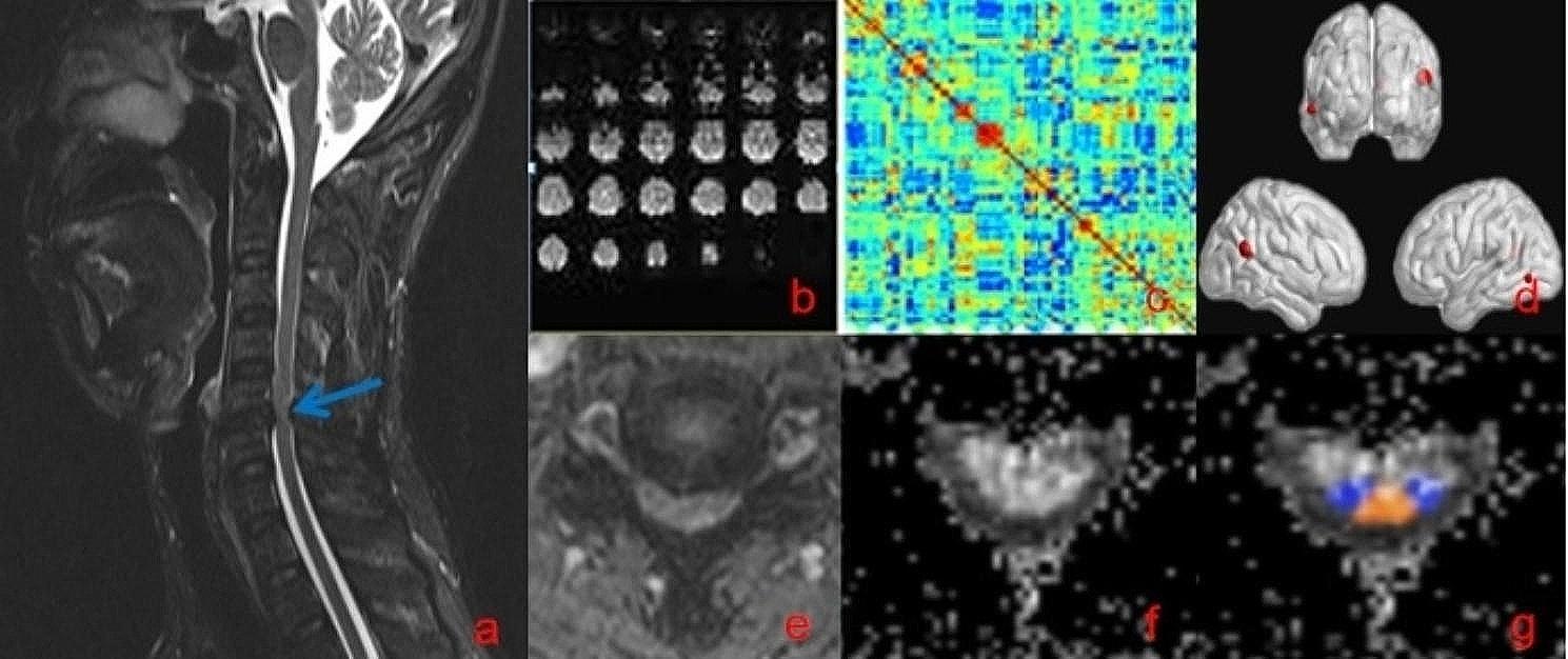
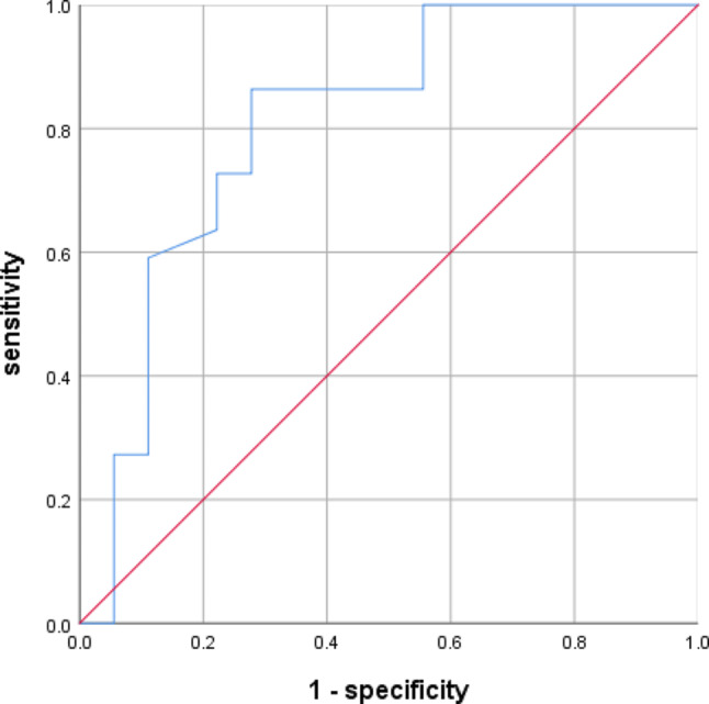
Methods: Forty patients with CSM (22 mild-moderate CSM, 18 severe CSM) and 25 healthy controls (HCs) were recruited for rs-fMRI and cervical spinal cord diffusion tensor imaging (DTI) scans. DTI at the spinal cord (level C2/3) with fractional anisotropy (FA) and degree centrality (DC) were recorded. Then one-way analysis of covariance (ANCOVA) was conducted to detect the group differences in the DC and FA values across the three groups. Pearson correlation analysis was then separately performed between JOA with FA and DC.
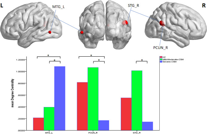
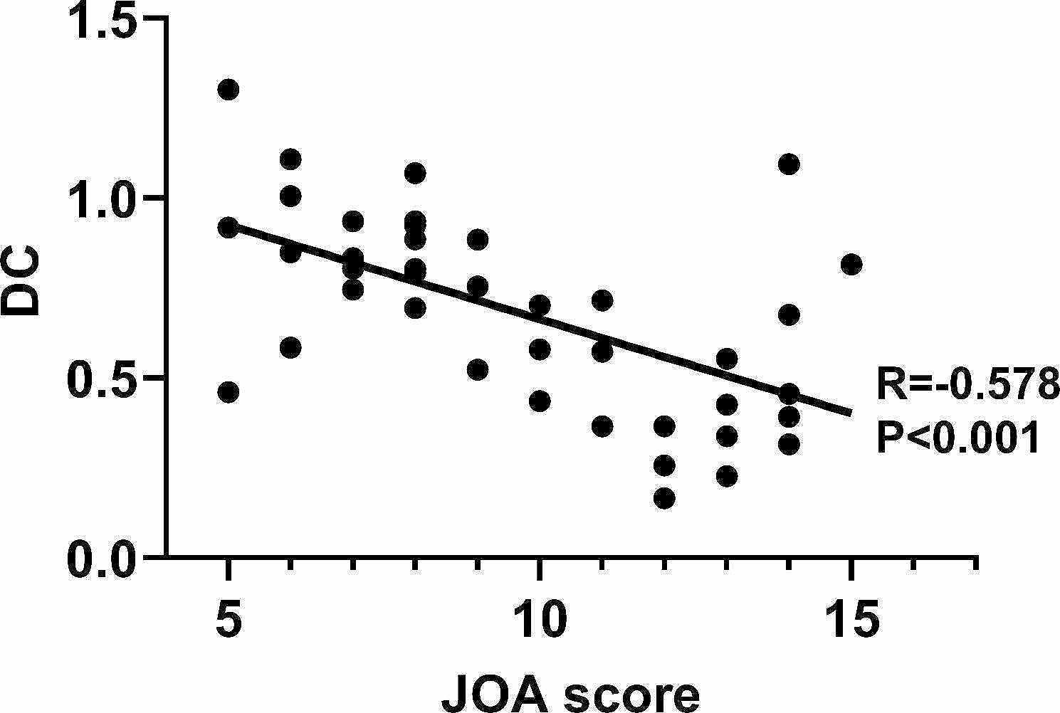
Results: Among them, degree centrality value of left middle temporal gyrus exhibited a progressive increase in CSM groups compared with HCs, the DC value in severe CSM group was higher compared with mild-moderate CSM group. (P < 0.05), and the DC values of the right superior temporal gyrus and precuneus showed a decrease after increase. Among them, DC values in the area of precuneus in severe CSM group were significantly lower than those in mild-moderate CSM and HCs. (P < 0.05). The fractional anisotropy (FA) values of the level C2/3 showed a progressive decrease in different clinical stages, that severe CSM group was the lowest, significantly lower than those in mild-moderate CSM and HCs (P < 0.05). There was negative correlation between DC value of left middle temporal gyrus and JOA scores (P < 0.001), and the FA values of dorsal column in the level C2/3 positively correlated with the JOA scores (P < 0.001).
Conclusion: Structural and functional changes have taken place in the cervical spinal cord and brain of CSM patients. The Brain reorganization plays an important role in maintaining the symptoms and signs of CSM, aberrant DC values in the left middle temporal gyrus may be the possible mechanism of inconsistency between imaging findings and clinical symptoms. Degree centrality is a potentially useful prognostic functional biomarker in cervical spondylotic myelopathy.
Keywords: Brain plasticity; Cervical spondylotic myelopathy; Degree centrality; Resting-state functional magnetic resonance imaging.
© 2024. The Author(s).
Conflict of interest statement
The authors declare no competing interests.
Figures


Similar articles
-
Feasibility of diffusion tensor imaging in cervical spondylotic myelopathy using MUSE sequence.Spine J. 2024 Aug;24(8):1352-1360. doi: 10.1016/j.spinee.2024.03.015. Epub 2024 Mar 29. Spine J. 2024. PMID: 38556218
-
Contribution of changes in the spinal cord and brain to the onset and progression of hand clumsiness symptoms in cervical spondylotic myelopathy.J Neurosurg Spine. 2024 Jun 21;41(3):396-406. doi: 10.3171/2024.4.SPINE231238. Print 2024 Sep 1. J Neurosurg Spine. 2024. PMID: 38905708
-
A preliminary study of 3.0-T magnetic resonance diffusion tensor imaging in cervical spondylotic myelopathy.Eur Spine J. 2018 Aug;27(8):1839-1845. doi: 10.1007/s00586-018-5579-z. Epub 2018 Apr 4. Eur Spine J. 2018. PMID: 29619562
-
Beyond the aging spine - a systematic review of functional changes in the human brain in cervical spondylotic myelopathy.Geroscience. 2024 Apr;46(2):1421-1450. doi: 10.1007/s11357-023-00954-8. Epub 2023 Oct 6. Geroscience. 2024. PMID: 37801201 Free PMC article.
-
Utility of Diffusion Tensor Imaging for Prognosis and Management of Cervical Spondylotic Myelopathy: A PRISMA Review.World Neurosurg. 2024 Oct;190:88-98. doi: 10.1016/j.wneu.2024.07.032. Epub 2024 Jul 8. World Neurosurg. 2024. PMID: 38986943 Review.
References
-
- Zhao R, Guo X, Wang Y, Song Y, Su Q, Sun H et al. Functional MRI evidence for primary motor cortex plasticity contributes to the disease’s severity and prognosis of cervical spondylotic myelopathy patients. Eur Radiol. 2022. - PubMed
MeSH terms
Grants and funding
- 81460329/National Natural Science Fund of China
- 81460329/National Natural Science Fund of China
- 20192ACBL20039、GJJ2200110、202310454/Natural Science Foundation Jiangxi Province
- 20192ACBL20039、GJJ2200110、202310454/Natural Science Foundation Jiangxi Province
- YC2023-B078/Special Funds for Graduate Student Innovation in Jiangxi Province
LinkOut - more resources
Full Text Sources
Medical
Miscellaneous
