

Loss of β-arrestin2 aggravated condylar cartilage degeneration at the early stage of temporomandibular joint osteoarthritis
- PMID: 38844905
- PMCID: PMC11154996
- DOI: 10.1186/s12891-024-07558-z
Loss of β-arrestin2 aggravated condylar cartilage degeneration at the early stage of temporomandibular joint osteoarthritis
Abstract
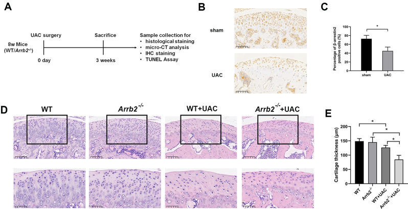
Objective: Temporomandibular joint osteoarthritis (TMJOA) is a chronic degenerative joint disorder characterized by extracellular matrix degeneration and inflammatory response of condylar cartilage. β-arrestin2 is an important regulator of inflammation response, while its role in TMJOA remains unknown. The objective of this study was to investigate the role of β-arrestin2 in the development of TMJOA at the early stage and the underlying mechanism.
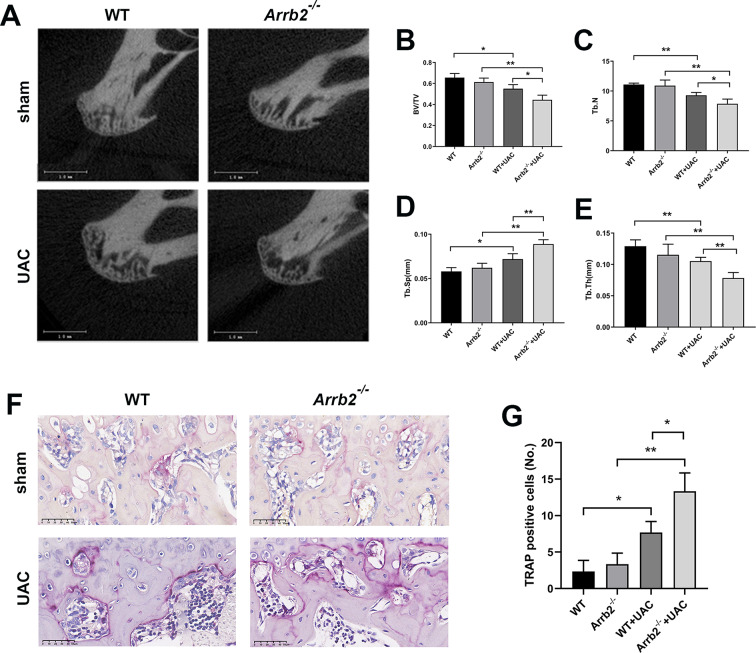
Methods: A unilateral anterior crossbite (UAC) model was established on eight-week-old wild-type (WT) and β-arrestin2 deficiency mice to simulate the progression of TMJOA. Hematoxylin-eosin (HE) staining and microcomputed tomography (micro-CT) analysis were used for histological and radiographic assessment. Immunohistochemistry was performed to detect the expression of inflammatory and degradative cytokines, as well as autophagy related factors. Terminal-deoxynucleotidyl transferase mediated nick end labeling (TUNEL) assay was carried out to assess chondrocyte apoptosis.
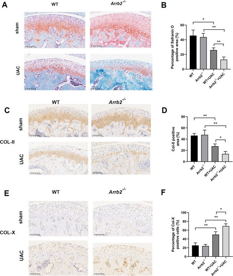
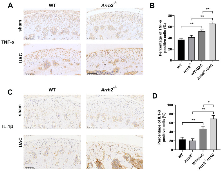
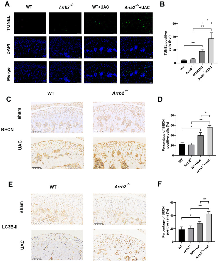
Results: The loss of β-arrestin2 aggravated cartilage degeneration and subchondral bone destruction in the model of TMJOA at the early stage. Furthermore, in UAC groups, the expressions of degradative (Col-X) and inflammatory (TNF-α and IL-1β) factors in condylar cartilage were increased in β-arrestin2 null mice compared with WT mice. Moreover, the loss of β-arrestin2 promoted apoptosis and autophagic process of chondrocytes at the early stage of TMJOA.
Conclusion: In conclusion, we demonstrated for the first time that β-arrestin2 plays a protective role in the development of TMJOA at the early stage, probably by inhibiting apoptosis and autophagic process of chondrocytes. Therefore, β-arrestin2 might be a potential therapeutic target for TMJOA, providing a new insight for the treatment of TMJOA at the early stage.
Keywords: Apoptosis; Autophagy; Cartilage degeneration; Inflammation; Temporomandibular joint osteoarthritis; β-arrestin2.
© 2024. The Author(s).
Conflict of interest statement
The authors declare no competing interests.
Figures
Similar articles
-
Ectodysplasin-A deficiency exacerbates TMJOA by upregulating ATF4/Ihh signaling in mice.Osteoarthritis Cartilage. 2025 Aug;33(8):965-979. doi: 10.1016/j.joca.2025.02.789. Epub 2025 Mar 24. Osteoarthritis Cartilage. 2025. PMID: 40139647
-
Syndecan-4 inhibition attenuates cartilage degeneration in temporomandibular joint osteoarthritis.J Oral Rehabil. 2024 Nov;51(11):2324-2335. doi: 10.1111/joor.13829. Epub 2024 Aug 5. J Oral Rehabil. 2024. PMID: 39101668
-
Matrix Stiffening-Induced SLIT3 Activation in Chondrocytes Drives Cartilage Degradation and Angiogenesis in Temporomandibular Joint Osteoarthritis.FASEB J. 2025 Aug 31;39(16):e70936. doi: 10.1096/fj.202501278RR. FASEB J. 2025. PMID: 40817785
-
Synovial microenvironment in temporomandibular joint osteoarthritis: crosstalk with chondrocytes and potential therapeutic targets.Life Sci. 2024 Oct 1;354:122947. doi: 10.1016/j.lfs.2024.122947. Epub 2024 Aug 8. Life Sci. 2024. PMID: 39117138 Review.
-
An Updated View on Temporomandibular Joint Degeneration: Insights from the Cell Subsets of Mandibular Condylar Cartilage.Stem Cells Dev. 2022 Aug;31(15-16):445-459. doi: 10.1089/scd.2021.0324. Epub 2022 Feb 18. Stem Cells Dev. 2022. PMID: 35044232 Review.
Cited by
-
Artificial Cell-Derived Vesicles: Extracellular Vesicle Mimetics for Chondrocyte Restoration in TMJOA Therapy.Int J Nanomedicine. 2025 Apr 26;20:5393-5405. doi: 10.2147/IJN.S508449. eCollection 2025. Int J Nanomedicine. 2025. PMID: 40313864 Free PMC article.
-
Different effects of abnormal mechanical stress on temporomandibular joint cartilage, subchondral bone, and discs.Front Physiol. 2025 Apr 3;16:1539342. doi: 10.3389/fphys.2025.1539342. eCollection 2025. Front Physiol. 2025. PMID: 40247923 Free PMC article. Review.
References
-
- Ma Y, He F, Chen X, Zhou S, He R, Liu Q et al. Low-frequency pulsed electromagnetic fields alleviate the condylar cartilage degeneration and synovitis at the early stage of temporomandibular joint osteoarthritis. J Oral Rehabil. 2023. - PubMed
-
- Wieckiewicz M, Paradowska A, Kawala B, Wieckiewicz W. SAPHO Syndrome as a possible cause of Masticatory System Anomalies - a review of the literature. Adv Clin EXPERIMENTAL Med. 2011;20(4):521–5.
MeSH terms
Substances
Grants and funding
LinkOut - more resources
Full Text Sources
Medical
Molecular Biology Databases
