

In vivo non-contact regions of proximal scaphoid in six extreme wrist positions
- PMID: 38844912
- PMCID: PMC11155112
- DOI: 10.1186/s12891-024-07561-4
In vivo non-contact regions of proximal scaphoid in six extreme wrist positions
Abstract
Introduction: Fractures of the scaphoid are the most common carpal injuries, account for 80-90% of all carpal fractures. 5-15% nonunion of scaphoid fractures were reported even with adequate primary treatment, which probably progresses to osteoarthritic changes several decades later. Researches regarding to scaphoid physiological characteristic in vitro and in vivo and kinds of trials in clinical practice are being kept on going, which contribute much to our clinical practice. With the advancing wrist arthroscopy, 3D-print patient-specific drill guide, and intraoperative fluoroscopic guidance, dorsal approach (mini-invasive and percutaneous technique) is being popular, through which we can implant the screw in good coincidence with biomechanics and with less disturbing tenuous blood supply of the scaphoid. Investigating the noncontact area of the dorsal proximal scaphoid in different wrist positions can facilitate preoperatively estimating insert point of the screw.
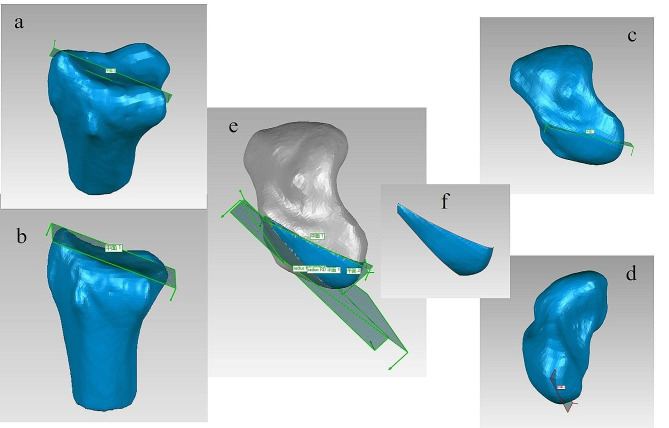
Materials and methods: Eight volunteers were recruited to accept CT scans in six extreme wrist positions. The images of DICOM mode were imput into the Mimics analytical system, the segmented scaphoid, lunate and radius were exported in mode of ASCII STL and were opened in the software of Geomagic studio. We created four planes based on anatomic markers on the surface of the radius and scaphoid to confine the proximal scaphoid to form the so-called non-contact regions. We measured and compared the areas in six targeted positions.
Results: Amidst six extreme wrist positions, area of the non-contact region in extreme dorsal extension (59.81 ± 26.46 mm2) was significantly the smallest, and it in extreme palmar flexion significantly was largest (170.51 ± 30.44 mm2). The non-contact regions increased in order of dorsal extension, supination, ulnar deviation, radial deviation, pronation and palmar flexion. As for two-group comparison, the non-contact region showed significantly larger (p < 0.05) in palmar flexion than the others except for in pronation individually, and in radial deviation (p < 0.05) than in dorsal extension.
Conclusions: Sufficient space was available for the screw started from the dorsal approach despite the wrist positions.
Keywords: In vivo; Non-contact regions; Scaphoid; Wrist position.
© 2024. The Author(s).
Conflict of interest statement
The author declare that there is no conflict of interest.
Figures

Similar articles
-
In vivo kinematics of the scaphoid, lunate, capitate, and third metacarpal in extreme wrist flexion and extension.J Hand Surg Am. 2013 Feb;38(2):278-88. doi: 10.1016/j.jhsa.2012.10.035. Epub 2012 Dec 23. J Hand Surg Am. 2013. PMID: 23266007 Free PMC article.
-
Changes in contact site of the radiocarpal joint and lengths of the carpal ligaments in forearm rotation: an in vivo study.J Hand Surg Am. 2013 Apr;38(4):712-20. doi: 10.1016/j.jhsa.2013.01.021. Epub 2013 Mar 6. J Hand Surg Am. 2013. PMID: 23474161
-
The Palpable Scaphoid Surface Area in Various Wrist Positions.J Hand Surg Am. 2015 Oct;40(10):2039-44. doi: 10.1016/j.jhsa.2015.06.121. Epub 2015 Aug 22. J Hand Surg Am. 2015. PMID: 26307024
-
Trans-scaphoid perilunate fracture dislocations: results of screw fixation of the scaphoid and lunotriquetral repair with a dorsal approach.J Hand Surg Am. 2005 Nov;30(6):1145-52. doi: 10.1016/j.jhsa.2005.07.007. J Hand Surg Am. 2005. PMID: 16344169
-
Computer Modelling of Wrist Biomechanics: Translation into Specific Tasks and Injuries.Curr Rheumatol Rev. 2020;16(3):178-183. doi: 10.2174/1573397115666190119095311. Curr Rheumatol Rev. 2020. PMID: 30659546 Review.
References
-
- Berger RA, Garcia-Elias M. General anatomy of the wrist. In: An KN, Berger RA, Cooney WP, editors Biomechanics of the wrist Joint. New York: Springer-; 10.1007/978-1-4612-3208-7_1. pp:1991:1–22.
-
- Buckwalter JA, Einhorn TA, Simon SR, editors. Orthopaedic Basic Science: Biology and Biomechanics of the Musculoskeletal System. 2. Rosemont, IL: American Academy of Orthopaedic Surgeons; 2000.
-
- Claes LE, Heigele CA, Neidlinger-Wilke C, Kaspar D, Seidl W, Margevicius KJ, Augat P. Effects of mechanical factors on the fracture healing process. Clin Orthop Relat Res. 1998;(355 Suppl):S132-47. 10.1097/00003086-199810001-00015. PMID: 9917634. - PubMed
MeSH terms
LinkOut - more resources
Full Text Sources
Research Materials
