

DNA methylation profiles of ovarian cysts resemble ovarian tissues but not endometrial tissues
- PMID: 38844959
- PMCID: PMC11155058
- DOI: 10.1186/s13048-024-01440-1
DNA methylation profiles of ovarian cysts resemble ovarian tissues but not endometrial tissues
Abstract
Introduction: Endometriosis is a heritable, complex chronic inflammatory disease, for which much of the causal pathogenic mechanism remain unknown.Despite the high prevalence of ovarian chocolate cyst, its origin is still under debate.
Methods: Prevailing retrograde menstruation model predicts that ectopic endometrial cells migrate and develop into ovarian chocolate cyst. However, other models were also proposed. Genome-wide association studies (GWASs) have proved successful in identifying common genetic variants of moderate effects for various complex diseases.
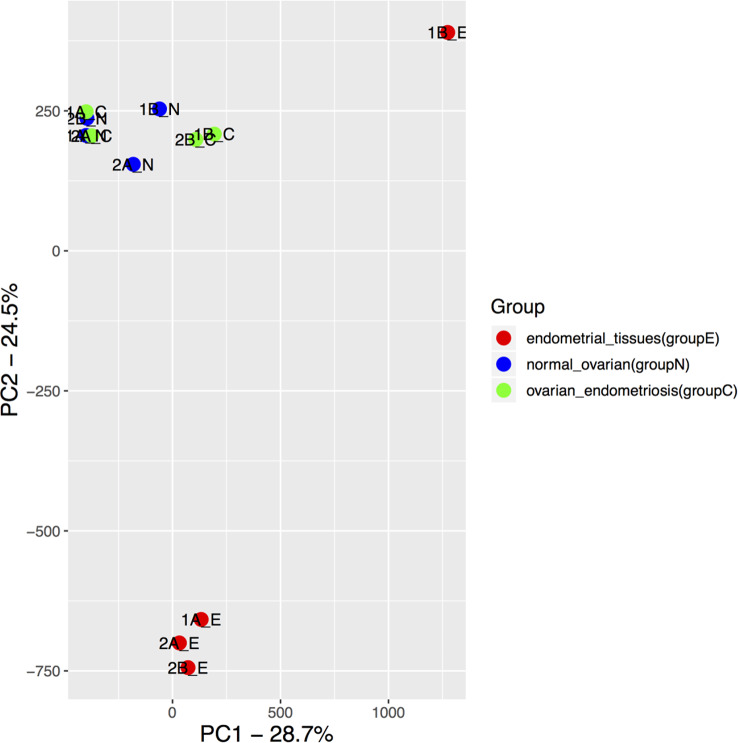
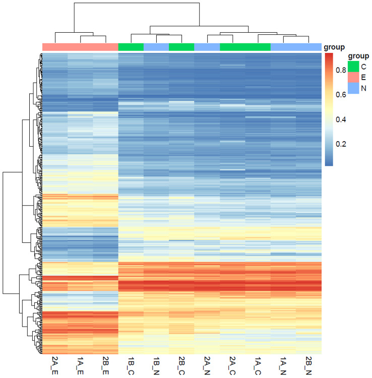
Results: A growing body of evidence shows that the remodeling of retrograde endometrial tissues to the ectopic endometriotic lesions involves multiple epigenetic alterations, such as DNA methylation, histone modification, and microRNA expression.Because DNA methylation states exhibit a tissue specific pattern, we profiled the DNA methylation for ovarian cysts and paired eutopic endometrial and ovarian tissues from four patients. Surprisingly, DNA methylation profiles showed the ovarian cysts were closely grouped with normal ovarian but not endometrial tissues.
Conclusions: These results suggested alterative origin of ovarian cysts or strong epigenetic reprogramming of infiltrating endometrial cells after seeding the ovarian tissue. The data provide contributing to the pathogenesis and pathophysiology of endometriosis.
Keywords: DNA methylation; Ovarian endometriosis; Tissue of origin.
© 2024. The Author(s).
Conflict of interest statement
The authors declare no competing interests.
Figures


Similar articles
-
miR-196b targets c-myc and Bcl-2 expression, inhibits proliferation and induces apoptosis in endometriotic stromal cells.Hum Reprod. 2013 Mar;28(3):750-61. doi: 10.1093/humrep/des446. Epub 2013 Jan 4. Hum Reprod. 2013. PMID: 23293219
-
Hypomethylation of the GSTM1 promoter is associated with ovarian endometriosis.Hum Reprod. 2019 May 1;34(5):804-812. doi: 10.1093/humrep/dez039. Hum Reprod. 2019. PMID: 30989213
-
RUNX3 is inactivated by promoter hypermethylation in malignant transformation of ovarian endometriosis.Oncol Rep. 2014 Dec;32(6):2580-8. doi: 10.3892/or.2014.3524. Epub 2014 Oct 3. Oncol Rep. 2014. PMID: 25333219
-
DNA methylation alterations-potential cause of endometriosis pathogenesis or a reflection of tissue heterogeneity?Biol Reprod. 2018 Aug 1;99(2):273-282. doi: 10.1093/biolre/ioy067. Biol Reprod. 2018. PMID: 29796617 Review.
-
Endometrial anomalies in women with endometriosis.Ann N Y Acad Sci. 2001 Sep;943:131-47. doi: 10.1111/j.1749-6632.2001.tb03797.x. Ann N Y Acad Sci. 2001. PMID: 11594534 Review.
References
MeSH terms
Grants and funding
LinkOut - more resources
Full Text Sources
Medical
