. 2024 Jun 6;26(1):94.
doi: 10.1186/s13058-024-01847-0.

NSUN2/YBX1 promotes the progression of breast cancer by enhancing HGH1 mRNA stability through m5C methylation

Affiliations

NSUN2/YBX1 promotes the progression of breast cancer by enhancing HGH1 mRNA stability through m5C methylation

Xuran Zhang et al. Breast Cancer Res. .

Abstract

Background: RNA m5C methylation has been extensively implicated in the occurrence and development of tumors. As the main methyltransferase, NSUN2 plays a crucial regulatory role across diverse tumor types. However, the precise impact of NSUN2-mediated m5C modification on breast cancer (BC) remains unclear. Our study aims to elucidate the molecular mechanism underlying how NSUN2 regulates the target gene HGH1 (also known as FAM203) through m5C modification, thereby promoting BC progression. Additionally, this study targets at preliminarily clarifying the biological roles of NSUN2 and HGH1 in BC.

Methods: Tumor and adjacent tissues from 5 BC patients were collected, and the m5C modification target HGH1 in BC was screened through RNA sequencing (RNA-seq) and single-base resolution m5C methylation sequencing (RNA-BisSeq). Methylation RNA immunoprecipitation-qPCR (MeRIP-qPCR) and RNA-binding protein immunoprecipitation-qPCR (RIP-qPCR) confirmed that the methylation molecules NSUN2 and YBX1 specifically recognized and bound to HGH1 through m5C modification. In addition, proteomics, co-immunoprecipitation (co-IP), and Ribosome sequencing (Ribo-Seq) were used to explore the biological role of HGH1 in BC.

Results: As the main m5C methylation molecule, NSUN2 is abnormally overexpressed in BC and increases the overall level of RNA m5C. Knocking down NSUN2 can inhibit BC progression in vitro or in vivo. Combined RNA-seq and RNA-BisSeq analysis identified HGH1 as a potential target of abnormal m5C modifications. We clarified the mechanism by which NSUN2 regulates HGH1 expression through m5C modification, a process that involves interactions with the YBX1 protein, which collectively impacts mRNA stability and protein synthesis. Furthermore, this study is the first to reveal the binding interaction between HGH1 and the translation elongation factor EEF2, providing a comprehensive understanding of its ability to regulate transcript translation efficiency and protein synthesis in BC cells.

Conclusions: This study preliminarily clarifies the regulatory role of the NSUN2-YBX1-m5C-HGH1 axis from post-transcriptional modification to protein translation, revealing the key role of abnormal RNA m5C modification in BC and suggesting that HGH1 may be a new epigenetic biomarker and potential therapeutic target for BC.

Keywords: Breast cancer; HGH1; NSUN2; RNA 5-methylcytosinine; Translation efficiency.


Conflict of interest statement

The authors declare no competing interests.

Figures
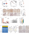
Fig. 1
Fig. 1
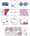
Higher NSUN2 expression in breast cancer samples. (A) NSUN2 expression in normal tissues and BC tissues from the TCGA dataset. (B) Plot of the overall survival probability of BC patients with high or low NSUN2 expression from the TCGA dataset. (C) NSUN2 mRNA expression in five clinical BC and adjacent tissue samples assessed by RNA-Seq (n = 5). (D) NSUN2 expression in five BC paraffin tissue samples. The samples were prepared and stained as described in the Materials and Methods (all images, 200×; n = 8). (E) LC-MS/TOF analysis of NSUN2 protein expression in five BC and adjacent tissue samples (n = 5). (F) Western blot analysis of NSUN2 expression in five breast cell lines. (G) The mammary fat pads of BALB/c nude mice (n = 5 per group) were surgically injected into the mammary fat pads with MCF7 cells, the tumors were weighed, and the tumor volume was measured. (H) Representative images of NSUN2 and Ki-67 expression in different groups of CDX tumors after IHC staining (all images, 200×). Data are mean ± SD (Standard deviation) of three independent experiments for (E) and (F). *p ≤ 0.05, **p ≤ 0.01, **p ≤ 0.001, ****p ≤ 0.0001 by t test of the indicated pairs or by log-rank (Mantel-Cox) test
Fig. 2
Fig. 2
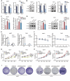
NSUN2-mediated promotion of breast cancer progression relies on methyltransferase enzymatic activity. (A) and (B) Cellular proliferation rates of BC cells with NSUN2 knockdown, NSUN2-WT or NSUN2-DM determined by CCK-8 assays and (C) and (D) colony formation assays. (E) and (F) Transwell assays were used to investigate the cell migration and invasion ability of NSUN2-knockdown, NSUN2-WT or NSUN2-DM cells. (G) FITC and PE fluorescence dyes were used to detect changes in cell apoptosis after NSUN2 knockdown by flow cytometry. Data are mean ± SD of three independent experiments for (E-G). **p ≤ 0.01, ***p ≤ 0.001, ****p ≤ 0.0001 by t test
Fig. 3
Fig. 3
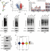
Screening of genes with abnormal m5C hypermethylation. (A) m5C dot blot assays using mRNA from MCF7 and T47D cells with or without NSUN2 knockdown. Methylene blue staining was used as a loading control. (B) Volcano plot showing differentially expressed m5C sites between tumor and adjacent tissues (n = 5). (C) Different proportions of m5C modifications in mRNA regions between BC tissues and adjacent tissues. (D) KEGG analysis revealed that m5C-hypermethylated genes with high expression levels in BC tissues were enriched in oncogenic signaling pathways. (E) Overlap of target genes from RNA-Seq and RNA-BisSeq of BC tissues (n = 5). The horizontal dashed line represents the threshold of the adjusted p-value of 0.05, while the vertical line represents the threshold of a log2-fold change greater than 1.5. (F) Clustering analysis heatmap showing the display level of different tissue samples. (G) Database analysis revealed an increase in HGH1 expression in tumor tissue. (H) Survival analysis showed that the group with high expression of HGH1 had a worse prognosis. (I) and (J) RT-qPCR and IHC experiments proved that HGH1 was expressed at higher levels in BC tissues than in paired adjacent tissues. Data are mean ± SD of three independent experiments for (I). *p ≤ 0.05, **p ≤ 0.01, ****p ≤ 0.0001 by t test or by log-rank (Mantel-Cox) test
Fig. 4
Fig. 4
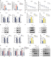
Suppression of HGH1 delays the progression of breast cancer. (A) CCK-8 assays were used to detect BC cell proliferation rates after interference with siHGH1 or oeHGH1. (B) Colony formation experiments detected the colony formation ability of BC cells after interference with shHGH1 or oeHGH1. (C) Transwell assays were used to assess the migration and invasion ability of BC cells after HGH1 knockdown. (D) FITC and PE fluorescence dyes were used to detect changes in cell apoptosis by flow cytometry after HGH1 knockdown. (E) PI fluorescence dye was used to detect cell cycle alterations by flow cytometry upon HGH1 knockdown or HGH1 overexpression. (F) The mammary fat pads of BALB/c nude mice (n = 5 per group) were surgically injected into the mammary fat pads with MCF7 cells, the tumors were weighed, and the tumor volume was measured. (G) Representative images of HGH1 and Ki-67 expression in different groups of CDX tumors after IHC staining (all images 200×) ns. no significant; Data are mean ± SD of three independent experiments for (C-E). *p ≤ 0.05, **p ≤ 0.01, ***p ≤ 0.001, ****p ≤ 0.0001 by t test
Fig. 5
Fig. 5
HGH1 affects translation efficiency in BC cells. (A) STRING database analysis showing a protein interaction relationship between EEF2 and HGH1. (B) and (C) Volcano plot and histogram of LC-MS/TOF data showing the high expression of EEF2 and HGH1 in BC tissues. (D) and (E) MCF7 and T47D cells transfected with siEEF2 (D) or siHGH1 (E) were treated with 1 µM puromycin for 1 h, and whole-cell lysates were subjected to western blot analysis with an anti-puromycin antibody. (F) Proteins were used in large excess to capture EEF2 from MCF7 cells by the anti-HGH1-protein A/G magnetic beads complex. The eluted material was analyzed by western blot and immunoblotting with anti-EEF2, anti-HGH1, and anti-GAPDH antibodies. (G) The average translation efficiency (TE) of MCF7 cells was analyzed via Ribo-Seq. ns. no significant; Data are mean ± SD of three independent experiments for (C) and (G). **p ≤ 0.01, ***p ≤ 0.001, ****p ≤ 0.0001 by t test
Fig. 6
Fig. 6
NSUN2-mediated mRNA m5C methylation promotes HGH1 expression (A) RT-qPCR assays were used to measure the expression of HGH1 in BC cell lines after NSUN2 knockdown, and (B) Western blot analysis was used to confirm the protein level. (C) and (D) RT-qPCR and Western blot assays were used to assess alterations in HGH1 expression after overexpression of NSUN2-WT or NSUN2-DM. (E) MeRIP-qPCR was used to measure the level of m5C on HGH1 mRNA after NSUN2 knockout in BC cells. (F) Changes in the response to actinomycin D-mediated RNA synthesis inhibition and mRNA transcription interference were detected after NSUN2 knockdown. (G) and (H) CCK-8 experiments confirmed the influence of NSUN2 and HGH1 on the proliferation of BC cells and their downstream relationship. (I) A colony formation assay was used to determine the effect of restoring the expression of the downstream gene HGH1 in the NSUN2-knockdown cell line on the expansion ability of BC cells. ns. no significant; Data are mean ± SD of three independent experiments. *p ≤ 0.05, **p ≤ 0.01, ***p ≤ 0.001, ****p ≤ 0.0001 by t test
Fig. 7
Fig. 7
YBX1 is an important molecule of m5C regulating HGH1 expression in breast cancer. (A) YBX1 antibodies were used for RIP-qPCR to measure the level of YBX1 binding to HGH1 mRNA in the presence of NSUN2-WT. (B) Changes in the response to actinomycin D-mediated RNA synthesis inhibition and mRNA transcription interference after NSUN2 knockdown were detected. (C) RT-qPCR detection of the changes in the expression of the target gene HGH1 in the MCF7 and T47D cell lines after knockdown of siYBX1 and (D) overexpression of YBX1-WT and YBX1-Mut. (E) and (F) Western blot analysis of changes in protein levels. (G) and (H) RT-qPCR and Western blot experiments were performed to detect the synergistic effect of NSUN2 and YBX1 on the expression of the target gene HGH1. ns. no significant; Data are mean ± SD of three independent experiments. *p ≤ 0.05, **p ≤ 0.01, ***p ≤ 0.001, ****p ≤ 0.0001 by t test
Fig. 8
Fig. 8
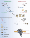
The mechanism diagram of this study

Similar articles

Cited by

References

    1. Sung H, et al. Global Cancer statistics 2020: GLOBOCAN estimates of incidence and Mortality Worldwide for 36 cancers in 185 countries. Cancer J Clin. 2021;71(3):209–49. doi: 10.3322/caac.21660. - DOI - PubMed
    1. Danenberg E, et al. Breast tumor microenvironment structures are associated with genomic features and clinical outcome. Nat Genet. 2022;54(5):660–9. doi: 10.1038/s41588-022-01041-y. - DOI - PMC - PubMed
    1. NICE, Early and locally advanced breast cancer: diagnosis and management, in Early and locally advanced breast cancer: diagnosis and management. 2024: London.
    1. Westcott PMK, et al. Mismatch repair deficiency is not sufficient to elicit tumor immunogenicity. Nat Genet. 2023;55(10):1686–95. doi: 10.1038/s41588-023-01499-4. - DOI - PMC - PubMed
    1. Chen X et al. Mechanisms and strategies to overcome PD-1/PD-L1 Blockade Resistance in Triple-negative breast Cancer. Cancers (Basel), 2022. 15(1). - PMC - PubMed

Publication types

MeSH terms