

Ferroptosis: principles and significance in health and disease
- PMID: 38844964
- PMCID: PMC11157757
- DOI: 10.1186/s13045-024-01564-3
Ferroptosis: principles and significance in health and disease
Abstract
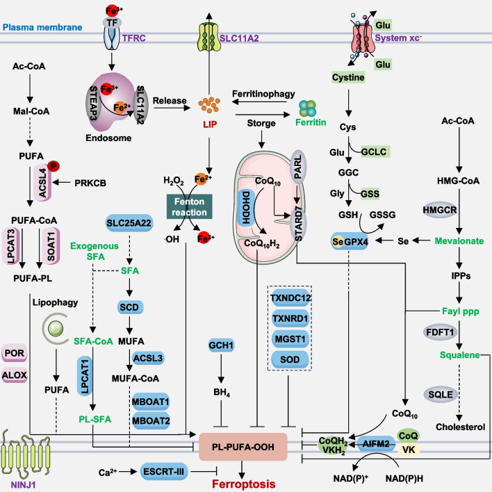
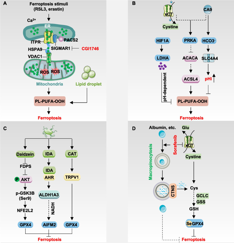
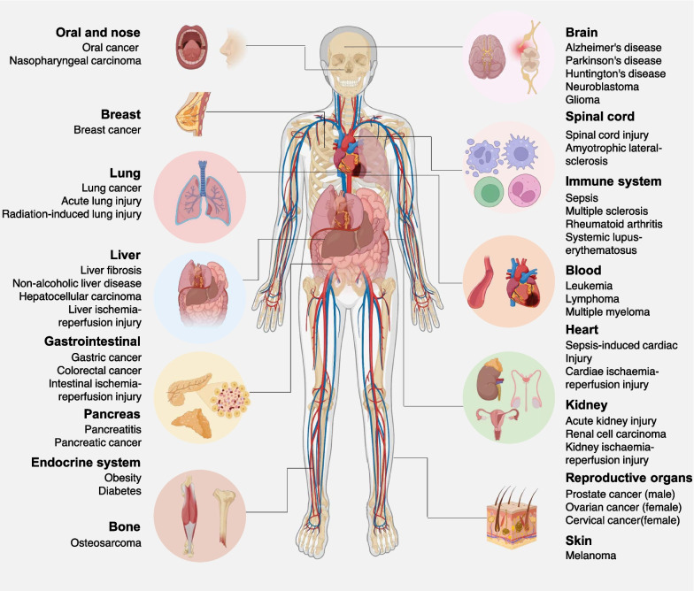
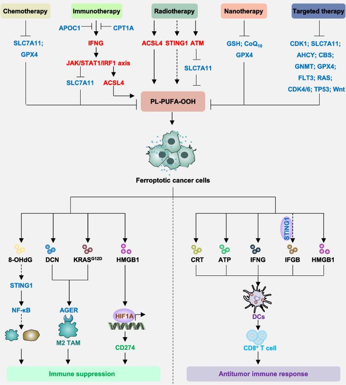
Ferroptosis, an iron-dependent form of cell death characterized by uncontrolled lipid peroxidation, is governed by molecular networks involving diverse molecules and organelles. Since its recognition as a non-apoptotic cell death pathway in 2012, ferroptosis has emerged as a crucial mechanism in numerous physiological and pathological contexts, leading to significant therapeutic advancements across a wide range of diseases. This review summarizes the fundamental molecular mechanisms and regulatory pathways underlying ferroptosis, including both GPX4-dependent and -independent antioxidant mechanisms. Additionally, we examine the involvement of ferroptosis in various pathological conditions, including cancer, neurodegenerative diseases, sepsis, ischemia-reperfusion injury, autoimmune disorders, and metabolic disorders. Specifically, we explore the role of ferroptosis in response to chemotherapy, radiotherapy, immunotherapy, nanotherapy, and targeted therapy. Furthermore, we discuss pharmacological strategies for modulating ferroptosis and potential biomarkers for monitoring this process. Lastly, we elucidate the interplay between ferroptosis and other forms of regulated cell death. Such insights hold promise for advancing our understanding of ferroptosis in the context of human health and disease.
Keywords: Biomarker; Cancer therapy; Ferroptosis; Human disease; Immunity.
© 2024. The Author(s).
Conflict of interest statement
The authors declare that they have no competing interests.
Figures
Similar articles
-
MicroRNA-mediated regulation of Ferroptosis: Implications for disease pathogenesis and therapeutic interventions.Cell Signal. 2025 Jan;125:111503. doi: 10.1016/j.cellsig.2024.111503. Epub 2024 Nov 5. Cell Signal. 2025. PMID: 39510403 Review.
-
Lipid metabolism in ferroptosis: mechanistic insights and therapeutic potential.Front Immunol. 2025 Mar 11;16:1545339. doi: 10.3389/fimmu.2025.1545339. eCollection 2025. Front Immunol. 2025. PMID: 40134420 Free PMC article. Review.
-
GPX4, ferroptosis, and diseases.Biomed Pharmacother. 2024 May;174:116512. doi: 10.1016/j.biopha.2024.116512. Epub 2024 Apr 3. Biomed Pharmacother. 2024. PMID: 38574617 Review.
-
Targeting ferroptosis by natural products in pathophysiological conditions.Arch Toxicol. 2024 Oct;98(10):3191-3208. doi: 10.1007/s00204-024-03812-4. Epub 2024 Jul 10. Arch Toxicol. 2024. PMID: 38987487 Review.
-
Non-coding RNA: A key regulator in the Glutathione-GPX4 pathway of ferroptosis.Noncoding RNA Res. 2024 May 20;9(4):1222-1234. doi: 10.1016/j.ncrna.2024.05.007. eCollection 2024 Dec. Noncoding RNA Res. 2024. PMID: 39036600 Free PMC article. Review.
Cited by
-
The molecular mechanism and therapeutic landscape of copper and cuproptosis in cancer.Signal Transduct Target Ther. 2025 May 9;10(1):149. doi: 10.1038/s41392-025-02192-0. Signal Transduct Target Ther. 2025. PMID: 40341098 Free PMC article. Review.
-
Molecular characteristics and immune microenvironment of gastrointestinal stromal tumours: targets for therapeutic strategies.Front Oncol. 2024 Jul 12;14:1405727. doi: 10.3389/fonc.2024.1405727. eCollection 2024. Front Oncol. 2024. PMID: 39070147 Free PMC article. Review.
-
A Novel Ferroptosis-Related Gene Prognosis Signature and Identifying Atorvastatin as a Potential Therapeutic Agent for Hepatocellular Carcinoma.Curr Issues Mol Biol. 2025 Mar 18;47(3):201. doi: 10.3390/cimb47030201. Curr Issues Mol Biol. 2025. PMID: 40136455 Free PMC article.
-
Inhibition of Setd7 protects against cardiomyocyte hypertrophy via inhibiting lipid oxidation.Acta Pharmacol Sin. 2025 Aug 12. doi: 10.1038/s41401-025-01626-3. Online ahead of print. Acta Pharmacol Sin. 2025. PMID: 40797115
-
Ferroptosis: a potential target for non-surgical treatment of laryngeal cancer.Eur Arch Otorhinolaryngol. 2025 Mar 14. doi: 10.1007/s00405-025-09279-y. Online ahead of print. Eur Arch Otorhinolaryngol. 2025. PMID: 40087171 Review.
References
Publication types
MeSH terms
LinkOut - more resources
Full Text Sources
