. 2024 Jun 6;22(1):544.
doi: 10.1186/s12967-024-05361-x.

Deficiency of SDHC promotes metastasis by reprogramming fatty acid metabolism in colorectal cancer

Affiliations

Deficiency of SDHC promotes metastasis by reprogramming fatty acid metabolism in colorectal cancer

Zhuoyu Ding et al. J Transl Med. .

Abstract

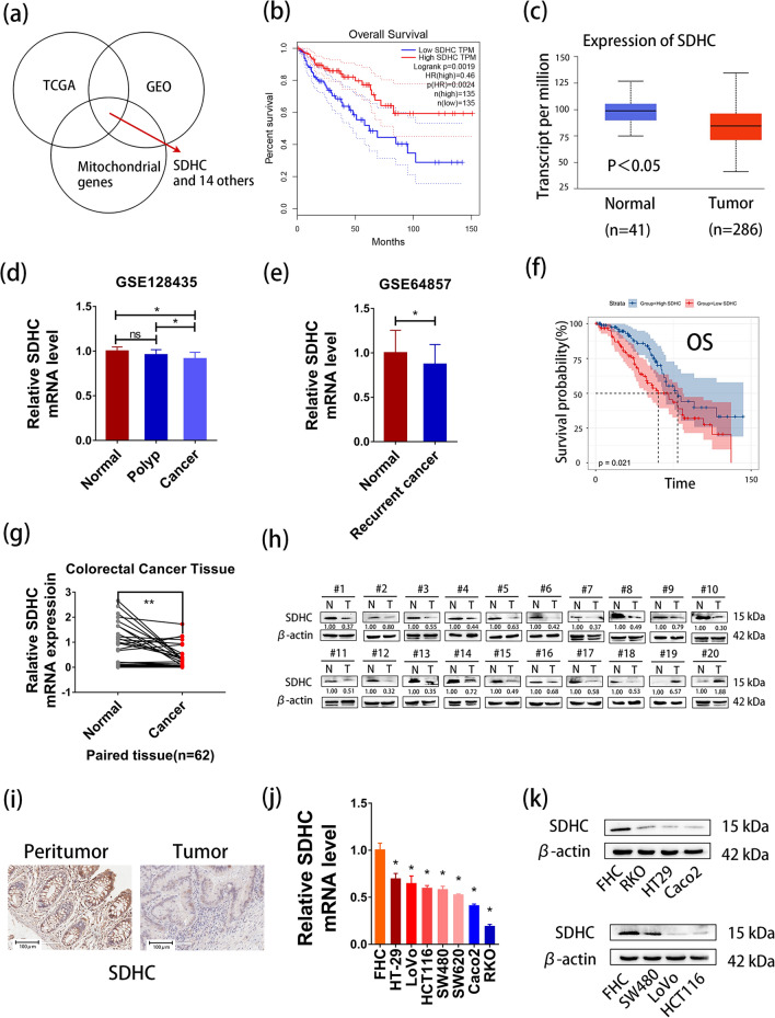
Background: Several studies have demonstrated a strong correlation between impaired Succinate dehydrogenase (SDH) function and the advancement of tumors. As a subunit of SDH, succinate dehydrogenase complex subunit C (SDHC) has been revealed to play tumor suppressive roles in several cancers, while its specific role in colorectal cancer (CRC) still needs further investigation.

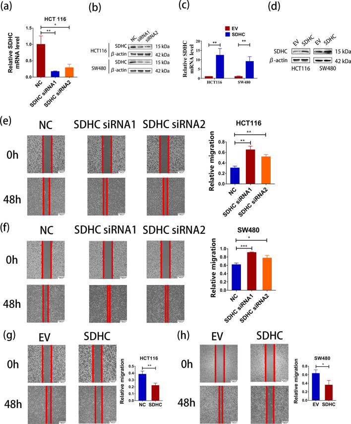
Methods: Online database were utilized to investigate the expression of SDHC in colorectal cancer and to assess its correlation with patient prognosis. Cell metastasis was assessed using transwell and wound healing assays, while tumor metastasis was studied in a nude mice model in vivo. Drug screening and RNA sequencing were carried out to reveal the tumor suppressor mechanism of SDHC. Triglycerides, neutral lipids and fatty acid oxidation were measured using the Triglyceride Assay Kit, BODIPY 493/503 and Colorimetric Fatty Acid Oxidation Rate Assay Kit, respectively. The expression levels of enzymes involved in fatty acid metabolism and the PI3K/AKT signaling pathway were determined by quantitative real-time PCR and western blot.

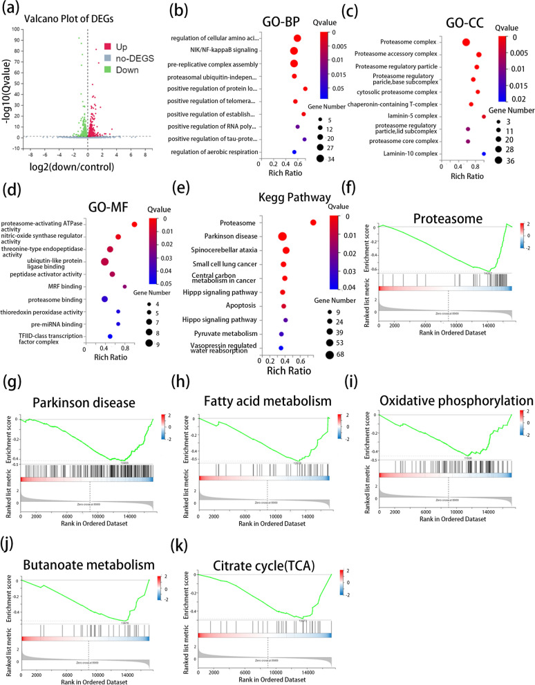
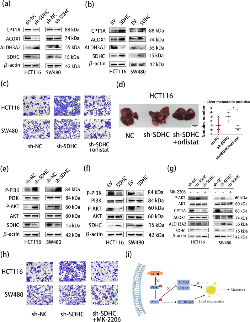
Results: Downregulation of SDHC was found to be closely associated with a poor prognosis in CRC. SDHC knockdown promoted CRC metastasis both in vitro and in vivo. Through drug screening and Gene set enrichment analysis, it was discovered that SDHC downregulation was positively associated with the fatty acid metabolism pathways significantly. The effects of SDHC silencing on metastasis were reversed when fatty acid synthesis was blocked. Subsequent experiments revealed that SDHC silencing activated the PI3K/AKT signaling axis, leading to lipid accumulation by upregulating the expression of aldehyde dehydrogenase 3 family member A2 (ALDH3A2) and reduction of fatty acid oxidation rate by suppressing the expression of acyl-coenzyme A oxidase 1 (ACOX1) and carnitine palmitoyltransferase 1A (CPT1A).

Conclusions: SDHC deficiency could potentially enhance CRC metastasis by modulating the PI3K/AKT pathways and reprogramming lipid metabolism.

Keywords: Colorectal cancer; Fatty acid metabolism; Metastasis; SDHC.


Conflict of interest statement

The authors declare no competing interests.

Figures

Fig. 1
Fig. 1
Expression and prognostic value of genes in colorectal cancer. a Intersection of differential genes in GEO, TCGA and mitochondrial genes. The dataset in GEO includes GSE81582, GSE77953, GSE77199 and GSE41258. b Low expression of SDHC was found to be associated with poor Overall Survival in CRC by GEPIA. c Low expression of SDHC in CRC tissues compared to normal tissues by UALCAN. d Low expression of SDHC in CRC tissues compared to normal tissues or polyps in GSE128435. e Low expression of SDHC in recurrent CRC compared to normal tissues in GSE64857. f Low expression of SDHC was found to be associated with poor Overall Survival in CRC in GSE17538. g qRT-PCR analysis of SDHC expression in 62 CRC patient samples. h Western blot analysis of SDHC expression in CRC tissues compared with adjacent normal tissues. i Immunohistochemistry analysis of SDHC expression in CRC tissues compared with adjacent normal tissues. j qRT-PCR analysis of SDHC expression in CRC cells and normal colon cells. k Western blot analysis of SDHC expression in CRC cells and normal colon cells. Data are presented as the mean ± SD and analyzed by Student’s t-test. *P < 0.05, **P < 0.01, ***P < 0.001
Fig. 2
Fig. 2
SDHC contributed to CRC metastasis and in vitro. a SDHC levels in HCT116 cells were analyzed by qPCR after transfection with siRNA negative control (NC), SDHC siRNA1 and SDHC siRNA2. b SDHC levels in HCT116 and SW480 cells were analyzed by western blot after transfection with NC, SDHC siRNA1 and SDHC siRNA2. c The overexpression of SDHC levels in HCT116 cells were analyzed by qPCR. d The overexpression of SDHC levels in HCT116 and SW480 cells were analyzed by western blot. e Wound healing assays showed that SDHC knockdown could promote the healing of scratches in HCT116 cells. f Overexpression of SDHC repressed the healing of scratches in HCT116 cells. g Wound healing assays showed that SDHC knockdown could promote the healing of scratches in SW480 cells. h Overexpression of SDHC repressed the healing of scratches in SW480 cells. Data are presented as the mean ± SD and analyzed by Student’s t-test. *P < 0.05, **P < 0.01, ***P < 0.001
Fig. 3
Fig. 3
SDHC contributed to CRC metastasis in vitro. a Transwell assays revealed that SDHC knockdown promoted HCT116 and SW480 cells invasion. b Transwell assays revealed that overexpression of SDHC inhibited HCT116 and SW480 cells invasion. c Livers were excised from tumor-bearing mice and metastatic nodules were determined. Left panel shows representative liver tissues with metastatic nodules, the red arrows indicated the tumor nodules. Right panel, analysis of liver nodule numbers. Each dot denotes an animal. d Lung tumors were shown. The black arrows indicated the tumor nodules. And the number of metastasis in the lung was determined. Data are presented as the mean ± SD and analyzed by Student’s t-test. *P < 0.05, **P < 0.01, ***P < 0.001
Fig. 4
Fig. 4
Bioinformatics analysis. a The volcano plot represents the DEGs. b GO annotations of the DEGs. The Bulb map presents enrichment scores of the top 10 significantly enriched GO terms in biological processes. c GO annotations of the DEGs. The Bulb map presents enrichment scores of the top 10 significantly enriched GO terms in cellular components. d GO annotations of the DEGs. The Bulb map presents the enrichment scores of the top 10 significantly enriched GO terms in molecular functions. e KEGG pathway enrichment analysis of DEGs. f Proteasome pathway enriched by GSEA. g Parkinson disease pathway enriched by GSEA. h Fatty acid metabolism pathway enriched by GSEA. i Oxidative phosphorylation pathway enriched by GSEA. j butanoate metabolism pathway enriched by GSEA. k Citrate cycle (TCA cycle) pathway enriched by GSEA. Data are presented as the mean ± SD and analyzed by Student’s t-test. *P < 0.05, **P < 0.01, ***P < 0.001
Fig. 5
Fig. 5
Lipid accumulation was mediated by SDHC silencing. a A log10-IC50 plot of the screening results. A log10 scale of IC50 values of the drugs against HCT116-sh-SDHC and control cells was plotted. Drugs with selectivity index (SI) > 2 were selected and marked as synthetic lethality candidates. b The content of neutral lipids in HCT116 (NC/siRNA1/siRNA2) and SW480 (NC/siRNA1/siRNA2) cells were measured by Confocal microscope. c The content of neutral lipids in HCT116 (EV/SDHC) and SW480 (EV/SDHC) cells were measured by Confocal microscope. d The content of neutral lipids in HCT116 (NC/siRNA1/siRNA2) and SW480 (NC/siRNA1/siRNA2) cells were measured by flow cytometry. e The content of neutral lipids in HCT116 (EV/SDHC) and SW480 (EV/SDHC) cells were measured by flow cytometry. f The silencing of SDHC enhanced the triglyceride levels in HCT116 and SW480 cells. g SDHC overexpression decreased the triglyceride levels in HCT116 and SW480 cells. h The silencing of SDHC decreased the FAO rate in HCT116 and SW480 cells. i SDHC overexpression increased the FAO rate in HCT116 and SW480 cells. Data are presented as the mean ± SD and analyzed by Student’s t-test. *P < 0.05, **P < 0.01, ***P < 0.001
Fig. 6
Fig. 6
SDHC reprogramed fatty acid metabolism by regulating the PI3K/AKT pathways. a Western blotting was used to demonstrate the effects of silencing SDHC on the expression of critical enzymes involved in lipid metabolism, namely ACOX1, CPT1A, and ALDH3A2. b Western blotting was used to demonstrate the effects of overexpression SDHC on the expression of critical enzymes involved in lipid metabolism. c Migration assay analysis of orlistat in HCT116 and SW480 cells with SDHC decreased. d The effect of orlistat on liver metastasis formation in nude mice bearing HCT116 cells with SDHC decreased. e Western blotting reveals the levels of PI3K, p-PI3K, AKT and p-AKT in HCT116 and SW480 cells with SDHC decreased and control. f Western blotting demonstrates the levels of PI3K, p-PI3K, AKT and p-AKT in HCT116 and SW480 cells with SDHC overexpress and control. g Western blotting demonstrates the levels of AKT, p-AKT, ACOX1, CPT1A and ALDH3A2 in HCT116-sh-SDHC and SW480-sh-SDHC cells treated with MK-2206. h Migration assay analysis of MK-2206 in HCT116 and SW480 cells with SDHC decreased. i Schematic depiction of the regulation on fatty acid metabolism by SDHC in CRC cells. Data are presented as the mean ± SD and analyzed by Student’s t-test. *P < 0.05, **P < 0.01, ***P < 0.001

Similar articles

References

    1. Sung H, Ferlay J, Siegel RL, Laversanne M, Soerjomataram I, Jemal A, et al. Global cancer statistics 2020: GLOBOCAN estimates of incidence and mortality worldwide for 36 cancers in 185 countries. CA Cancer J Clin. 2021;71(3):209–49. 10.3322/caac.21660 - DOI - PubMed
    1. Siegel RL, Miller KD, Goding Sauer A, Fedewa SA, Butterly LF, Anderson JC, et al. Colorectal cancer statistics, 2020. CA Cancer J Clin. 2020;70(3):145–64. 10.3322/caac.21601 - DOI - PubMed
    1. Eng C, Kiuru M, Fernandez M, Aaltonen L. A role for mitochondrial enzymes in inherited neoplasia and beyond. Nat Rev Cancer. 2003;3(3):193–202. 10.1038/nrc1013 - DOI - PubMed
    1. Bardella C, Pollard P, Tomlinson I. SDH mutations in cancer. Biochem Biophys Acta. 2011;1807(11):1432–43. - PubMed
    1. Ishii T, Yasuda K, Akatsuka A, Hino O, Hartman P, Ishii N. A mutation in the SDHC gene of complex II increases oxidative stress, resulting in apoptosis and tumorigenesis. Can Res. 2005;65(1):203–9.10.1158/0008-5472.203.65.1 - DOI - PubMed

MeSH terms

Substances