

Effects of super-enhancers in cancer metastasis: mechanisms and therapeutic targets
- PMID: 38844984
- PMCID: PMC11157854
- DOI: 10.1186/s12943-024-02033-8
Effects of super-enhancers in cancer metastasis: mechanisms and therapeutic targets
Abstract
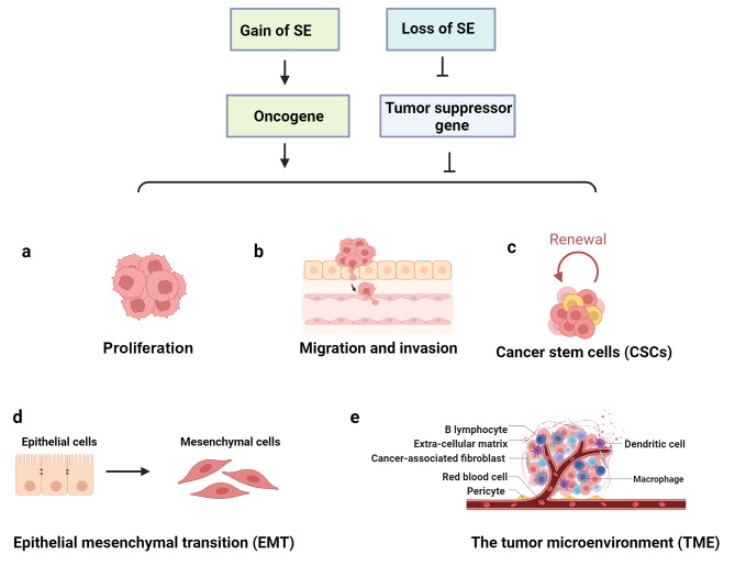
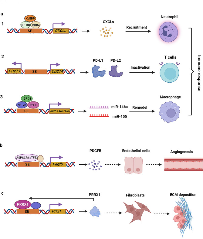
Metastasis remains the principal cause of cancer-related lethality despite advancements in cancer treatment. Dysfunctional epigenetic alterations are crucial in the metastatic cascade. Among these, super-enhancers (SEs), emerging as new epigenetic regulators, consist of large clusters of regulatory elements that drive the high-level expression of genes essential for the oncogenic process, upon which cancer cells develop a profound dependency. These SE-driven oncogenes play an important role in regulating various facets of metastasis, including the promotion of tumor proliferation in primary and distal metastatic organs, facilitating cellular migration and invasion into the vasculature, triggering epithelial-mesenchymal transition, enhancing cancer stem cell-like properties, circumventing immune detection, and adapting to the heterogeneity of metastatic niches. This heavy reliance on SE-mediated transcription delineates a vulnerable target for therapeutic intervention in cancer cells. In this article, we review current insights into the characteristics, identification methodologies, formation, and activation mechanisms of SEs. We also elaborate the oncogenic roles and regulatory functions of SEs in the context of cancer metastasis. Ultimately, we discuss the potential of SEs as novel therapeutic targets and their implications in clinical oncology, offering insights into future directions for innovative cancer treatment strategies.
Keywords: Metastasis; Molecular mechanisms; Super-enhancers; Therapeutic targets.
© 2024. The Author(s).
Conflict of interest statement
The authors declare no competing interests.
Figures
Similar articles
-
EphA2 super-enhancer promotes tumor progression by recruiting FOSL2 and TCF7L2 to activate the target gene EphA2.Cell Death Dis. 2021 Mar 12;12(3):264. doi: 10.1038/s41419-021-03538-6. Cell Death Dis. 2021. PMID: 33712565 Free PMC article.
-
Oncogenic super-enhancer formation in tumorigenesis and its molecular mechanisms.Exp Mol Med. 2020 May;52(5):713-723. doi: 10.1038/s12276-020-0428-7. Epub 2020 May 7. Exp Mol Med. 2020. PMID: 32382065 Free PMC article. Review.
-
[Research progress of super enhancer in cancer].Yi Chuan. 2019 Jan 20;41(1):41-51. doi: 10.16288/j.yczz.18-152. Yi Chuan. 2019. PMID: 30686784 Review. Chinese.
-
The Emerging Role of Super-enhancers as Therapeutic Targets in The Digestive System Tumors.Int J Biol Sci. 2023 Jan 22;19(4):1036-1048. doi: 10.7150/ijbs.78535. eCollection 2023. Int J Biol Sci. 2023. PMID: 36923930 Free PMC article. Review.
-
The emerging role of super enhancer-derived noncoding RNAs in human cancer.Theranostics. 2020 Sep 2;10(24):11049-11062. doi: 10.7150/thno.49168. eCollection 2020. Theranostics. 2020. PMID: 33042269 Free PMC article. Review.
Cited by
-
Recent insights on the impact of SWELL1 on metabolic syndromes.Front Pharmacol. 2025 Mar 21;16:1552176. doi: 10.3389/fphar.2025.1552176. eCollection 2025. Front Pharmacol. 2025. PMID: 40191429 Free PMC article. Review.
-
Enhancer regulation in cancer: from epigenetics to m6A RNA modification.Arch Pharm Res. 2025 Aug 19. doi: 10.1007/s12272-025-01561-1. Online ahead of print. Arch Pharm Res. 2025. PMID: 40830299 Review.
-
Targeting c-Met in Cancer Therapy: Unravelling Structure-activity Relationships and Docking Insights for Enhanced Anticancer Drug Design.Curr Top Med Chem. 2025;25(4):409-433. doi: 10.2174/0115680266331025241015084546. Curr Top Med Chem. 2025. PMID: 39484763 Review.
-
New insights into the dule roles CDK12 in human cancers: Mechanisms and interventions for cancer therapy.J Pharm Anal. 2025 Jul;15(7):101173. doi: 10.1016/j.jpha.2024.101173. Epub 2024 Dec 28. J Pharm Anal. 2025. PMID: 40747342 Free PMC article. Review.
-
Non-Coding RNAs and Innate Immune Responses in Cancer.Biomedicines. 2024 Sep 11;12(9):2072. doi: 10.3390/biomedicines12092072. Biomedicines. 2024. PMID: 39335585 Free PMC article. Review.
References
Publication types
MeSH terms
Grants and funding
- 20224BAB216121/Natural Science Foundation of Jiangxi Province, China
- 20232BAB216137/Natural Science Foundation of Jiangxi Province, China
- 20212ACB216002/Natural Science Foundation of Jiangxi Province, China
- 82003797/National Natural Science Foundation of China
- 82111530101/National Natural Science Foundation of China
LinkOut - more resources
Full Text Sources
Medical
