. 2024 Jun 6:28:0031.
doi: 10.34133/bmr.0031. eCollection 2024.

Recent Advances in Functional Hydrogel for Repair of Abdominal Wall Defects: A Review

Affiliations

Recent Advances in Functional Hydrogel for Repair of Abdominal Wall Defects: A Review

Ye Liu et al. Biomater Res. .

Abstract

The abdominal wall plays a crucial role in safeguarding the internal organs of the body, serving as an essential protective barrier. Defects in the abdominal wall are common due to surgery, infection, or trauma. Complex defects have limited self-healing capacity and require external intervention. Traditional treatments have drawbacks, and biomaterials have not fully achieved the desired outcomes. Hydrogel has emerged as a promising strategy that is extensively studied and applied in promoting tissue regeneration by filling or repairing damaged tissue due to its unique properties. This review summarizes the five prominent properties and advances in using hydrogels to enhance the healing and repair of abdominal wall defects: (a) good biocompatibility with host tissues that reduces adverse reactions and immune responses while supporting cell adhesion migration proliferation; (b) tunable mechanical properties matching those of the abdominal wall that adapt to normal movement deformations while reducing tissue stress, thereby influencing regulating cell behavior tissue regeneration; (c) drug carriers continuously delivering drugs and bioactive molecules to sites optimizing healing processes enhancing tissue regeneration; (d) promotion of cell interactions by simulating hydrated extracellular matrix environments, providing physical support, space, and cues for cell migration, adhesion, and proliferation; (e) easy manipulation and application in surgical procedures, allowing precise placement and close adhesion to the defective abdominal wall, providing mechanical support. Additionally, the advances of hydrogels for repairing defects in the abdominal wall are also mentioned. Finally, an overview is provided on the current obstacles and constraints faced by hydrogels, along with potential prospects in the repair of abdominal wall defects.


Conflict of interest statement

Competing interests: The authors declare that they have no competing interests.

Figures
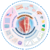
Fig. 1.
Fig. 1.
Illustrations of hydrogel with 5 prominent properties applied to repair abdominal wall defects.
Fig. 2.
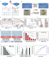
Fig. 2.
The excellent tunable mechanical properties of hydrogels. (A) Schematic of the CS/PAM hydrogel with molecular network prepared by UV treatment. (B) Schematic and photographs of the alkali treatment mechanism of CS/PAM and CSA/PAM hydrogels. (C) CS/PAM, CS/PAM- pf, CPF, and CAPF frequency scans and (D) strain sweep. (E) Commercial PP mesh, PF, CPF, and CAPF tensile strength–strain curves in transverse and longitudinal directions and (F) 16 N cm−1 elongation. (G) Mechanism of adhesion of CAPFAH hydrogels to wet tissue. (H) Hydrogel shear strength after multiple adhesion. (I) Adhesion strength of hydrogel with time after multiple adhesions. Reproduced from [93] with permission from John Wiley and Sons, Copyright 2023. (J) Cy5.5@ECM and Cy5.5@ECMB composites’ residual fluorescence. (K) Cy5.5@ECM and Cy5.5@ECMB composite explants’ wall thickness. (L) Cy5.5@ECMB composite fluorescence loss and explant thickness. Reproduced from [102] with permission from the Royal Society of Chemistry, Copyright 2021.
Fig. 3.
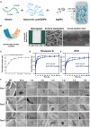
Fig. 3.
Drug carrier is one of the excellent properties of hydrogel. (A) A multifunctional hydrogel was synthesized for promoting abdominal wall defect repair based on dopamine-modified hyaluronic acid, gelatin, and nano-silver. Reproduced from [105] with permission from Elsevier, Copyright 2022. (B) An illustration, macrographs, and FESEM image of the GO-PCL/NAC-CS-PCL scaffolds. (C) NAC cumulative release from the scaffolds is time-dependent. Reproduced from [106] with permission from Dove Medical Press, Copyright 2021. (D) In vitro release pharmacological profile of Rhodamine B and (E) VEGF by simple XG hydrogel and XG/TPEG hydrogel carriers. Reproduced from [107] with permission from the Royal Society of Chemistry, Copyright 2020. (F) Scanning electron microscopy view of ADSCs (indicated by red arrows) cultured on scaffolds to show cell morphology. Reproduced from [108] with permission from Frontiers Media S.A, Copyright 2021.
Fig. 4.
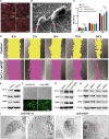
Fig. 4.
Biomimetic property is one of the excellent properties of hydrogel. (A) hGFs co-cultured with Gel 1 to 3 fluorescence images. Reproduced from [79] with permission from John Wiley and Sons, Copyright 2021. (B) Morphology of human dermal fibroblasts cultured on 0.1% GO-PCL/NAC-CS-PCL scaffold. Cell pseudopods are denoted by red arrows. (C) CCK-8 assay of human dermal fibroblasts. Reproduced from [106] with permission from Dove Medical Press, Copyright 2021. (D) Scratches healing experiments with images of hGF migration at different times. Reproduced from [79] with permission from John Wiley and Sons, Copyright 2021. (E) Western blot to examine the expression of HIF-1α, VEGF, and α-SMA in hypoxia-induced ADSCs. (F) Transfection of ADSCs with lenti-HIF-1α and lenti-VEGF for 72 h. (G) Western blotting to examine the expression and analysis of HIF-1α, VEGF, and α-SMA in lenti-HIF-1α and lenti-VEGF-transfected ADSCs. (H) Western blot to examine the expression and analysis of HIF-1α, VEGF, and α-SMA after HIF-1α siRNA knockdown of ADSCs. (I) WPBs (red triangles) were visible by TEM after transfection of lenti-HIF-1α and lenti-VEGF with ADSCs. Reproduced from [108] with permission from Frontiers Media S.A, Copyright 2021.
Fig. 5.
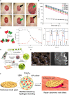
Fig. 5.
Simplicity of the method using hydrogels. (A) An in situ injection of chitosan-hyaluronic acid hydrogel was applied for abdominal wall repair. Reproduced from [123] with permission from Springer Nature, Copyright 2017. (B) WBPU dispersion and A-WBPU ink flow viscosity profiles according to shear rate increment. Reproduced from [124] with permission from John Wiley and Sons, Copyright 2022. (C) The 4-arm-PEG-CHO/CMCS hydrogel for macroscopic self-repair and rheological recovery testing. Reproduced from [79] with permission from John Wiley and Sons, Copyright 2021. (D) A novel composite scaffold (DFO-HPMs-PADM) was constructed by spraying DFO-loaded HPMs on the surface of PADM. Reproduced from [95] with permission from the Royal Society of Chemistry, Copyright 2018. (E) Optical micrographs and SEM images of AH-PPM. Reproduced from [125] with permission from Elsevier, Copyright 2022. (F) A dECM-based patch adhered to the injured tissue for abdominal wall defect repair. Reproduced from [126] with permission from the American Chemical Society, Copyright 2023.
Fig. 6.
Fig. 6.
Advances of hydrogel for abdominal wall repair. (A) A rat model was used to evaluate the effect of abdominal wall defect repair. (B) HE, MT, collagen, and anti-CD68 antibody stain of treated abdominal wall defects. Reproduced from [126] with permission from the American Chemical Society, Copyright 2023. (C) Immunofluorescence of CD31/α-SMA of Janus porous hydrogel. Reproduced from [83] with permission from John Wiley and Sons, Copyright 2022. (D) Image of Masson trichrome staining of PP mesh, CAPF, and CAPFAH at 14, 30, and 150 days after implantation. Reproduced from [93] with permission from John Wiley and Sons, Copyright 2023. (E) Self-assembly of natural antimicrobial peptide jelly-1 (J-1) in sodium adenosine diphosphate (ADP) solution to form the J-1-ADP hydrogel for prevention of postoperative adhesions. Reproduced from [139] with permission from the American Chemical Society, Copyright 2022. (F) In vivo prevention of visceral effectiveness of adhesion formation. Reproduced from [83] with permission from John Wiley and Sons, Copyright 2022.

Similar articles

Cited by

References

    1. Heniford BT, Park A, Ramshaw BJ, Voeller G. Laparoscopic repair of ventral hernias nine years' experience with 850 consecutive hernias. Ann Surg. 2003;238(3):391–399. - PMC - PubMed
    1. Mathes SJ, Steinwald PM, Foster RD, Hoffman WY, Anthony JP. Complex abdominal wall reconstruction: A comparison of flap and mesh closure. Ann Surg. 2000;232(4):586–594. - PMC - PubMed
    1. Rathore A, Murherjee I, Kumar P. Traumatic abdominal wall injuries: A review. Minerva Chir. 2013;68(3):221–232. - PubMed
    1. Gillion J, Sanders D, Miserez M, Muysoms F. The economic burden of incisional ventral hernia repair: A multicentric cost analysis. Hernia. 2016;20(6):819–830. - PubMed
    1. Beadles CA, Meagher AD, Charles AG. Trends in emergent hernia repair in the United States. JAMA Surg. 2015;150(3):194–200. - PubMed

LinkOut - more resources