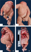
Craniorachischisis in a 33-week-old Female Fetus: A Case Report
- PMID: 38846167
- PMCID: PMC11151135
- DOI: 10.47895/amp.vi0.6712
Craniorachischisis in a 33-week-old Female Fetus: A Case Report
Abstract
We report the case of a 33-week-old female fetus born with craniorachischisis to a gravida 5, para 4 (3104) mother with no previous history of conceiving a child with a neural tube defect. Craniorachischisis is characterized by anencephaly and an open defect extending from the brain to the spine and is the most severe and fatal type of neural tube defect. Although the cause of neural tube defects is hypothesized to be multifactorial and is usually sporadic, the risk is increased in neonates born to mothers with a family history or a previous pregnancy with neural tube defect, both of which are not present in the index case. This case is unique in that only during the fifth pregnancy did the couple conceive a child with a neural tube defect, emphasizing that folic acid supplementation, the sole preventive measure proven to decrease the risk of neural tube defects, remains to be important in the periconceptual period for all women of childbearing age.
Keywords: autopsy; case report; congenital abnormalities; craniorachischisis; neural tube defects.
© 2024 Acta Medica Philippina.
Conflict of interest statement
All authors declared no conflicts of interest.
Figures


Similar articles
-
Craniorachischisis with Exencephaly.Fetal Pediatr Pathol. 2021 Oct;40(5):501-504. doi: 10.1080/15513815.2020.1716282. Epub 2020 Jan 27. Fetal Pediatr Pathol. 2021. PMID: 31986946
-
Craniorachischisis Totalis: A Detailed Case Report.Iran J Child Neurol. 2025;19(2):149-153. doi: 10.22037/ijcn.v19i2.44334. Epub 2025 Mar 11. Iran J Child Neurol. 2025. PMID: 40231282 Free PMC article.
-
Pre-conception Folic Acid and Multivitamin Supplementation for the Primary and Secondary Prevention of Neural Tube Defects and Other Folic Acid-Sensitive Congenital Anomalies.J Obstet Gynaecol Can. 2015 Jun;37(6):534-52. doi: 10.1016/s1701-2163(15)30230-9. J Obstet Gynaecol Can. 2015. PMID: 26334606 English, French.
-
[Craniorachischisis in conjoined "diprosopus" twins. Case report and review of the literature].Ann Pathol. 1989;9(5):346-50. Ann Pathol. 1989. PMID: 2692575 Review. French.
-
Concomitant craniorachischisis and omphalocele in a male fetus: prenatal magnetic resonance imaging findings and literature review.Taiwan J Obstet Gynecol. 2009 Sep;48(3):286-91. doi: 10.1016/S1028-4559(09)60306-5. Taiwan J Obstet Gynecol. 2009. PMID: 19797022 Review.
References
-
- Centers for Disease Control and Prevention ; Key findings: Global burden of neural tube defects [Internet]. 2017. [cited 2022 Aug]. Available from: https://www.cdc.gov/ncbddd/birthdefectscount/features/kf-neutral-tube-de...
-
- Ostrea Jr. EM. Prevention of fetal neural tube defect with folic acid supplementation. Editorial. Acta Med Philipp. 2022;56(5):4-5. doi: 10.47895/amp.v56i5.5539 - DOI
Publication types
LinkOut - more resources
Full Text Sources
