

Optic Nerve Schwannoma: A Report of a Rare Case From India and Literature Review
- PMID: 38846181
- PMCID: PMC11156426
- DOI: 10.7759/cureus.59824
Optic Nerve Schwannoma: A Report of a Rare Case From India and Literature Review
Abstract
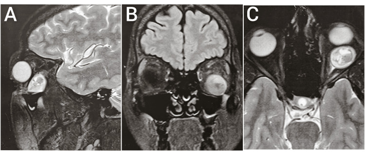
Optic nerve schwannoma is a very rarely occurring tumor described in the literature. It is due to the fact that the optic nerve is myelinated by oligodendrocytes. Schwannomas are tumors of the peripheral nervous system, hence optic nerve schwannoma is a rare phenomenon. A 34-year-old patient presented in the outpatient department with complaints of gradual painless protrusion of the left eye (LE) for the past one year. There was no history of diminution of vision. On examination, vision in both eyes was 6/6, anterior segment examination in both eyes was normal, and pupils were central, circular, and reacting to light. Intraocular pressure was measured on a noncontact tonometer and was within normal range. Both eyes' optic disc, fundus, and visual fields were normal. On inspection, axial proptosis was noted in the LE. Proptosis measurement (on Hertel exophthalmometer) in the right eye was 17 mm and in the left eye was 21 mm. MRI of the orbit without contrast was done and showed a well-defined, soft tissue lesion of the optic nerve in the intraconal compartment of the left orbit. Surgical excision of the tumor was done by lateral orbitotomy approach and the tumor was removed in total. Histopathological examination of the mass revealed a benign spindle cell neoplasm suggestive of schwannoma. Postoperatively, proptosis was resolved, 17 mm both in the right and left eye (on Hertel exophthalmometer), and vision in LE remained unchanged (6/6). Postoperatively, intraocular pressure (on noncontact tonometer) was within normal range, and the optic disc, fundus, and visual fields were normal.
Keywords: case report; india; optic nerve sheath; orbital tumors; schwannoma.
Copyright © 2024, Lune et al.
Conflict of interest statement
The authors have declared that no competing interests exist.
Figures


Similar articles
-
Primary optic nerve sheath schwannoma: a case report.Br J Neurosurg. 2023 Oct;37(5):1333-1335. doi: 10.1080/02688697.2020.1869181. Epub 2021 Jan 8. Br J Neurosurg. 2023. PMID: 33416410 Review.
-
Primary optic nerve sheath schwannoma: A case report and literature review.Radiol Case Rep. 2023 Sep 18;18(11):4211-4213. doi: 10.1016/j.radcr.2023.08.085. eCollection 2023 Nov. Radiol Case Rep. 2023. PMID: 37745769 Free PMC article.
-
Is primary optic nerve sheath schwannoma a misnomer? Report of two cases and literature review.Orbit. 2019 Oct;38(5):419-423. doi: 10.1080/01676830.2018.1545239. Epub 2018 Nov 16. Orbit. 2019. PMID: 30444169 Review.
-
Orbital oculomotor nerve schwannoma extending to the cavernous sinus: a rare cause of proptosis.J Ophthalmic Vis Res. 2014 Oct-Dec;9(4):514-6. doi: 10.4103/2008-322X.150833. J Ophthalmic Vis Res. 2014. PMID: 25709780 Free PMC article.
-
Orbital intraconal abducens nerve schwannoma: An interdisciplinary approach for management.Natl J Maxillofac Surg. 2022 May-Aug;13(2):302-306. doi: 10.4103/njms.njms_360_21. Epub 2022 Jun 15. Natl J Maxillofac Surg. 2022. PMID: 36051799 Free PMC article.
Cited by
-
Clinico-radiological features of optic nerve sheath schwannoma: Review and illustrative case.Eur J Ophthalmol. 2025 Mar;35(2):456-465. doi: 10.1177/11206721241287575. Epub 2024 Sep 28. Eur J Ophthalmol. 2025. PMID: 39340435 Free PMC article. Review.
-
Optic nerve sheath schwannoma: illustrative case.J Neurosurg Case Lessons. 2025 Jun 30;9(26):CASE25156. doi: 10.3171/CASE25156. Print 2025 Jun 30. J Neurosurg Case Lessons. 2025. PMID: 40587901 Free PMC article.
References
-
- Schwannomas and their pathogenesis. Hilton DA, Hanemann CO. https://doi.org/10.1111/bpa.12125. Brain Pathol. 2014;24:205–220. - PMC - PubMed
-
- Primary optic nerve sheath schwannoma: a case report and literature review. Benzalim M, Ondima H, Alj S. https://doi.org/10.1016/j.radcr.2023.08.085. Radiol Case Rep. 2023;18:4211–4213. - PMC - PubMed
-
- Primary optic nerve sheath schwannoma: a case report. Sharma A, Singh D, Saran R. https://doi.org/10.1080/02688697.2020.1869181. Br J Neurosurg. 2023;37:1333–1335. - PubMed
-
- Is primary optic nerve sheath schwannoma a misnomer? Report of two cases and literature review. Kashkouli MB, Abdolalizadeh P, Jafari S, Shahrzad S, Karimi N. https://doi.org/10.1080/01676830.2018.1545239. Orbit. 2019;38:419–423. - PubMed
-
- Orbital schwannoma: a clinicopathologic study. Pushker N, Khurana S, Kashyap S, et al. https://doi.org/10.1007/s10792-014-9973-1. Int Ophthalmol. 2015;35:481–486. - PubMed
Publication types
LinkOut - more resources
Full Text Sources
Miscellaneous