

Open Gastrostomy of a Gastric Leiomyoma Proximal to the Gastroesophageal Junction: A Case Report
- PMID: 38846219
- PMCID: PMC11155490
- DOI: 10.7759/cureus.59810
Open Gastrostomy of a Gastric Leiomyoma Proximal to the Gastroesophageal Junction: A Case Report
Abstract
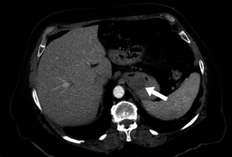
Gastric leiomyomas are benign, submucosal tumors found incidentally on unrelated imaging or during autopsy. The majority of leiomyomas are asymptomatic; however, patients can develop central ulcerations on the lesions leading to upper gastrointestinal (GI) bleeding. A 75-year-old female, with a past medical history of hypertension, hyperlipidemia, and a cerebrovascular accident, presented with complaints of melena, near-syncope events, lightheadedness, weakness, and hematemesis. A computed tomography (CT) of the abdomen with contrast found a heterogeneous low-attenuation mass of 4×4×3 cm3 within the gastric fundus and near the gastroesophageal (GE) junction. After an open gastrostomy and excisional biopsy, the mass was identified as a leiomyoma. This case report reviews the presentation, diagnostic assessments, and treatment of a gastric leiomyoma in a complex location proximal to the gastroesophageal junction. Gastric leiomyomas should be considered as a differential diagnosis for patients presenting with an upper gastrointestinal bleed.
Keywords: gastrointestinal bleed; laparoscopy; open surgery; smooth muscle tumors; spindle cell tumor; surgical intervention.
Copyright © 2024, Jones et al.
Conflict of interest statement
The authors have declared that no competing interests exist.
Figures

Similar articles
-
Gastric Leiomyoma Near the Gastroesophageal Junction Causing Massive Gastrointestinal Bleeding.Cureus. 2023 Nov 6;15(11):e48374. doi: 10.7759/cureus.48374. eCollection 2023 Nov. Cureus. 2023. PMID: 38060747 Free PMC article.
-
Ulcerated gastric leiomyoma causing massive upper gastrointestinal bleeding: A case report.Mol Clin Oncol. 2018 May;8(5):671-674. doi: 10.3892/mco.2018.1597. Epub 2018 Mar 26. Mol Clin Oncol. 2018. PMID: 29725533 Free PMC article.
-
Robotic-assisted endoluminal gastric leiomyoma resection: a novel surgical technique for benign gastroesophageal junction tumors.Chin Clin Oncol. 2024 Feb;13(1):6. doi: 10.21037/cco-23-112. Epub 2024 Feb 1. Chin Clin Oncol. 2024. PMID: 38372060
-
Laparoscopic excision of leiomyomas in the esophageal and gastric wall.Surg Technol Int. 2007;16:82-8. Surg Technol Int. 2007. PMID: 17429773 Review.
-
Bleeding jejunal leiomyoma: a new approach.Am J Gastroenterol. 1995 Jan;90(1):131-3. Am J Gastroenterol. 1995. PMID: 7801914 Review.
References
-
- Florence AM, Fatehi M. Statpearls [Internet. 30855861. Treasure Island, FL: StatPearls Publishing; 2023. Leiomyoma.
-
- Gastric leiomyoma and its management: a rare occurrence. Naz S, Afzal M, Sarwar H, Shakeel O, Rehman S. https://www.iomcworld.org/open-access/gastric-leiomyoma-and-its-manageme... Oncol Cancer Case Rep. 2019;5:1000154.
-
- Gastric true leiomyoma: computed tomographic findings and pathological correlation. Lee MJ, Lim JS, Kwon JE, et al. J Comput Assist Tomogr. 2007;31:204–208. - PubMed
Publication types
LinkOut - more resources
Full Text Sources
Miscellaneous