

Conservative Management of Odontogenic Fibromyxoma of the Maxilla: A Case Report
- PMID: 38846229
- PMCID: PMC11153981
- DOI: 10.7759/cureus.59763
Conservative Management of Odontogenic Fibromyxoma of the Maxilla: A Case Report
Abstract
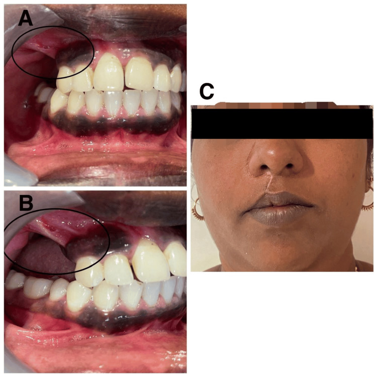
Odontogenic fibromyxoma typically presents as painless swelling in the jaw, and clinically, it grows slowly, becoming benign and asymptomatic. It causes the cortical plates to expand gradually, which leads to mobility and drifting of the teeth. Root resorption is also common. The tumor is locally aggressive in nature. It is also known to have a high recurrence rate. We present the case of a 30-year-old female patient who was diagnosed and treated for odontogenic fibromyxoma of the maxilla conservatively with enucleation. The radiograph showed a multilocular lesion, which can be confused with ameloblastoma, aneurysmal bone cyst, or odontogenic keratocyst. Hence, with proper clinical, radiographic, and histopathological examination, a correct diagnosis can be made and adequate treatment can be planned.
Keywords: conservative management; maxilla; myxofibroma; odontogenic fibromyxoma; odontogenic tumour; odotogenic myxoma.
Copyright © 2024, Hurkat et al.
Conflict of interest statement
The authors have declared that no competing interests exist.
Figures


Similar articles
-
Mandibular Odontogenic Fibromyxoma: A Case Report of Clinical and Radiographic Presentations.Cureus. 2025 Apr 9;17(4):e81981. doi: 10.7759/cureus.81981. eCollection 2025 Apr. Cureus. 2025. PMID: 40351933 Free PMC article.
-
Odontogenic fibromyxoma of maxilla: a rare case report.Case Rep Dent. 2013;2013:345479. doi: 10.1155/2013/345479. Epub 2013 Mar 4. Case Rep Dent. 2013. PMID: 23533825 Free PMC article.
-
Odontogenic fibromyxoma: A case report in myasthenia gravis patient and review of the literature.Int J Surg Case Rep. 2022 Jul;96:107306. doi: 10.1016/j.ijscr.2022.107306. Epub 2022 Jun 17. Int J Surg Case Rep. 2022. PMID: 35728373 Free PMC article.
-
Odontogenic myxoma: An updated analysis of 1,692 cases reported in the literature.Oral Dis. 2019 Apr;25(3):676-683. doi: 10.1111/odi.12875. Epub 2018 Jun 8. Oral Dis. 2019. PMID: 29683236 Review.
-
Odontogenic myxofibroma: a concise review of the literature with emphasis on the surgical approach.Med Oral Patol Oral Cir Bucal. 2015 Jan 1;20(1):e1-6. doi: 10.4317/medoral.19842. Med Oral Patol Oral Cir Bucal. 2015. PMID: 25129249 Free PMC article. Review.
Cited by
-
Conservative Management of Odontogenic Myxoma - A Case Report.Ann Maxillofac Surg. 2024 Jul-Dec;14(2):224-227. doi: 10.4103/ams.ams_38_24. Epub 2024 Aug 30. Ann Maxillofac Surg. 2024. PMID: 39957876 Free PMC article.
-
Mandibular Odontogenic Fibromyxoma: A Case Report of Clinical and Radiographic Presentations.Cureus. 2025 Apr 9;17(4):e81981. doi: 10.7759/cureus.81981. eCollection 2025 Apr. Cureus. 2025. PMID: 40351933 Free PMC article.
References
-
- Myxoma of the jaw bones. Ghosh BC, Huvos AG, Gerold FP, Miller TR. Cancer. 1973;31:1–3. - PubMed
-
- Odontogenic myxofibroma: report of two cases. Gormley MB, Mallin RE, Solomon M, Jarrett W, Bromberg B. https://pubmed.ncbi.nlm.nih.gov/1055192/ J Oral Surg. 1975;33:356–359. - PubMed
-
- Odontogenic myxoma involving maxilla: a case report. Alok A, Hasan K, Singh S, Bhattacharya PT. J Indian Acad Oral Med Radiol. 2019;31:70–73.
Publication types
LinkOut - more resources
Full Text Sources