

doi: 10.21037/qims-23-1772.
Epub 2024 May 8.
Multimodality imaging applications in the diagnosis of and surgical treatment strategy for intravenous leiomyomatosis: a case description and literature analysis
Affiliations
- PMID: 38846306
- PMCID: PMC11151228
- DOI: 10.21037/qims-23-1772
Item in Clipboard
Multimodality imaging applications in the diagnosis of and surgical treatment strategy for intravenous leiomyomatosis: a case description and literature analysis
Quant Imaging Med Surg.
.
No abstract available
Keywords: Cardiac; cine; diastole; mass; systole.
Conflict of interest statement
Conflicts of Interest: All authors have completed the ICMJE uniform disclosure form (available at https://qims.amegroups.com/article/view/10.21037/qims-23-1772/coif). The authors have no conflicts of interest to declare.
Figures

Contrast-enhanced computed tomography scans. The coronal reconstruction (A), sagittal reconstruction (B), and volume rendering (C) images showed a cord-like mass (arrows) in the lumen of the left ovarian vein, extending to the inferior vena cava and right atrium. Varicosity and dilatation of the left genital vein were observed (triangles). No significant enhancement was observed in the enlarged uterus.
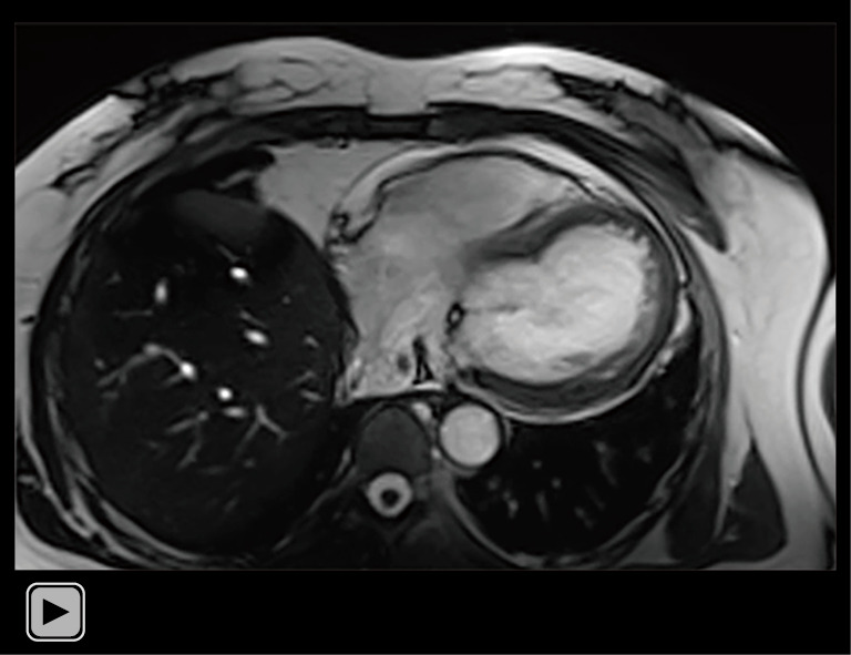
Cardiac magnetic resonance imaging cine of the four chambers revealed that the mass in the atrium traversed the outflow tract from the right atrium to the right ventricle during diastole, and returned during systole.

Sagittal cardiac magnetic resonance imaging cine revealed that the mass ascended from the inferior vena cava into the right atrium, exhibiting regular movement with the cardiac cycle and protruding into the right ventricle during diastole.

Gadolinium-enhanced magnetic resonance imaging scans. The enhancement of the cord-like mass was more discernible on the magnetic resonance images than the computed tomography scans due to the superior soft tissue contrast (arrows), and linear filling defects were also prominently visible. An inhomogeneously enhanced, enlarged soft tissue mass was identified on the anterior wall of the uterus (triangles). (A) Coronal reconstruction. (B) Oblique-sagittal reconstruction. (C) Maximum intensity projection in magnetic resonance angiography.
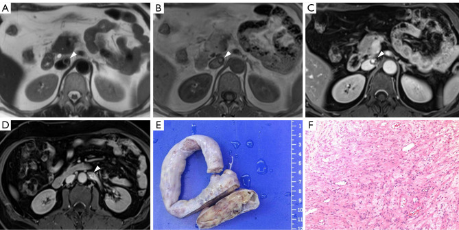
Magnetic resonance axial images and pathological images. (A) Axial T2-weighted imaging showing a lesion with low signal intensity in the IVC (triangle). (B) On the axial T1-weighted images, the lesion in the IVC exhibited moderate signal intensity (triangle). (C) The enhanced axial T1-weighted fat-suppressed images demonstrated pronounced enhancement of the lesion in the IVC, characterized by a sieve-pore appearance (triangle). (D) On the axial T1-weighted fat-suppressed enhanced images, a lesion in the left renal vein demonstrated marked enhancement, resembling a sieve pore (triangle). (E) A gross pathological specimen showed a cord-like mass. (F) The histopathological analysis confirmed the cord-like mass to be intravenous leiomyomatosis (hematoxylin and eosin staining, ×100 magnification). IVC, inferior vena cava.
Similar articles
-
Multimodality Evaluation of Intravenous Leiomyomatosis: A Rare, Benign but Potentially Life-Threatening Tumor.Am J Case Rep. 2015 Nov 7;16:794-800. doi: 10.12659/ajcr.894939. Am J Case Rep. 2015. PMID: 26546569 Free PMC article.
-
Surgical Strategy for Intravenous Cardiac Leiomyomatosis.Heart Lung Circ. 2021 Feb;30(2):240-246. doi: 10.1016/j.hlc.2020.07.006. Epub 2020 Aug 21. Heart Lung Circ. 2021. PMID: 32830033 Review.
-
Intravenous leiomyomatosis: A case study and literature review.Radiol Case Rep. 2022 Sep 7;17(11):4203-4208. doi: 10.1016/j.radcr.2022.08.020. eCollection 2022 Nov. Radiol Case Rep. 2022. PMID: 36105826 Free PMC article.
-
Simultaneous high-resolution cardiac T1 mapping and cine imaging using model-based iterative image reconstruction.Magn Reson Med. 2019 Feb;81(2):1080-1091. doi: 10.1002/mrm.27474. Epub 2018 Sep 5. Magn Reson Med. 2019. PMID: 30183094
-
Massive pelvic recurrence of uterine leiomyomatosis with intracaval-intracardiac extension: video case report and literature review.BMC Surg. 2017 Nov 29;17(1):118. doi: 10.1186/s12893-017-0306-y. BMC Surg. 2017. PMID: 29187188 Free PMC article. Review.
Cited by
-
Ultrasound diagnostic value and clinical analysis of 61 uterine intravenous leiomyomatosis cases.Quant Imaging Med Surg. 2025 Apr 1;15(4):3347-3359. doi: 10.21037/qims-24-1724. Epub 2025 Mar 28. Quant Imaging Med Surg. 2025. PMID: 40235766 Free PMC article.
References
-
- Birch-Hirschfeld FV. Lehrbuch der Pathologischen Anatomie. 5th ed. Leipzig (Germany): FCW Vogel, 1896.
-
- Durck H. Ueber ien Kontinvierlich durch die entere Holhlvene in das Herz vorwachsendes: fibromyom des uterus. Munch Med Wochenschr 1907;54:1154-5.
LinkOut - more resources
Full Text Sources