Coronoid fractures and complex elbow instability: current concepts
- PMID: 38846340
- PMCID: PMC11152979
- DOI: 10.52965/001c.118439
Coronoid fractures and complex elbow instability: current concepts
Abstract
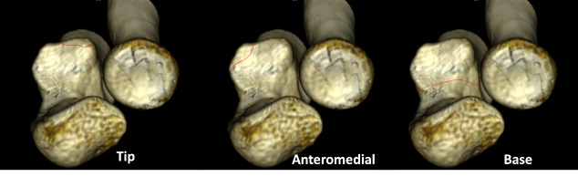
Fractures of the coronoid process typically occur as part of more complex injury patterns, such as terrible triads, trans-olecranon fracture-dislocations, posteromedial rotatory injuries or Monteggia-like lesions. Each pattern is associated with a specific type of coronoid fracture with regard to shape and size and specific soft-tissue lesions. O' Driscoll classification incorporates those associations identifying three major types of fractures: tip, anteromedial facet, and basal fractures. The objective of this study is to review the most common types of complex elbow instability, identify the indications for coronoid fixation and guide the appropriate management. Tip fractures as those seen in terrible triads can conditionally left untreated provided that elbow stability has been restored after radial head fixation and ligaments repair. Anteromedial facet fractures benefit from a buttress plate, while large basilar fractures can be effectively secured with posteroanterior screws. Coronoid reconstruction with a graft should be considered in post-traumatic cases of chronic coronoid deficiency.
Keywords: anteromedial facet; coronoid; elbow instability; terrible triad; trans-olecranon fracture dislocation.
Conflict of interest statement
The authors have no relevant financial or non-financial interests to disclose.
Figures
References
-
- Articular Surface Area of the Coronoid Process and Radial Head in Elbow Extension: Surface Ratio in Cadavers and a Computed Tomography Study In Vivo. Shin S. H., Jeon I. H., Kim H. J.., et al. 2010J Hand Surg. 35(7):1120–1125. doi: 10.1016/j.jhsa.2010.04.002. doi: 10.1016/j.jhsa.2010.04.002. - DOI - DOI - PubMed
-
- Coronoid process of the ulna: paleopathologic and anatomic study with imaging correlation. Emphasis on the anteromedial “facet.”. Weber M. F. V. D. L., Barbosa D. M., Belentani C., Ramos P. M. N., Trudell D., Resnick D. 2009Skeletal Radiol. 38(1):61–67. doi: 10.1007/s00256-008-0556-y. doi: 10.1007/s00256-008-0556-y. - DOI - DOI - PubMed
LinkOut - more resources
Full Text Sources