

Molecular characterization of SARS-CoV-2 nucleocapsid protein
- PMID: 38846351
- PMCID: PMC11153676
- DOI: 10.3389/fcimb.2024.1415885
Molecular characterization of SARS-CoV-2 nucleocapsid protein
Abstract
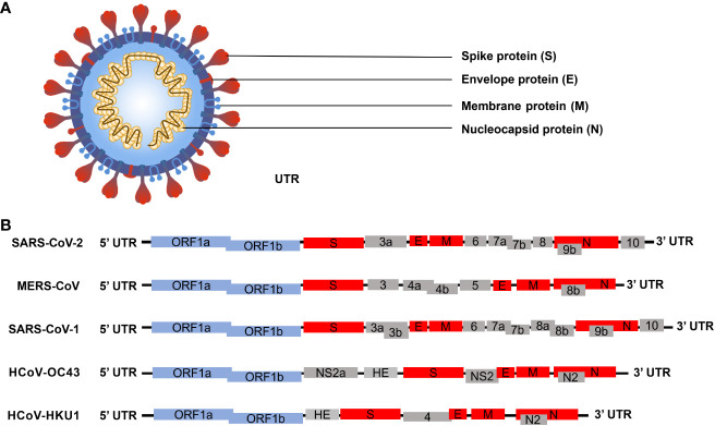
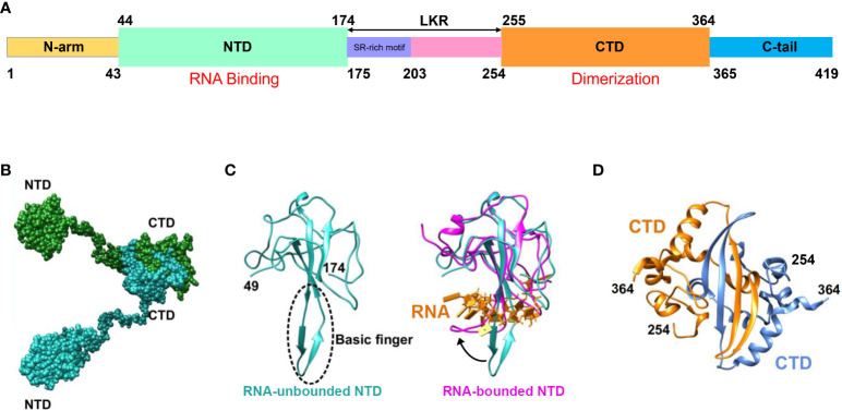
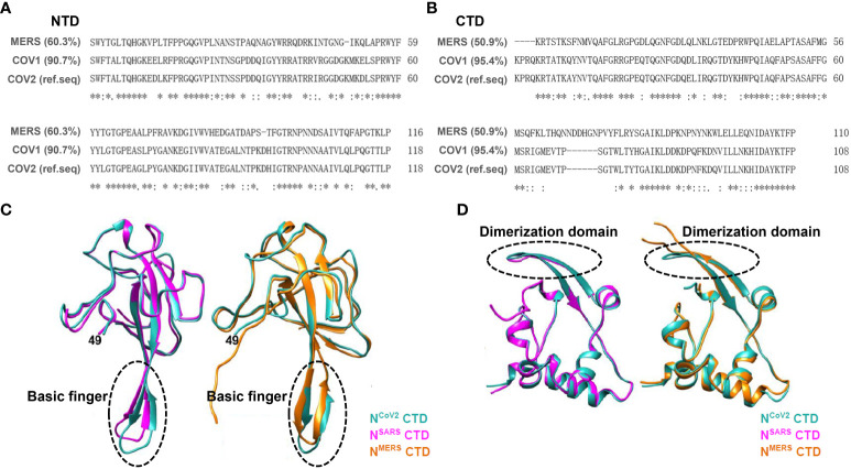
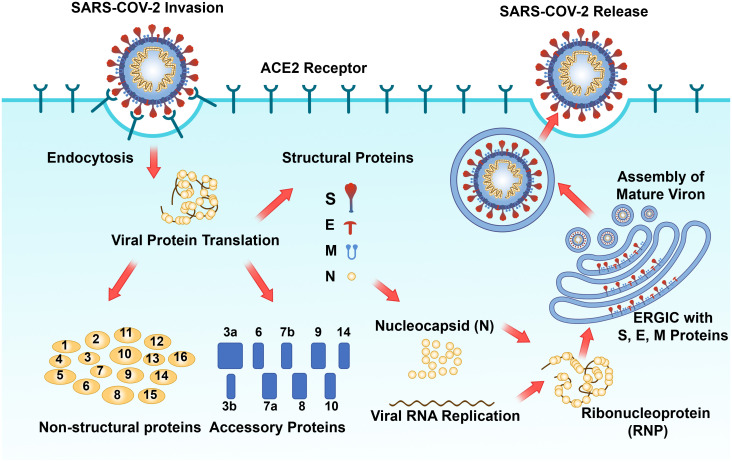
Corona Virus Disease 2019 (COVID-19) is a highly prevalent and potent infectious disease caused by severe acute respiratory syndrome coronavirus 2 (SARS-CoV-2). Until now, the world is still endeavoring to develop new ways to diagnose and treat COVID-19. At present, the clinical prevention and treatment of COVID-19 mainly targets the spike protein on the surface of SRAS-CoV-2. However, with the continuous emergence of SARS-CoV-2 Variants of concern (VOC), targeting the spike protein therapy shows a high degree of limitation. The Nucleocapsid Protein (N protein) of SARS-CoV-2 is highly conserved in virus evolution and is involved in the key process of viral infection and assembly. It is the most expressed viral structural protein after SARS-CoV-2 infection in humans and has high immunogenicity. Therefore, N protein as the key factor of virus infection and replication in basic research and clinical application has great potential research value. This article reviews the research progress on the structure and biological function of SARS-CoV-2 N protein, the diagnosis and drug research of targeting N protein, in order to promote researchers' further understanding of SARS-CoV-2 N protein, and lay a theoretical foundation for the possible outbreak of new and sudden coronavirus infectious diseases in the future.
Keywords: COVID-19; SARS-CoV-2; clinical application; diagnostics; nucleocapsid protein.
Copyright © 2024 Huang, Chen, Chen, Huang, Li, Li, Jin, Zhang, Pan, Du, Liu and Liu.
Conflict of interest statement
The authors declare that the research was conducted in the absence of any commercial or financial relationships that could be construed as a potential conflict of interest.
Figures

Similar articles
-
SARS-CoV-2, the pandemic coronavirus: Molecular and structural insights.J Basic Microbiol. 2021 Mar;61(3):180-202. doi: 10.1002/jobm.202000537. Epub 2021 Jan 18. J Basic Microbiol. 2021. PMID: 33460172 Free PMC article. Review.
-
Evolution of SARS-CoV-2 Envelope, Membrane, Nucleocapsid, and Spike Structural Proteins from the Beginning of the Pandemic to September 2020: A Global and Regional Approach by Epidemiological Week.Viruses. 2021 Feb 4;13(2):243. doi: 10.3390/v13020243. Viruses. 2021. PMID: 33557213 Free PMC article.
-
Modulation of biophysical properties of nucleocapsid protein in the mutant spectrum of SARS-CoV-2.Elife. 2024 Jun 28;13:RP94836. doi: 10.7554/eLife.94836. Elife. 2024. PMID: 38941236 Free PMC article.
-
Identification of SARS-CoV-2 Nucleocapsid and Spike T-Cell Epitopes for Assessing T-Cell Immunity.J Virol. 2021 Feb 24;95(6):e02002-20. doi: 10.1128/JVI.02002-20. Print 2021 Feb 24. J Virol. 2021. PMID: 33443088 Free PMC article.
-
Properties of Coronavirus and SARS-CoV-2.Malays J Pathol. 2020 Apr;42(1):3-11. Malays J Pathol. 2020. PMID: 32342926 Review.
Cited by
-
Universal Bacterium-Vectored COVID-19 Vaccine Expressing Early SARS-CoV-2 Conserved Proteins Cross-Protects Against Late Variants in Hamsters.Vaccines (Basel). 2025 Jun 12;13(6):633. doi: 10.3390/vaccines13060633. Vaccines (Basel). 2025. PMID: 40573964 Free PMC article.
-
Diagnostic Utility of SARS-CoV-2 Nucleocapsid Antigenemia: A Meta-analysis.Open Forum Infect Dis. 2024 Oct 2;11(10):ofae561. doi: 10.1093/ofid/ofae561. eCollection 2024 Oct. Open Forum Infect Dis. 2024. PMID: 39431150 Free PMC article.
-
Structural basis for the participation of the SARS-CoV-2 nucleocapsid protein in the template switch mechanism and genomic RNA reorganization.J Biol Chem. 2024 Nov;300(11):107834. doi: 10.1016/j.jbc.2024.107834. Epub 2024 Sep 27. J Biol Chem. 2024. PMID: 39343000 Free PMC article. Review.
-
Research Progress on the Structure and Function, Immune Escape Mechanism, Antiviral Drug Development Methods, and Clinical Use of SARS-CoV-2 Mpro.Molecules. 2025 Jan 16;30(2):351. doi: 10.3390/molecules30020351. Molecules. 2025. PMID: 39860219 Free PMC article. Review.
-
The Role of TDP-43 in SARS-CoV-2-Related Neurodegenerative Changes.Viruses. 2025 May 19;17(5):724. doi: 10.3390/v17050724. Viruses. 2025. PMID: 40431734 Free PMC article. Review.
References
-
- Afkhami S., D'agostino M. R., Zhang A., Stacey H. D., Marzok A., Kang A., et al. . (2022). Respiratory mucosal delivery of next-generation covid-19 vaccine provides robust protection against both ancestral and variant strains of sars-cov-2. Cell 185, 896–915 E19. doi: 10.1016/j.cell.2022.02.005 - DOI - PMC - PubMed
-
- Ahn J. Y., Lee J., Suh Y. S., Song Y. G., Choi Y. J., Lee K. H., et al. . (2022). Safety and immunogenicity of two recombinant dna covid-19 vaccines containing the coding regions of the spike or spike and nucleocapsid proteins: an interim analysis of two open-label, non-randomised, phase 1 trials in healthy adults. Lancet Microbe 3, E173–E183. doi: 10.1016/S2666-5247(21)00358-X - DOI - PMC - PubMed
Publication types
MeSH terms
Substances
Supplementary concepts
LinkOut - more resources
Full Text Sources
Medical
Miscellaneous