. 2024 May 3;15(22):8311-8322.
doi: 10.1039/d4sc01294a. eCollection 2024 Jun 5.

Clinically used drug arsenic trioxide targets XIAP and overcomes apoptosis resistance in an organoid-based preclinical cancer model

Affiliations

Clinically used drug arsenic trioxide targets XIAP and overcomes apoptosis resistance in an organoid-based preclinical cancer model

Liwa Shi et al. Chem Sci. .

Abstract

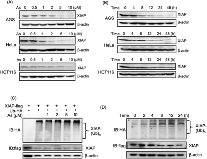
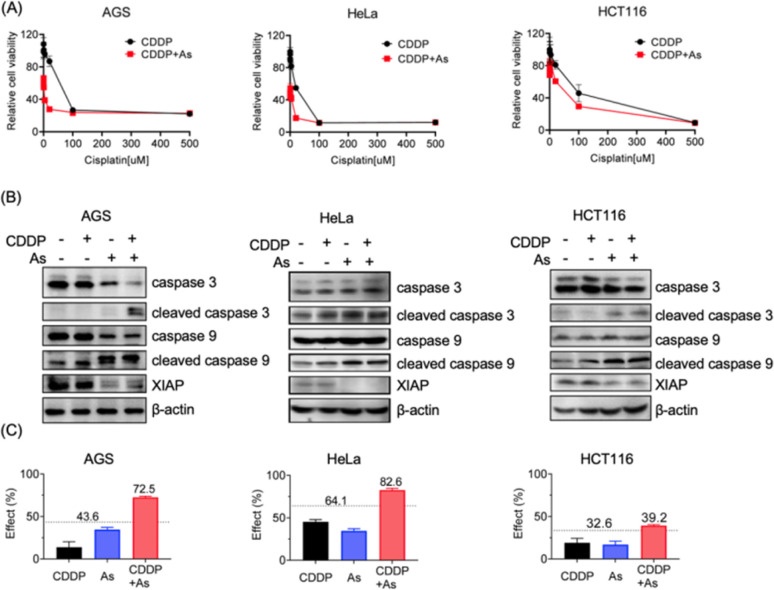
Drug resistance in tumor cells remains a persistent clinical challenge in the pursuit of effective anticancer therapy. XIAP, a member of the inhibitor of apoptosis protein (IAP) family, suppresses apoptosis via its Baculovirus IAP Repeat (BIR) domains and is responsible for drug resistance in various human cancers. Therefore, XIAP has attracted significant attention as a potential therapeutic target. However, no XIAP inhibitor is available for clinical use to date. In this study, we surprisingly observed that arsenic trioxide (ATO) induced a rapid depletion of XIAP in different cancer cells. Mechanistic studies revealed that arsenic attacked the cysteine residues of BIR domains and directly bound to XIAP, resulting in the release of zinc ions from this protein. Arsenic-XIAP binding suppressed the normal anti-apoptosis functions of BIR domains, and led to the ubiquitination-dependent degradation of XIAP. Importantly, we further demonstrate that arsenic sensitized a variety of apoptosis-resistant cancer cells, including patient-derived colon cancer organoids, to the chemotherapy drug using cisplatin as a showcase. These findings suggest that targeting XIAP with ATO offers an attractive strategy for combating apoptosis-resistant cancers in clinical practice.


Conflict of interest statement

There are no conflicts to declare.

Figures

Fig. 1
Fig. 1. Arsenic trioxide induces depletion of XIAP in different cancer cells. (A) Arsenic induces depletion of XIAP protein in a dose-dependent manner. Gradient arsenic trioxide was added into the cell cultured medium for 48 h. (B) Arsenic induces depletion of XIAP protein in a time-dependent manner. In the case of HCT116, it is noteworthy that approximately 30% of XIAP expression was still observable after 12 h of arsenic treatment. This observation could potentially be attributed to the inherent resistance of the HCT116 cell line to arsenic. XIAP levels were revealed by immunoblotting with anti-XIAP antibody. (C) Arsenic induces ubiquitination of XIAP in a dose-dependent manner. (D) Arsenic induces ubiquitination of XIAP in a time-dependent manner.
Fig. 2
Fig. 2. Arsenic binds to BIR domains of XIAP directly. (A) Representative CETSA blot for arsenic–XIAP binding in cell. HEK293t cells expressing XIAP–Flag were pretreated with arsenic for 4 h following indicated heat shocks. Soluble Flag-fused XIAP full-length protein in the sample supernatant was revealed by immunoblotting with anti-Flag antibody. The melting temperature shift (ΔTm) between treated and control samples was measured based on CETSA melt curves. (B) Representative CETSA blot for arsenic–BIR1–BIR2–BIR3 binding in cells. HEK293t cells expressing BIR1–BIR2–BIR3–Flag were pretreated with arsenic for 4 h following indicated heat shocks. Soluble Flag-fused XIAP truncated protein (BIR1–BIR2–BIR3) in the sample supernatant was revealed by immunoblotting with anti-Flag antibody. (C–E) MALDI-TOF mass spectrometry for monitoring the interaction between arsenic and BIR domains of XIAP. Apo-form BIR1 (20–99), BIR2 (120–240) and BIR3 (241–356) were pre-incubated with or without the gradient arsenic trioxide. The molar ratios of binding between As3+ and BIR domains were determined to be 1 : 1. (F) UV absorption spectra of apo-BIR3 upon addition of gradient arsenic as indicated.
Fig. 3
Fig. 3. Arsenic induces Zn2+ release from BIR domains and a conformational change in XIAP. (A) UV-vis absorption spectra of 100 μM PAR and Zn-bound BIR1, BIR2 and BIR3 protein mixture with or without addition of arsenic trioxide in the presence of 5 mM GSH; (B) CD spectra of the Zn- and As-bound BIR1/BIR2/BIR3 domain of XIAP. (C) Analytical gel filtration analysis of the apo-, Zn- and As-bound BIR3 domain of XIAP. The indicated apo-BIR3 at a concentration of 20 μM was incubated with zinc or arsenic and applied to a Superdex 200 gel filtration column. *Impurity or fluctuation of the absorbance due to buffer.
Fig. 4
Fig. 4. Arsenic inhibits the anti-apoptosis activity of all three BIR domains. (A) Flag pull down assay to assess XIAP-caspase-3/-9 interaction. (B) Western blot analysis of pro-caspase-3 and cleaved caspase-3 in cell lysates. β-Actin served as the loading control. Zn-BIR2 or As-BIR2 was added to the reactions. Cytochrome c and dATP were used to induce the processing of pro-caspase-3 in vitro. Reactions were stopped by using additional 5× SDS loading buffer. (C) Western blot analysis of pro-caspase-9 and cleaved caspase-9 in cell lysates. β-Actin served as the loading control. Zn-BIR3 or As-BIR3 was added to the reactions. Cytochrome c and dATP were used to induce the processing of pro-caspase-9 in vitro. Reactions were stopped by using additional 5× SDS loading buffer. (D) Representative immunoblots of AGS cells showing the effect of arsenic on the TGFβ-induced NF-κB activation. XIAP-mediated NF-κB signaling in AGS cells was initiated by addition of TGFβ into the cultured medium. Phosphorated and total p65 were revealed by immunoblotting with anti-p-p65 and anti-p65 antibodies, respectively.
Fig. 5
Fig. 5. Arsenic sensitizes cancer cells with intrinsic drug resistance to cisplatin via XIAP targeting. (A) Cell viability of cancer cells upon the treatment of cisplatin with or without arsenic. Cancer cells AGS, HeLa and HCT116 were cultured for 48 h with various concentrations of cisplatin (500, 100, 20, 4, 0.8, 0.16, 0.032, and 0 μM) with or without arsenic. The percentage of viable cells relative to the control was examined by MTT assay (mean + SE, n = 3). (B) Representative immunoblots showing the protein level of endogenous caspase-3, caspase-9 and XIAP in AGS, HeLa and HCT116 upon the treatment with cisplatin and arsenic. (C) The Bliss independence model indicates a synergistic effect of the arsenic (2 μM) and cisplatin (20 μM) combination. The Bliss independence threshold was labelled and is shown as a dotted line.
Fig. 6
Fig. 6. Arsenic sensitizes cancer cells with acquired drug resistance to cisplatin targeting via XIAP targeting. (A) Representative immunoblots showing the protein level of endogenous XIAP in H3B and H3B-R cells. (B) Quantitative real-time PCR analysis of XIAP expression in H3B and H3B-R cells. (C) Arsenic induces depletion of XIAP protein in a dose-dependent manner in H3B-R cells. (D) Arsenic induces depletion of XIAP protein in a time-dependent manner in H3B-R cells. XIAP levels were revealed by immunoblotting with anti-XIAP antibody. (E) Cell viability of H3B-R cells upon the treatment with cisplatin with or without arsenic (mean + SE, n = 3). (F) The Bliss independence model indicates a synergistic effect of the arsenic (2 μM) and cisplatin (64 μM) combination. The Bliss independence threshold was labelled and is shown as a dotted line.
Fig. 7
Fig. 7. Arsenic overcomes apoptosis resistance in patient-derived colon cancer organoids. (A) Schematic overview for the assessment of drug sensitivity in a patient-derived colon cancer organoid model. (B) Cell viability of patient-derived colon cancer organoids from four donors. Patient-derived cancer organoids were cultured with various concentrations of cisplatin as indicated, with or without 1 μM arsenic. The cell viability relative to that of the control was examined by using a 3D cell viability assay kit (mean + SE, n = 3). (C) The Bliss independence model indicates a synergistic effect for the arsenic (1 μM) and cisplatin (12.5 μM) combination. The Bliss independence threshold was labeled and is shown as a dotted line. (D) Representative images for the formation of colon cancer organoids with or without the treatment with arsenic, cisplatin and their combination.
Fig. 8
Fig. 8. Proposed mechanism by which the arsenic-based drug overcomes apoptosis resistance in cancer cells via targeting XIAP. Arsenic (As3+) replaces Zn2+ from the BIR domains and disrupts XIAP anti-apoptosis functions via a dual-action mechanism, (a) blocking the anti-apoptosis functions of solo BIR domains and (b) inducing the ubiquitination-mediated degradation of XIAP whole protein.

Similar articles

Cited by

References

    1. Fulda S. Vucic D. Targeting IAP proteins for therapeutic intervention in cancer. Nat. Rev. Drug Discovery. 2012;11:109–124. doi: 10.1038/nrd3627. - DOI - PubMed
    1. Eckelman B. P. Salvesen G. S. Scott F. L. Human inhibitor of apoptosis proteins: why XIAP is the black sheep of the family. EMBO Rep. 2006;7:988–994. doi: 10.1038/sj.embor.7400795. - DOI - PMC - PubMed
    1. Schimmer A. D. Dalili S. Batey R. A. Riedl S. J. Targeting XIAP for the treatment of malignancy. Cell Death Differ. 2006;13:179–188. doi: 10.1038/sj.cdd.4401826. - DOI - PubMed
    1. Kesavardhana S. Malireddi R. K. S. Kanneganti T. D. Caspases in Cell Death, Inflammation, and Pyroptosis. Annu. Rev. Immunol. 2020;38:567–595. doi: 10.1146/annurev-immunol-073119-095439. - DOI - PMC - PubMed
    1. Burstein E. Ganesh L. Dick R. D. van De Sluis B. Wilkinson J. C. Klomp L. W. Wijmenga C. Brewer G. J. Nabel G. J. Duckett C. S. A novel role for XIAP in copper homeostasis through regulation of MURR1. EMBO J. 2004;23:244–254. doi: 10.1038/sj.emboj.7600031. - DOI - PMC - PubMed