

False-Negative PET/CT scan with Metastatic Sebaceous Carcinoma: An Important Diagnostic Consideration
- PMID: 38846664
- PMCID: PMC11152212
- DOI: 10.12890/2024_004491
False-Negative PET/CT scan with Metastatic Sebaceous Carcinoma: An Important Diagnostic Consideration
Abstract
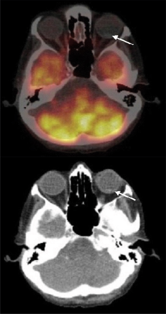
Positron emission tomography (PET) has gained widespread acceptance as a valuable diagnostic tool for cancer. It is rare for a PET/CT scan to overlook the presence of metastatic disease. Sebaceous carcinoma is an uncommon malignant tumour that typically originates in the skin of the eyelid. In this case report, we present a unique case involving a metastatic sebaceous carcinoma that was not initially detected by a PET/CT scan in an 88-year-old female. Therefore, clinicians must maintain a heightened awareness of sebaceous carcinoma and exercise caution when making decisions solely based on PET scan results. It is crucial to recognise this potential limitation of PET scans in sebaceous carcinoma and consider further diagnostic approaches to ensure timely and accurate detection of sebaceous carcinoma.
Learning points: PET scans may miss slow-growing tumours such as sebaceous carcinoma.A high index of suspicion for sebaceous carcinoma is crucial, even with negative PET scans. Additional diagnostic approaches might be necessary for accurate detection.Sebaceous carcinoma is a rare but aggressive cancer. Diagnosis can be challenging due to its varied presentations.
Keywords: False-negative PET/CT; diagnostic limitations; rare cancer; sebaceous carcinoma.
© EFIM 2024.
Conflict of interest statement
Conflicts of Interests: The Authors declare that there are no competing interests.
Figures
References
-
- Kyllo RL, Brady LK, Hurst EA. Sebaceous carcinoma: review of the literature. Dermatol Surg. 2015;41:1–15. - PubMed
-
- Owen JL, Kibbi N, Worley B, Kelm RC, Wang JV, Barker CA, et al. Sebaceous carcinoma: evidence-based clinical practice guidelines. Lancet Oncol. 2019;20:e699–e714. - PubMed
-
- Tripathi R, Chen Z, Li L, Bordeaux JS. Incidence and survival of sebaceous carcinoma in the United States. J Am Acad Dermatol. 2016;75:1210–1215. - PubMed
-
- Kumabe A, Kawase T, Miura K, Tada Y, Masubuchi T, Kamata SE, et al. Sebaceous carcinoma of the parotid gland: F-18 FDG PET/CT findings. Clin Nucl Med. 2010;35:260–262. - PubMed
-
- Ferreira I, Wiedemeyer K, Demetter P, Adams DJ, Arends MJ, Brenn T. Update on the pathology, genetics and somatic landscape of sebaceous tumours. Histopathology. 2020;75:640–649. - PubMed
LinkOut - more resources
Full Text Sources