. 2024 May 23:15:1253072.
doi: 10.3389/fimmu.2024.1253072. eCollection 2024.

Exosomal ROR1 in peritoneal fluid identifies peritoneal disseminated PDAC and is associated with poor survival

Affiliations

Exosomal ROR1 in peritoneal fluid identifies peritoneal disseminated PDAC and is associated with poor survival

Anke Mittelstädt et al. Front Immunol. .

Abstract

Background: Pancreatic ductal adenocarcinoma (PDAC) is one of the deadliest forms of cancer and peritoneal dissemination is one major cause for this poor prognosis. Exosomes have emerged as promising biomarkers for gastrointestinal cancers and can be found in all kinds of bodily fluids, also in peritoneal fluid (PF). This is a unique sample due to its closeness to gastrointestinal malignancies. The receptor tyrosine kinase-like orphan receptor 1 (ROR1) has been identified as a potential biomarker in human cancers and represents a promising target for an immunotherapy approach, which could be considered for future treatment strategies. Here we prospectively analyzed the exosomal surface protein ROR1 (exo-ROR1) in PF in localized PDAC patients (PER-) on the one hand and peritoneal disseminated tumor stages (PER+) on the other hand followed by the correlation of exo-ROR1 with clinical-pathological parameters.

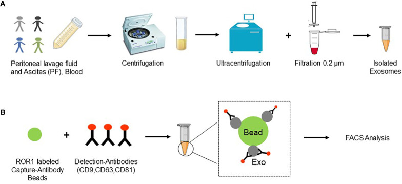
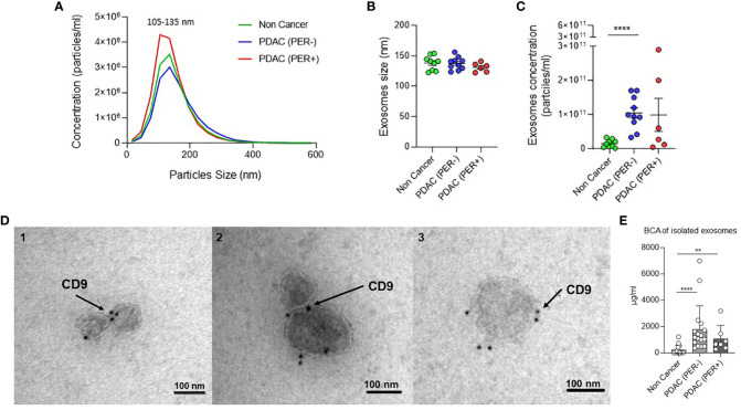
Methods: Exosomes were isolated from PF and plasma samples of non-cancerous (NC) (n = 15), chronic pancreatitis (CP) (n = 4), localized PDAC (PER-) (n = 18) and peritoneal disseminated PDAC (PER+) (n = 9) patients and the surface protein ROR1 was detected via FACS analysis. Additionally, soluble ROR1 in PF was analyzed. ROR1 expression in tissue was investigated using western blots (WB), qPCR, and immunohistochemistry (IHC). Exosome isolation was proven by Nano Tracking Analysis (NTA), WB, Transmission electron microscopy (TEM), and BCA protein assay. The results were correlated with clinical data and survival analysis was performed.

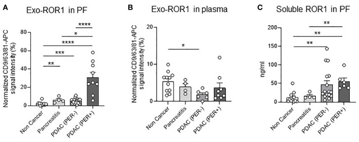
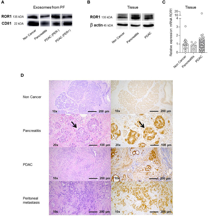
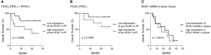
Results: PDAC (PER+) patients have the highest exo-ROR1 values in PF and can be discriminated from NC (p <0.0001), PDAC (PER-) (p <0.0001), and CP (p = 0.0112). PDAC (PER-) can be discriminated from NC (p = 0.0003). In plasma, exo-ROR1 is not able to distinguish between the groups. While there is no expression of ROR1 in the exocrine pancreatic tissue, PDAC and peritoneal metastasis show expression of ROR1. High exo-ROR1 expression in PF is associated with lower overall survival (p = 0.0482).

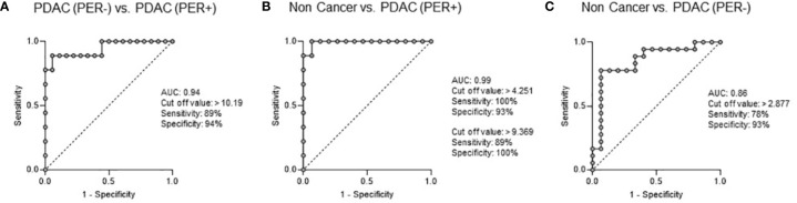
Conclusion: With exo-ROR1 in PF we found a promising diagnostic and prognostic biomarker possibly discriminating between NC, PDAC (PER-) and PDAC (PER+) and might shed light on future diagnostic and therapeutic concepts in PDAC.

Keywords: PDAC; ROR1; biomarker; exosomes; peritoneal carcinomatosis; peritoneal fluid; peritoneal lavage; targeted therapy.


Conflict of interest statement

The authors declare that the research was conducted in the absence of any commercial or financial relationships that could be construed as a potential conflict of interest.

Figures

Figure 1
Figure 1
Experimental layout. (A) Samples (PF and blood) were collected and several centrifugation steps and two ultracentrifugation steps were performed. Additionally, samples were filtered through a 0.2 µm filter and finally exosomes were collected in 500 µl of PBS. (B) Exosomes were incubated with ROR1 labeled capture antibody beads and following with detection antibodies for the three known exosomal markers CD9, CD63, and CD81. Finally, FACS analysis was performed. Created with BioRender.com.
Figure 2
Figure 2
Exo-ROR1 in peritoneal fluid (PF) and plasma and soluble ROR1 in PF. (A) Values of normalized CD9/63/81-APC signal intensity of exo-ROR1 (%) in PF. Non Cancer: n=15, Pancreatitis: n=4, PDAC (PER-): n=18, PDAC (PER+): n=9. *p<0.05, **p<0.01, ***p<0.001, ****p<0.0001. (B) Values of normalized CD9/63/81-APC signal intensity of exo-ROR1 (%) in plasma. Non Cancer: n=11, Pancreatitis: n=4, PDAC (PER-): n=6, PDAC (PER+): n=8. *p<0.05. (C) ELISA: Concentrations of soluble ROR1 (ng/ml) in PF. Non Cancer: n=11, Pancreatitis: n=4, PDAC (PER-): n=16, PDAC (PER+): n=6. **p<0.01.
Figure 3
Figure 3
ROC Curve Analysis: Exo-ROR1 in PF. (A) PDAC (PER-) vs. PDAC (PER+). AUC: 0.94, cut off value: >10.19, sensitivity: 89%, specificity: 94%. PDAC (PER-): n=18, PDAC (PER+): n=9. (B) Non Cancer vs. PDAC (PER+). AUC: 0.99, cut off value: >4.251, sensitivity: 100%, specificity: 93%; cut off value: >9.369, sensitivity: 89%, specificity: 100%. Non Cancer: n=15, PDAC (PER+): n=9. (C) Non Cancer vs. PDAC (PER-). AUC: 0.86, cut off value: >2.877, sensitivity: 78%, specificity: 93%. Non Cancer: n=15, PDAC (PER-): n=18.
Figure 4
Figure 4
Western Blots (WB), qPCR and Immunhistochemistry (IHC) of exosomes in PF and lysed tissue. (A) Western Blot with ROR1 and CD81 of isolated exosomes from PF. Both proteins are expressed on the exosomes of all different groups (Non Cancer, Pancreatitis, PDAC (PER-), PDAC (PER+). For uncropped WB refer to Supplementary Figure S2 . (B) Western Blot of lysed tissue from Non Cancer, Pancreatitis, PDAC. ROR1 and also β-Actin as loading control is expressed in all three groups. For uncropped WB refer to Supplementary Figure S2 . (C) qPCR analysis and relative ROR1 expression of pancreatic tissue. ROR1 is expressed on NC, CP and PDAC tissue showing no significant differences between the groups but a slightly higher expression in the PDAC tissue. (D) Immunohistochemistry (IHC) and HE staining of non-cancerous exocrine pancreatic tissue, CP, PDAC and peritoneal metastasis. The exocrine pancreas is ROR1 negative. In the CP tissue ROR1 positive islet cells are shown in the higher magnification (arrows). The fibrotic tissue is negative. In PDAC and the peritoneal metastasis the morphologic tumor cells are ROR1 positive.
Figure 5
Figure 5
Survival Analysis. (A) Survival analysis of all PDAC patients with and without peritoneal carcinomatosis (PDAC (PER+) + PDAC (PER-)). p=0.0482, n=27. (B) Survival analysis of only PDAC (PER-) patients without peritoneal carcinomatosis. p=0.1699, n=18. Blue: High expression of exo-ROR1, Black: Low expression of exo-ROR1. (C) Survival analysis according to ROR1 mRNA in tumor tissue. p=0.8414, n=25. Blue: High expression of ROR1 mRNA, Black: Low expression of ROR1 mRNA.
Figure 6
Figure 6
Verification of exosome isolation. (A) Nanotracking Analysis (NTA): Exosome concentration and size distribution. Green: Non Cancer n=8, Blue: PDAC (PER-) n=9, Red: PDAC (PER+) n=6. (B) Nanotracking Analysis (NTA): Exosomes size distribution by ZetaView analysis showing the mode size of exosomes in 1 ml PF from Non Cancer, PDAC (PER-), and PDAC (PER+) patients. Green: Non Cancer n=8, Blue: PDAC (PER-) n=9, Red: PDAC (PER+) n=6. (C) Nanotracking Analysis (NTA): Exosome concentration by ZetaView analysis showing the number of exosomes per milliliter of PF derived from Non Cancer, PDAC (PER-), and PDAC (PER+) patients. ****p<0.0001. Green: Non Cancer n=8, Blue: PDAC (PER-) n=9, Red: PDAC (PER+) n=6. (D) Transmission Electron Microscopy (TEM) of isolated exosomes from three PF samples: 1=Non Cancer, 2=PDAC (PER-), 3=PDAC (PER+). Black dots: CD9-immunogold. (E) BCA protein concentration analysis of isolated exosomes (µg/ml): Non Cancer: n=15, PDAC (PER-): n=17, PDAC (PER+) n=7. **p<0.01.

References

    1. Siegel RL, Miller KD, Jemal A. Cancer statistics, 2020. CA Cancer J Clin. (2020) 70:7–30. doi: 10.3322/caac.21590 - DOI - PubMed
    1. Mokdad AA, Minter RM, Zhu H, Augustine MM, Porembka MR, Wang SC, et al. . Neoadjuvant therapy followed by resection versus upfront resection for resectable pancreatic cancer: A propensity score matched analysis. J Clin Oncol. (2017) 35:515–22. doi: 10.1200/JCO.2016.68.5081 - DOI - PubMed
    1. Uesaka K, Boku N, Fukutomi A, Okamura Y, Konishi M, Matsumoto I, et al. . Adjuvant chemotherapy of S-1 versus gemcitabine for resected pancreatic cancer: a phase 3, open-label, randomised, non-inferiority trial (JASPAC 01). Lancet. (2016) 388:248–57. doi: 10.1016/S0140-6736(16)30583-9 - DOI - PubMed
    1. Suenaga M, Fujii T, Kanda M, Takami H, Okumura N, Inokawa Y, et al. . Pattern of first recurrent lesions in pancreatic cancer: hepatic relapse is associated with dismal prognosis and portal vein invasion. Hepatogastroenterol. (2014) 61:1756–61. - PubMed
    1. Jones RP, Psarelli E-E, Jackson R, Ghaneh P, Halloran CM, Palmer DH, et al. . Patterns of recurrence after resection of pancreatic ductal adenocarcinoma: A secondary analysis of the ESPAC-4 randomized adjuvant chemotherapy trial. JAMA Surgery. (2019) 154:1038–48. doi: 10.1001/jamasurg.2019.3337 - DOI - PMC - PubMed

MeSH terms

Substances