. 2024 Jun;17(6):e011107.
doi: 10.1161/CIRCHEARTFAILURE.123.011107. Epub 2024 Jun 7.

Empagliflozin Improves Diastolic Function in HFpEF by Restabilizing the Mitochondrial Respiratory Chain

Affiliations

Empagliflozin Improves Diastolic Function in HFpEF by Restabilizing the Mitochondrial Respiratory Chain

Antje Schauer et al. Circ Heart Fail. 2024 Jun.

Abstract

Background: Clinical studies demonstrated beneficial effects of sodium-glucose-transporter 2 inhibitors on the risk of cardiovascular death in patients with heart failure with preserved ejection fraction (HFpEF). However, underlying processes for cardioprotection remain unclear. The present study focused on the impact of empagliflozin (Empa) on myocardial function in a rat model with established HFpEF and analyzed underlying molecular mechanisms.

Methods: Obese ZSF1 (Zucker fatty and spontaneously hypertensive) rats were randomized to standard care (HFpEF, n=18) or Empa (HFpEF/Empa, n=18). ZSF1 lean rats (con, n=18) served as healthy controls. Echocardiography was performed at baseline and after 4 and 8 weeks, respectively. After 8 weeks of treatment, hemodynamics were measured invasively, mitochondrial function was assessed and myocardial tissue was collected for either molecular and histological analyses or transmission electron microscopy.

Results: In HFpEF Empa significantly improved diastolic function (E/é: con: 17.5±2.8; HFpEF: 24.4±4.6; P<0.001 versus con; HFpEF/Empa: 19.4±3.2; P<0.001 versus HFpEF). This was accompanied by improved hemodynamics and calcium handling and by reduced inflammation, hypertrophy, and fibrosis. Proteomic analysis demonstrated major changes in proteins involved in mitochondrial oxidative phosphorylation. Cardiac mitochondrial respiration was significantly impaired in HFpEF but restored by Empa (Vmax complex IV: con: 0.18±0.07 mmol O2/s/mg; HFpEF: 0.13±0.05 mmol O2/s/mg; P<0.041 versus con; HFpEF/Empa: 0.21±0.05 mmol O2/s/mg; P=0.012 versus HFpEF) without alterations of mitochondrial content. The expression of cardiolipin, an essential stability/functionality-mediating phospholipid of the respiratory chain, was significantly decreased in HFpEF but reverted by Empa (con: 15.9±1.7 nmol/mg protein; HFpEF: 12.5±1.8 nmol/mg protein; P=0.002 versus con; HFpEF/Empa: 14.5±1.8 nmol/mg protein; P=0.03 versus HFpEF). Transmission electron microscopy revealed a reduced size of mitochondria in HFpEF, which was restored by Empa.

Conclusions: The study demonstrates beneficial effects of Empa on diastolic function, hemodynamics, inflammation, and cardiac remodeling in a rat model of HFpEF. These effects were mediated by improved mitochondrial respiratory capacity due to modulated cardiolipin and improved calcium handling.

Keywords: cardiolipins; empagliflozin; heart failure; hypertension; inflammation.


Conflict of interest statement

Disclosures Dr Winzer reports personal fees from Amarin, Amgen, AstraZeneca, Daiichi Sankyo, Bayer, Boehringer Ingelheim, CVRx, and Novartis for lectures and advisory board activities outside the submitted work. Dr Linke reports grants from Novartis, personal fees from Medtronic, Abbott, Edwards Lifesciences, Boston Scientific, AstraZeneca, Novartis, Pfizer, Abiomed, Bayer, Boehringer, and other from Picardia, Transverse Medical, Claret Medical, outside the submitted work. The other authors report no conflicts.

Figures

Figure 1.
Figure 1.
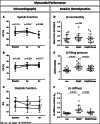
Myocardial performance displayed by echocardiographic and hemodynamic measurements. Ejection fraction (EF, A) and fractional shortening (FS, B) were preserved in all groups at all times (charts show data after 8 weeks of empagliflozin [Empa]). Time course of diastolic function (C) demonstrating significantly reduced early mitral inflow (E)/tissue Doppler mitral annulus velocity in early diastole (é) ratios already after 4 weeks after treatment start. Invasive hemodynamic measurements revealed comparable myocardial contractility between all groups (D), whereas comparison of left ventricular (LV) end-diastolic pressure (LVEDP, E) demonstrated a pressure-lowering impact of Empa on left ventricular conditions. LV stiffness was enhanced in heart failure with preserved ejection fraction (HFpEF) and significantly reduced in HFpEF/Empa (indicated by the stiffness constant βW, F). con denotes lean control group; HFpEF denotes obese control group; and HFpEF/Empa denotes obese treatment group. LV-Ees, left ventricular end-systolic elastance. *P<0.001 vs con; ‡P<0.05 vs con; and §P<0.001 vs HFpEF.
Figure 2.
Figure 2.
Effects of Empa on cardiac intracellular Ca2+ transient. A, Representative Ca2+ transients were detected in freshly isolated Fura-2AM loaded ventricular cardiomyocytes isolated from control, heart failure with preserved ejection fraction (HFpEF) and HFpEF/Empa at 2 Hz. B, Ca2+ transient amplitude. C, Transient peak velocity. D, Time constant τ of transient decay. E, Sarcoplasmic reticulum (SR) load. F, Fractional Ca2+ release. con denotes lean control group; HFpEF denotes obese control group; and HFpEF/Empa denotes obese treatment group (n=cells/N=animals: con (21/3), HFpEF (34/5), and HFpEF/Empa (26/4). Empa indicates empagliflozin.
Figure 3.
Figure 3.
Impact of Empa treatment on mitochondrial respiratory function and protein expression. Stimulation of complex I with Glutamat/Malat (Glut/Mal, A) or with octanoyl-carnitine (B) resulted in a reduced oxygen consumption in heart failure with preserved ejection fraction (HFpEF) compared with control, whereas Empa restored the oxygen consumption. Accordingly, stimulation of complex II with succinate (D) and uncoupling of mitochondria (E) revealed in reduced oxygen consumption in HFpEF and significantly enhanced respiration rates in HFpEF/Empa. Complex IV uncoupling caused a tendentially decreased oxygen consumption in HFpEF compared with control but significantly enhanced respiration in HFpEF/Empa (G). Compared with control, protein expression levels of complex I (C), complex II (F), and complex IV (H) were significantly decreased in both HFpEF groups, respectively, but not different from each other. Protein expression levels of porin (I), a marker for mitochondrial quantity, were similar between all groups. con denotes lean control group; HFpEF denotes obese control group; and HFpEF/Empa denotes obese treatment group. AU indicates arbitrary units; and Empa, empagliflozin.
Figure 4.
Figure 4.
Impact of Empa on left ventricular CL concentration, synthesis, and maturation. CL concentration was distinctly reduced in heart failure with preserved ejection fraction (HFpEF) but significantly enhanced by Empa treatment (A) as was tafazzin (B). CL synthase expression was highly upregulated (C), possibly following compensatory mechanisms. CL72:8 revealed to be the predominant isoform in the healthy control (D) and was significantly downregulated in HFpEF (E). In Empa-treated rats, the amount of CL72:8 was enhanced compared with untreated HFpEF (E). con denotes lean control group; HFpEF denotes obese control group; and HFpEF/Empa denotes obese treatment group. CL indicates cardiolipin; and Empa, empagliflozin.
Figure 5.
Figure 5.
Impact of Empa on cardiac mitochondrial size. Electron micrographs showing inter myofibrillar cardiac mitochondria (yellow arrow) localized in correspondence of sarcomeric Z-disks in the left ventricle of con (A and B), heart failure with preserved ejection fraction (HFpEF; C and D) and HFpEF/Empa (E and F). Mitochondrial area was assessed for 200 to 300 mitochondria per group and revealed significantly smaller mitochondrial areas in HFpEF compared with con (G). Empa treatment resulted in significantly enhanced cardiac mitochondrial size. The distribution graph illustrates the distribution frequency of the measured areas between the groups (H), with a shift toward higher sizes observed in HFpEF/Empa-treated compared with HFpEF. con denotes lean control group; HFpEF denotes obese control group; and HFpEF/Empa denotes obese treatment group. Empa indicates empagliflozin.
Figure 6.
Figure 6.
Impact of Empa treatment on left ventricular hypertrophy, fibrosis, and inflammation. Echo measurements performed after 8 weeks of Empa demonstrated reduced left ventricular (LV) mass (A), as well as reduced wall thickness of posterior (LVPW, B) and septal walls (C) compared with heart failure with preserved ejection fraction (HFpEF). Crosssectional areas (CSA) of cardiomyocytes were enhanced in HFpEF but reduced by Empa (D). LV mRNA levels of brain natriuretic peptide (BNP, E) were upregulated in HFpEF but significantly reduced after 8 weeks of Empa. Fibrotic remodeling in HFpEF was significantly reduced by Empa, as demonstrated by lower LV mRNA levels of Col1a1 (collagen type 1 a1, F), Col3a1 (collagen type 3 a 1, G) and lysyl oxidase (LOX, H). Accordingly, perivascular fibrosis was more pronounced in HFpEF but decreased in the Empa-treated group (I). Myocardial stiffness was revealed by the measurement of enhanced LV protein expression levels of detyrosinated tubulin (dTyr-tubulin, J) in HFpEF, which was slightly but nonsignificantly reduced by Empa. Protein expression of TNF-α (tumor necrosis factor alpha) was significantly elevated in LV tissue of HFpEF compared with con (K), whereas Empa treatment reduced TNF-α levels. Enhanced LV macrophage infiltration was analyzed by measuring CD68 on mRNA level (L) and by histological CD68 staining (M, CD68 positive cells seem brown and are indicated by yellow arrows). con denotes lean control group; HFpEF denotes obese control group; and HFpEF/Empa denotes obese treatment group. AU indicates arbitrary units; and Empa, empagliflozin.
Figure 7.
Figure 7.
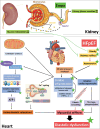
SGLT2 inhibition—mode of action in the kidney and suggested link to the beneficial impact on the pathophysiology of heart failure with preserved ejection fraction (HFpEF). Green stars represent alterations accomplished by empagliflozin (Empa), which positively influences the clinical picture. SARCO indicates sarco/endoplasmic reticulum Ca2+ ATPase; SGLT2, sodium-glucose linked transporter 2; SR, sarcoplasmic reticulum; and TNF-α, tumor necrosis factor α.

Comment in

References

    1. Adamczak DM, Oduah MT, Kiebalo T, Nartowicz S, Bęben M, Pochylski M, Ciepłucha A, Gwizdała A, Lesiak M, Straburzyńska-Migaj E. Heart failure with preserved ejection fraction: a concise review. Curr Cardiol Rep. 2020;22:82. doi: 10.1007/s11886-020-01349-3 - PMC - PubMed
    1. Bhatia RS, Tu JV, Lee DS, Austin PC, Fang J, Haouzi A, Gong Y, Liu PP. Outcome of heart failure with preserved ejection fraction in a population-based study. N Engl J Med. 2006;355:260–269. doi: 10.1056/NEJMoa051530 - PubMed
    1. Borlaug BA. Evaluation and management of heart failure with preserved ejection fraction. Nat Rev Cardiol. 2020;17:559–573. doi: 10.1038/s41569-020-0363-2 - PubMed
    1. Packer M, Butler J, Zannad F, Filippatos G, Ferreira JP, Pocock SJ, Carson P, Anand I, Doehner W, Haass M, et al. . Effect of empagliflozin on worsening heart failure events in patients with heart failure and preserved ejection fraction: EMPEROR-preserved trial. Circulation. 2021;144:1284–1294. doi: 10.1161/CIRCULATIONAHA.121.056824 - PMC - PubMed
    1. Anker SD, Butler J, Filippatos G, Ferreira JP, Bocchi E, Böhm M, Brunner-La Rocca HP, Choi DJ, Chopra V, Chuquiure-Valenzuela E, et al. ; EMPEROR-Preserved Trial Investigators. Empagliflozin in heart failure with a preserved ejection fraction. N Engl J Med. 2021;385:1451–1461. doi: 10.1056/NEJMoa2107038 - PubMed

MeSH terms