. 2024 Sep;26(9):101174.
doi: 10.1016/j.gim.2024.101174. Epub 2024 Jun 3.

Dominant missense variants in SREBF2 are associated with complex dermatological, neurological, and skeletal abnormalities

Collaborators, Affiliations

Dominant missense variants in SREBF2 are associated with complex dermatological, neurological, and skeletal abnormalities

Matthew J Moulton et al. Genet Med. 2024 Sep.

Abstract

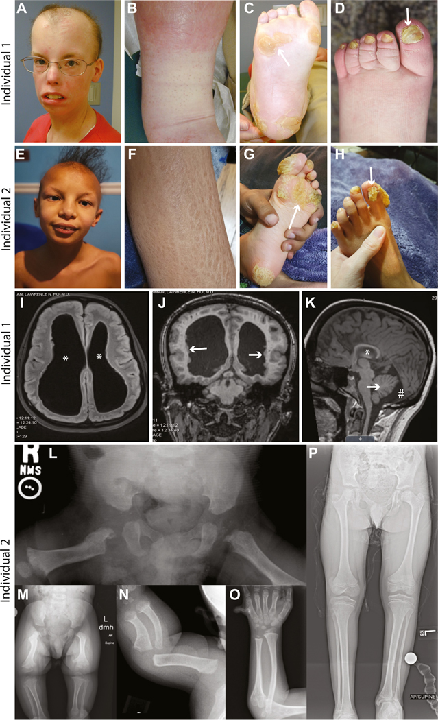
Purpose: We identified 2 individuals with de novo variants in SREBF2 that disrupt a conserved site 1 protease (S1P) cleavage motif required for processing SREBP2 into its mature transcription factor. These individuals exhibit complex phenotypic manifestations that partially overlap with sterol regulatory element binding proteins (SREBP) pathway-related disease phenotypes, but SREBF2-related disease has not been previously reported. Thus, we set out to assess the effects of SREBF2 variants on SREBP pathway activation.

Methods: We undertook ultrastructure and gene expression analyses using fibroblasts from an affected individual and utilized a fly model of lipid droplet (LD) formation to investigate the consequences of SREBF2 variants on SREBP pathway function.

Results: We observed reduced LD formation, endoplasmic reticulum expansion, accumulation of aberrant lysosomes, and deficits in SREBP2 target gene expression in fibroblasts from an affected individual, indicating that the SREBF2 variant inhibits SREBP pathway activation. Using our fly model, we discovered that SREBF2 variants fail to induce LD production and act in a dominant-negative manner, which can be rescued by overexpression of S1P.

Conclusion: Taken together, these data reveal a mechanism by which SREBF2 pathogenic variants that disrupt the S1P cleavage motif cause disease via dominant-negative antagonism of S1P, limiting the cleavage of S1P targets, including SREBP1 and SREBP2.

Keywords: Drosophila; Lipid droplet; S1P; SREBF1; SREBF2.


Conflict of interest statement

Conflict of Interest The authors declare no conflicts of interest.

Figures

Figure 1
Figure 1. Individuals harboring variants in SREBF2 p.(Arg519) exhibit dermatological, neurological, skeletal, and other abnormalities.
Individual 1 p.(Arg519His) and individual 2 p.(Arg519Gly) share phenotypes including scarring alopecia (A and E), scaly skin (B and F), hyperkeratosis (C and G), and abnormal nails (D and H). Individual 1 also has dilated cerebral ventricles (I, asterisks), periventricular nodular heterotopias (J, arrows), leukomalacia, hippocampal atrophy (K, asterisk), a mega-cisterna magna (K, pound symbol), and a small cerebellum (K, arrow). Short stature and an initial suggestion of metaphyseal dysplasia on a bone survey as a child that resolved with age (see supplemental clinical description) was also observed. Individual 2 also presents with bilateral enlargement of the lateral cerebral ventricles and other neurological findings (see supplemental clinical description). Radiographs of individual 2 demonstrate skeletal dysplasia. Anterior-posterior (AP) radiograph of the pelvis taken at 5 months shows right to left asymmetry of the proximal femur with chondrodysplasia punctata present in the hip region bilaterally (L). AP radiograph of the bilateral lower extremities taken at 22 months also demonstrates right to left asymmetry with metaphyseal changes noted throughout and absent mineralization of the capital femoral epiphyses (M). Left forearm taken at 5 months and 6 years 11 months, respectively (N and O), show metaphyseal widening with broad diaphysis, and bowing present in the younger image. The metaphyseal widening remains in the older image, but the diaphyseal changes appear resolved though slight bowing remains. Unilateral postaxial polydactyly was also present (O). Supine AP image of the lower extremity taken at 10 years 11 months (P), noting significant right to left asymmetry with leg length discrepancy.
Figure 2
Figure 2. Fibroblasts from individual 1 have limited lipid droplet (LD) accumulation, exhibit expanded endoplasmic reticula (ER), and accumulate lamellar inclusion bodies.
Fibroblasts from the unaffected father (A and B) and from Individual 1 (C and D) were subjected to transmission electron tomography analysis. LD accumulation is evident in the father’s fibroblasts (A and B, labeled with black arrowheads) but is severely limited in the cells from Individual 1 (C, quantified in E). Fibroblasts from individual 1 exhibit increased ER area compared with cells from the father (A and C, white arrowheads, quantified in F) and accumulate electron-dense lamellar inclusion bodies (C, white arrows; high magnification image in D), which are absent in the cells from the father (quantified in G). All quantifications are graphed as mean ± SEM, Welch’s t test, *P < .05, **P < .01, ****P < .0001.
Figure 3
Figure 3. Overexpression of SREBF2R519H or SREBF2R519G induces age-dependent neurological deficits in fly models.
Assessment of neurological function via climbing assay (A) of aged flies expressing SREBF2R519H and SREBF2R519G compared with nonvariant SREBF2 transcripts show an age-dependent increase in the time to climb, indicative of nervous system deficits (B). Neuronal function was also assessed using a bang sensitivity assay (C) showing an age-dependent increase in the time for variant-expressing flies to right themselves (D), further indicating nervous system defects with age. Finally, assessment of neurotransmission capacity via electroretinogram (ERG) was performed on flies expressing variant transcripts of SREBF2, in which a probe placed on the fly eye was used to measure neuronal response to light flashes (E). Synaptic transmission from the photoreceptors to the optic lobe is measured by the on and off transient phases and photoreceptor function is measured by the amplitude phase of the ERG recording. Age-dependent defects in neuronal function were observed in the on and off transients (F and G), indicating impaired synaptic transmission to the central brain in aged animals. All quantifications are graphed as mean ± SEM, One-way ANOVA with Dunnett’s multiple comparison correction, *P < .05, **P < .01, ***P < 0.001, ****P < 0.0001.
Figure 4
Figure 4. Fly SREBP and human SREBF2 overexpression induces glial lipid droplet formation, but SREBF2 variant overexpression fails to do so.
The fly compound eye is comprised of several hundred ommatidia in which photoreceptor neurons are surrounded by glia (A). Overexpression of fly SREBP in neurons induces glial LD formation (via Nile Red staining) as previously reported (B, solid white line outlines ommatidium, dotted white line outlines photoreceptor cells), but an empty control vector does not have this effect (C). Overexpression of nonvariant human SREBF2 in neurons induces glial LD formation to similar levels as fly SREBP (D). Neuronal overexpression of either the SREBF2R519H variant (E) or the SREBF2R519G variant (F) fails to induce LD formation, suggesting that the variants are loss-of-function alleles. Quantification of LDs from all genotypes tested is graphed in (G, n = 10 animals per genotype, aged 1–2 days post eclosion) as mean ± SEM, One-way ANOVA with Dunnett’s multiple comparison correction, ****P < .0001.
Figure 5
Figure 5. SREBF2 variants act in a dominant-negative manner by limiting the S1P enzyme pool required for SREBP protein activation.
Human SREBF2 overexpression in the fly photoreceptor neurons induces glial LD formation as visualized by Nile Red staining (A). However, co-overexpression of SREBF2R519H (B) or SREBF2R519G (C) with nonvariant SREBF2 severely limits glial LD formation, indicating that these variants act in a dominant-negative manner. Concomitant expression of the S1P encoding gene, MBTPS1, with the nonvariant SREBF2 (D) has no effect on LD formation, but expression together with SREBF2R519H (E) or SREBF2R519G (F) variants rescues the loss of glial LD production caused by the variants, revealing that the variants limit the function of S1P. Quantification of LDs from all genotypes tested is graphed in (G, n = 10 animals per genotype, aged 1–2 days post eclosion) as mean ± SEM, One-way ANOVA with Dunnett’s multiple comparison correction, ***P < .001, ****P < .0001.
Figure 6
Figure 6. Fibroblast gene expression analysis reveals defects in SREBP pathway activity caused by SREBF2 variants.
The activation of gene expression by SREBP proteins requires the cleavage of full-length SREBP proteins by S1P and S2P followed by dimerization of the SREBP transcription factor and its translocation to the nucleus (A). The cleaved, mature SREBP transcription factor recognizes and promotes the expression of genes containing Sterol Regulatory Elements (SRE) in their promoters, including genes involved in steroidogenesis and fatty acid production (B). Fibroblasts from individual 1 and unaffected parents were analyzed for gene expression of key SREBP-regulated genes via quantitative RT-PCR. We observed significant reductions in RNA from individual 1 in the SREBP-regulated genes: SREBF1, SREBF2, HMGCR, FASN, SCD, INSIG1, and LDLR (C and D) but no statistically significant changes in the genes encoding the SREBP cleavage enzymes, S1P (MBTPS1) and S2P (MBTPS2) (E). Quantifications (n = 3 replicates per genotype) are graphed as mean ± SEM, One-way ANOVA with Tukey multiple comparison correction after log transformation. ns, no statistically significant difference; *P < .05, **P < .01, ***P < .001.
Figure 7
Figure 7. Model diagram of proposed mechanism of disease-associated variants in SREBF2.
The cell maintains a pool of S1P cleavage enzyme capable of recognizing the RXXL motif in SREBP proteins for proper cleavage into its mature transcription factor. After cleavage, S1P dissociates from SREBP proteins and S1P can be recycled back into the available pool for additional cleavage of SREBP and other protein targets (A). Our data indicate that SREBF2 p.(Arg519) variants inhibit S1P-mediated cleavage of SREBP2 for transcription factor generation. We propose that this failed cleavage may cause an S1P-SREBP2 interaction to persist, or alter the ability of S1P to cleave SREBP2, subsequently causing a failure in S1P pool regeneration for cleavage of SREBP1, nonvariant SREBP2, and other targets of S1P (B). Thus, dominant-negative variants in SREBF2 may inhibit the proper production of any protein product requiring S1P, causing a broader array of symptoms than would be caused by loss of SREBP2 function alone.

References

    1. Jeon YG, Kim YY, Lee G, Kim JB. Physiological and pathological roles of lipogenesis. Nat Metab. 2023;5(5):735–759. 10.1038/s42255-023-00786-y - DOI - PubMed
    1. Horton JD, Goldstein JL, Brown MS. SREBPs: activators of the complete program of cholesterol and fatty acid synthesis in the liver. J Clin Invest. 2002;109(9):1125–1131. 10.1172/JCI15593 - DOI - PMC - PubMed
    1. Rawson RB. The SREBP pathway — insights from insigs and insects. Nat Rev Mol Cell Biol. 2003;4(8):631–640. 10.1038/nrm1174 - DOI - PubMed
    1. Shimano H, Sato R. SREBP-regulated lipid metabolism: convergent physiology — divergent pathophysiology. Nat Rev Endocrinol. 2017;13(12):710–730. 10.1038/nrendo.2017.91 - DOI - PubMed
    1. Brown MS, Goldstein JL. The SREBP pathway: regulation of cholesterol metabolism by proteolysis of a membrane-bound transcription factor. Cell. 1997;89(3):331–340. 10.1016/S0092-8674(00)80213-5 - DOI - PubMed

Substances

LinkOut - more resources