Review
. 2024 Jul;122(1):81-112.
doi: 10.1111/mmi.15283. Epub 2024 Jun 7.

Bacterial chromatin proteins, transcription, and DNA topology: Inseparable partners in the control of gene expression

Affiliations
Review

Bacterial chromatin proteins, transcription, and DNA topology: Inseparable partners in the control of gene expression

Christine M Hustmyer et al. Mol Microbiol. 2024 Jul.

Erratum in

Abstract

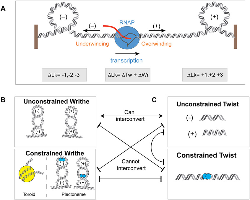
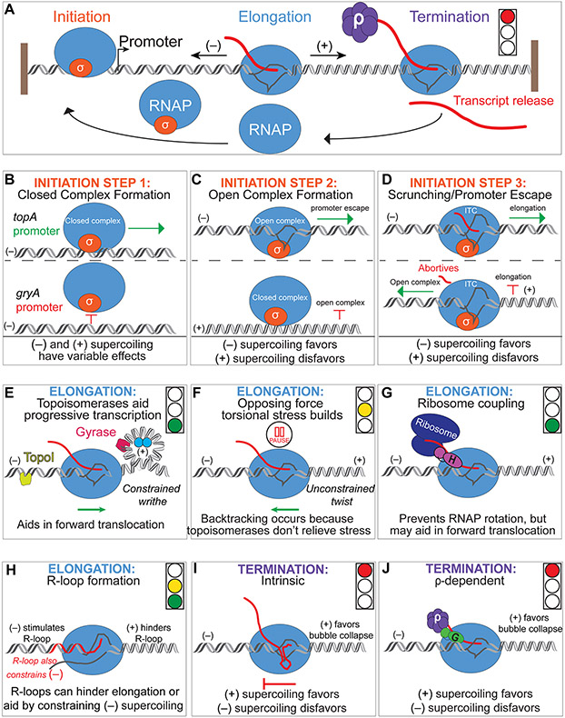
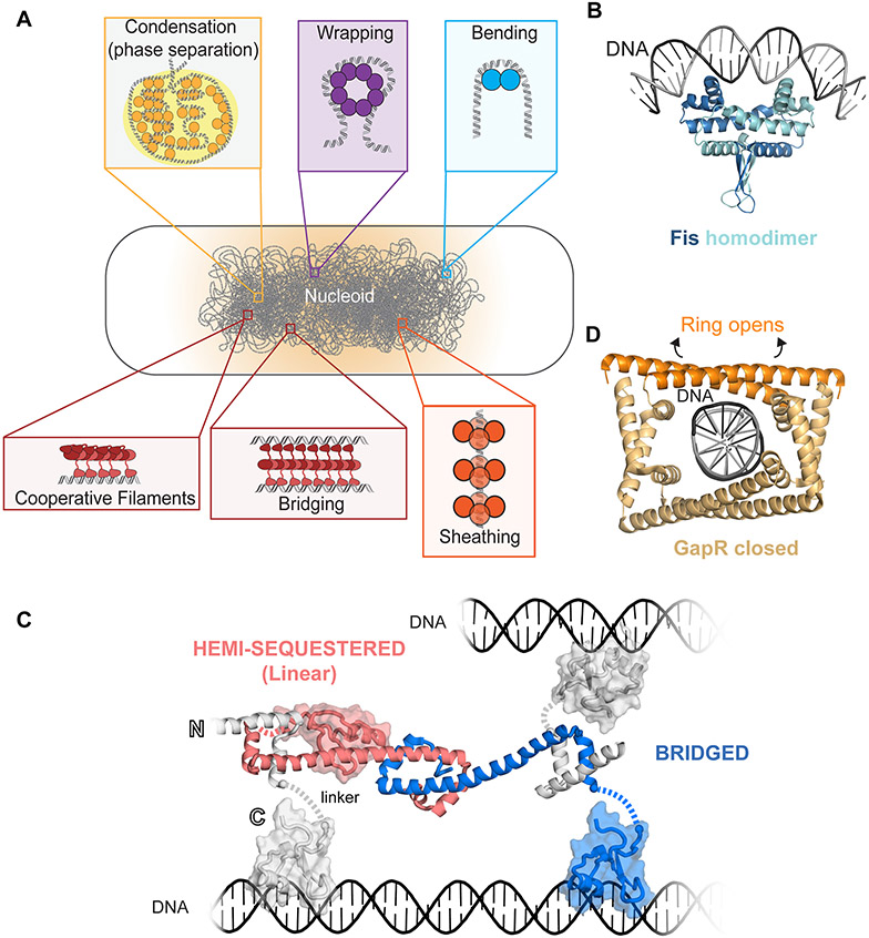
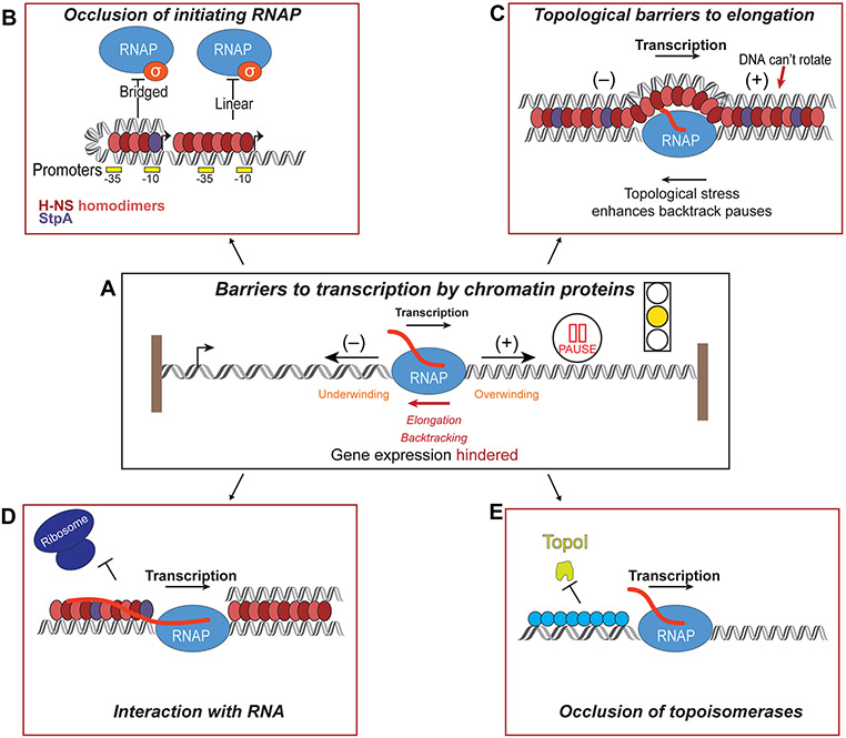
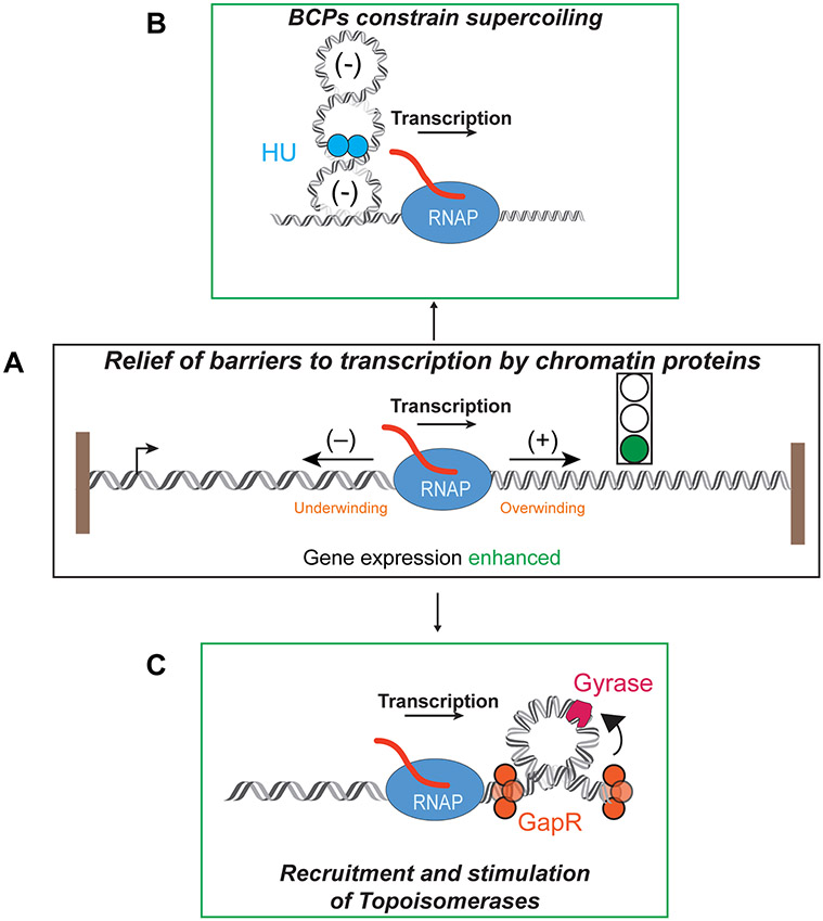
DNA in bacterial chromosomes is organized into higher-order structures by DNA-binding proteins called nucleoid-associated proteins (NAPs) or bacterial chromatin proteins (BCPs). BCPs often bind to or near DNA loci transcribed by RNA polymerase (RNAP) and can either increase or decrease gene expression. To understand the mechanisms by which BCPs alter transcription, one must consider both steric effects and the topological forces that arise when DNA deviates from its fully relaxed double-helical structure. Transcribing RNAP creates DNA negative (-) supercoils upstream and positive (+) supercoils downstream whenever RNAP and DNA are unable to rotate freely. This (-) and (+) supercoiling generates topological forces that resist forward translocation of DNA through RNAP unless the supercoiling is constrained by BCPs or relieved by topoisomerases. BCPs also may enhance topological stress and overall can either inhibit or aid transcription. Here, we review current understanding of how RNAP, BCPs, and DNA topology interplay to control gene expression.

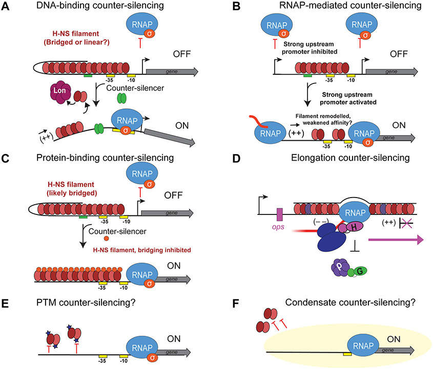
Keywords: H‐NS; bacterial chromatin; bridging; counter‐silencing; supercoiling; topological barriers; topology; transcription.


Figures

Figure 1.
Figure 1.. BCPs and RNAP constrain and generate supercoiling of DNA.
A) Torsional stress generated by RNAP (blue circle) generates diffusion of (−) supercoiling upstream and (+) supercoiling downstream as RNAP transcribes DNA (black and gray double helix) into mRNA (red). The change in linking number, or the supercoiled state of DNA, equals (Δtwist + Δwrithe), see also Figure 1B and C. The change in Lk relative to the relaxed DNA (ΔLk) can be positive [(+) supercoiling; ΔLk +1, +2, +3….] or negative [(−) supercoiling; ΔLk –1, –2, –3….]. B) Changes in supercoiling of DNA can be manifest by changes in writhe. Top: When unconstrained (not bound by a BCP, top), both (−) and (+) plectonemes can form and can interconvert into unconstrained twist (Figure 1C, top). Lower: (−) writhe can be constrained into toroids (left, yellow histone). (−) or (+) writhe can also be constrained in plectonemes by BCPs (two blue circles) which pin the apex of a plectoneme (left, (−) plectoneme) and bend DNA or by a BCP (two blue circles) which binds the cross-over point of a plectoneme (right, (+) plectoneme). Constrained writhe cannot interconvert with constrained or unconstrained twist. C) Changes in supercoiling of DNA can be manifest by changes in twist. (−) unconstrained twist (not bound by a BCP, top) is manifest by underwound DNA, whereas (+) unconstrained twist is formed by underwound DNA. Unconstrained twist can interconvert to unconstrained writhe. Constrained twist (lower) occurs when a BCP (two blue circles) binds twist and prevents diffusion of twist along DNA. Constrained twist cannot interconvert with unconstrained twist, unconstrained writhe, or constrained writhe.
Figure 2.
Figure 2.. Effects of supercoiling on each stage of transcription.
A) Steps of transcription. Transcription begins when RNAP (blue) and σ-factor (orange) contact promoters. RNAP can elongate and transcribe DNA into mRNA (red) while generating (+) supercoiling in front and (−) supercoiling behind. RNAP continues transcription until termination, for example by ρ (purple hexamer). RNAP and the nascent transcript will be released from DNA. RNAP is now free to bind another sigma factor to re-initiate. B) Initiation step 1: closed complex formation. Closed complex occurs when RNAP (blue) and σ-factor (orange) contact promoters. (−) and (+) supercoiling have variable effects on closed complex formation. For example, at the topA promoter, (−) supercoiling promotes closed complex formation, so RNAP can proceed to next step (Figure 2C). At the gryA promoter, (−) supercoiling prevents closed complex formation and initiation does not proceed. C) Initiation step 2: open complex formation. Open complex formation is favored when DNA is (−) supercoiled; initiation can proceed to promoter escape (Figure 2D). (+) supercoiling favors reversion to closed complex (Figure 2A). D) Initiation step 3: scrunching/promoter escape. (−) supercoiling promotes promoter escape and subsequent elongation, whereas (+) supercoiling favors reversion to closed complex, triggering release of abortive transcripts. E) RNAP elongation can occur without perturbation (green light) when topoisomerases such as Topoisomerase I (yellow) removes (−) supercoils generated by RNAP upstream and gyrase (dark pink) removes (+) supercoils downstream. BCPs (blue circles, bound to apex of (+) plectoneme) can constrain writhe, preventing supercoiling diffusion, and may recruit topoisomerases. F) In cases where topoisomerases cannot remove supercoils, torsional stress on RNAP builds. In cases where (+) twist is unconstrained (i.e. not bound by BCP), RNAP pauses and can backtrack, slowing transcription (yellow light). G) In some cases, the ribosome (dark blue) is coupled to transcription, which can promote efficient translocation of RNAP (green light). The ribosome may be coupled to RNAP by factors such as the NusG paralog RfaH (pink). H) R-loops (red RNA) form behind RNAP, where (−) supercoiling is stimulated. R-loops can both slow RNAP (yellow light) or may aid transcription (green light) if the R-loop constrains (−) supercoiling. I) Intrinsic termination occurs when a nascent RNA hairpin forms in the transcription bubble. (+) supercoiling upstream of RNAP favors termination by promoting collapse of the transcription bubble (red light). J) Rho-dependent termination occurs when ρ (purple hexamer) is recruited by a factor such as NusG (green). (+) supercoiling upstream of RNAP favors termination by promoting collapse of the transcription bubble (red light).
Figure 3.
Figure 3.. BCPs alter DNA structure.
A) Cartoon model of a hypothetical bacterial nucleoid and the BCPs that structure the chromosome, adapted from (Shen & Landick, 2019). Examples of BCP structures are shown in different colors (see also Table 1). Proteins (such as HU, IHF, Fis) can bend a dsDNA (blue). Proteins (such as HU, IHF) can wrap DNA around their outer surfaces (purple). Proteins (such as GapR, Bd0055) can sheath, or bind end-on to coat DNA inside their structures (orange). Proteins (such as H-NS) can form cooperative filaments on one side of a dsDNA, or bridge two dsDNAs with multiple contacts (red). Proteins (such as Dps) can oligomerize and aggregate, resulting in condensation and protein-DNA phase separated complexes (yellow). B) Structure of Fis homodimer (blue monomers) bound to bent DNA (PDB 4IHV; (Hancock et al., 2016)). C) Models of H-NS filaments in a hemi-sequestered (linear) filament (pink and gray monomer) and bridged filament (blue and gray monomers). In a bridged filament, biochemical evidence suggests H-NS binds to two duplexes of DNA (black). But in a hemi-sequestered filament, the C-terminal DNA binding domain is sequestered and inaccessible to bind two segments of DNA and thus binds only one dsDNA duplex (van der Valk et al., 2017). Model was created using structures of the H-NS CTD structure (PDB 1HNR; (Shindo et al., 1995)), the H-NS paralog Ler bound to DNA (PDB 2LEV; (Cordeiro et al., 2011)) and the H-NS N-terminus oligomerization domain structure (PDB 3NR7; (Arold et al., 2010)). A flexible linker connects the N- and C-terminus (dashed line). D) Structure of closed GapR homotetramer bound to DNA (PDB 6CG8; (Guo et al., 2018)). The dark orange α-helices can re-fold to open to bind (+) twisted DNA (Huang et al., 2020).
Figure 4.
Figure 4.. Barriers to transcription by BCPs.
A) Transcription can be impeded at the promoter and at the level of elongation. Cartoon model examples of mechanisms of transcription barriers. B) H-NS (dark red and light red monomers) can form both linear and bridged filaments on DNA. StpA (purple) can form constitutively bridged filaments with H-NS, bound to two dsDNAs. Both conformations of filaments can occlude RNAP from initiating transcription at promoters. C) H-NS-StpA filaments can bridge RNAP in vitro, promoting RNAP backtracking. Bridged H-NS filaments, unlike linear filaments, prevent DNA rotation and thus stimulate pausing. The topological trapping of RNAP by H-NS bridged filaments promotes premature transcription termination (Kotlajich et al., 2015). D) H-NS and StpA may also interact with nascent RNA (red), preventing RNAP rotation and forward translocation. An interaction with RNA could also block ribosomes (dark blue) from binding RNA. E) BCPs, such as HU (blue) can oligomerize to form a filament and occlude topoisomerases from targeting and relieving supercoiled DNA (Ghosh et al., 2014).
Figure 5.
Figure 5.. Relief of topological barriers to transcription by BCPs.
A) Transcription can be promoted by BCPs that mediate transcription through topological changes. B) Proteins, such as HU, Fis, and IHF, (blue two circles) can constrain supercoiling diffusion by constraining writhe or twist. These proteins act as topological homeostats. C) Proteins like GapR (orange) can bind to overtwisted (+) supercoiled DNA downstream of RNAP at highly transcribed genes. GapR can stimulate topoisomerase IV and gyrase (pink) to promote positive outcomes on transcription.
Figure 6.
Figure 6.. Counter-silencing of BCPs.
A) DNA-binding counter-silencing of H-NS filaments (red monomers) (unknown conformation) at promoters. H-NS can occlude RNAP (blue) from binding to promoters. A counter-silencer (green) can bind to DNA upstream of promoters to remodel H-NS filament in an unknown conformation. H-NS, when displaced is degraded by the Lon protease (burgundy hexamer). (+) supercoiling can also remodel the filament to activate transcription. B) RNAP counter-silencing of H-NS filaments at promoters. A strong, active upstream promoter can drive RNAP elongation into an H-NS filament. Elongation, perhaps mediated by upstream (+) supercoiling can counter-silence H-NS filaments. C) H-NS can be counter-silenced by protein-binding counter-silencers (orange) that bind to the N-terminus of H-NS and lock H-NS into a linear filament. D) H-NS filaments can be silenced at the level of elongation by the elongation factor RfaH (pink), which recruits the ribosome (dark blue) and excludes NusG-Rho binding. E) BCPs can be counter-silenced by post-translational modifications (stars on proteins), which may disrupt DNA binding modes of BCPs. F) Effects of BCPs on transcription could be counter-silenced by condensates (yellow cloud), which may exclude certain BCPs from accessing transcription complexes.

Similar articles

Cited by

References

    1. A P, and Weber SC (2019) Evidence for and against Liquid-Liquid Phase Separation in the Nucleus. Noncoding RNA 5. DOI: 10.3390/ncrna5040050. - DOI - PMC - PubMed
    1. Afflerbach H, Schroder O, and Wagner R (1999) Conformational changes of the upstream DNA mediated by H-NS and FIS regulate E. coli RrnB P1 promoter activity. J Mol Biol 286: 339–353. DOI: 10.1006/jmbi.1998.2494. - DOI - PubMed
    1. Aguilera A, and Garcia-Muse T (2012) R loops: from transcription byproducts to threats to genome stability. Mol Cell 46: 115–124. DOI: 10.1016/j.molcel.2012.04.009. - DOI - PubMed
    1. Albano M, Smits WK, Ho LT, Kraigher B, Mandic-Mulec I, Kuipers OP, and Dubnau D (2005) The Rok protein of Bacillus subtilis represses genes for cell surface and extracellular functions. J Bacteriol 187: 2010–2019. DOI: 10.1128/JB.187.6.2010-2019.2005. - DOI - PMC - PubMed
    1. Ali SS, Whitney JC, Stevenson J, Robinson H, Howell PL, and Navarre WW (2013) Structural insights into the regulation of foreign genes in Salmonella by the Hha/H-NS complex. J Biol Chem 288: 13356–13369. DOI: 10.1074/jbc.M113.455378. - DOI - PMC - PubMed

Publication types

MeSH terms

LinkOut - more resources