Review
. 2024 Jun 7;81(1):250.
doi: 10.1007/s00018-024-05286-0.

Redox regulation of UPR signalling and mitochondrial ER contact sites

Affiliations
Review

Redox regulation of UPR signalling and mitochondrial ER contact sites

Jose C Casas-Martinez et al. Cell Mol Life Sci. .

Abstract

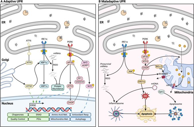
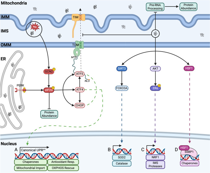
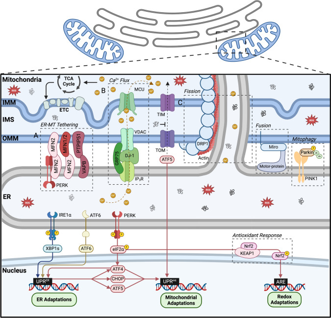
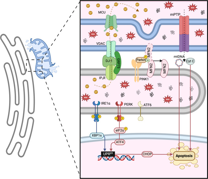
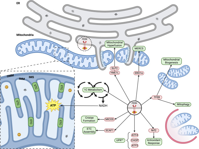
Mitochondria and the endoplasmic reticulum (ER) have a synergistic relationship and are key regulatory hubs in maintaining cell homeostasis. Communication between these organelles is mediated by mitochondria ER contact sites (MERCS), allowing the exchange of material and information, modulating calcium homeostasis, redox signalling, lipid transfer and the regulation of mitochondrial dynamics. MERCS are dynamic structures that allow cells to respond to changes in the intracellular environment under normal homeostatic conditions, while their assembly/disassembly are affected by pathophysiological conditions such as ageing and disease. Disruption of protein folding in the ER lumen can activate the Unfolded Protein Response (UPR), promoting the remodelling of ER membranes and MERCS formation. The UPR stress receptor kinases PERK and IRE1, are located at or close to MERCS. UPR signalling can be adaptive or maladaptive, depending on whether the disruption in protein folding or ER stress is transient or sustained. Adaptive UPR signalling via MERCS can increase mitochondrial calcium import, metabolism and dynamics, while maladaptive UPR signalling can result in excessive calcium import and activation of apoptotic pathways. Targeting UPR signalling and the assembly of MERCS is an attractive therapeutic approach for a range of age-related conditions such as neurodegeneration and sarcopenia. This review highlights the emerging evidence related to the role of redox mediated UPR activation in orchestrating inter-organelle communication between the ER and mitochondria, and ultimately the determination of cell function and fate.

Keywords: C. elegans; Contact-sites; Hormesis; Mitochondrial dynamics; Redox signalling; Skeletal muscle.


Conflict of interest statement

Not applicable.

Figures

Fig. 1
Fig. 1
The UPRER. A Adaptive UPRER. Following ER stress, BiP binds to misfolded proteins on the substrate-binding site and the ATPase domain dissociates from the transmembrane receptors, allowing allosteric activation of the UPRER regulators by oligomerisation and phosphorylation [14, 15]. (1) IRE1α RNase activity mediates unconventional splicing of XBP1 [–18], XBP1s translocates to the nucleus to promote expression of genes related to quality control [9, 19]. IRE1α also mediates the cleavage and degradation of mRNAs and microRNAs; regulated IRE1α-dependent decay (RIDD), decreasing the protein load in the ER lumen [20]. (2) PERK phosphorylates eIF2α [21], promoting rapid attenuation of global mRNA translation [22, 23]. Phosphorylated eIF2α also regulates the translation of the transcription factor ATF4 [24]. ATF4 regulates the feedback loop responsible for the restoration of protein synthesis. ATF4 induction of CHOP, upregulates the expression of GADD34 which forms a complex with PP1 to dephosphorylate eIF2α [25, 26]. (3) ATF6α translocates to the Golgi apparatus, where it is cleaved to generate ATF6f, which acts as a transcription factor that promotes the expression of ER chaperones [27, 28]. ATF6α promotes the expression of Xbp1 mRNA, enhancing the substrate load for IRE1α splicing [29]. B Maladaptive UPRER. Following prolonged ER stress the homeostatic capacity of the UPRER becomes saturated that can activate pro-apoptotic signalling. (1) IRE1α interacts with TRAF2 to promote a kinase signalling cascade that activates JNK [30, 31]. JNK promotes the oligomerisation of BAX and BAK on the mitochondrial membrane and the assembly of the apoptosome [32, 33]. RIDD can promote apoptosis by degrading essential cell-survival mRNAs such as the negative regulators of TXNIP, promoting the assembly of the inflammasome leading to apoptosis [34, 35]. (2) PERK-eIF2α induces the translation of ATF4, activation of CHOP and GADD34 [25, 26]. CHOP promotes the expression of PUMA, NOXA, BIM and BID, which induce the mitochondrial BCL-2 pro-apoptotic proteins. CHOP can also activate the translation of ERO1α, promoting the oxidation of the ER environment [36, 37]. PERK-ATF4-CHOP arm regulates IP3R-mediated Ca2+ leakage from the ER [38, 39]. Sustained and excessive Ca2+ transport from the ER to the mitochondria impairs mitochondrial metabolism and lead to opening of the mPTP and pro-apoptotic signalling [40, 41]
Fig. 2
Fig. 2
The UPRmt. Most proteins that constitute the mitochondrial proteome are synthesised in the cytoplasm, targeted and imported into mitochondria [125] via the TOM/TIM complex [126], perturbation of this trafficking can impair mitochondrial proteostasis and induce mitochondrial stress [127]. A The canonical axis of the UPRmt is controlled by the expression of ATF5, ATF4 and CHOP [132]. CHOP alleviates proteotoxic stress by inducing the expression of the mitochondrial chaperones HSP10 and HSP60 [134]. ATF5 is normally imported into mitochondria via TOM and TIM, where it is degraded by proteases [138]. Mitochondrial proteotoxic stress will perturb mitochondrial import efficiency, resulting in the activation of ATF5 by p-eIF2α and its translocation to the nucleus [139]. ATF5 promotes the transcription of genes related to chaperones, proteases and antioxidant proteins [137]. B The sirtuin axis of the UPRmt activates SIRT3, which deacetylates FOXO3A, promoting its translocation to the nucleus and transcription of SOD2 and catalase [142, 143]. C AKT mediates the ROS-dependant phosphorylation of ERα, which activates NRF1 and the IMS protease HTRA2 [144]. NRF1 stimulates mitochondrial respiration, proteasome activity and the IMS protease OMI. D Mitochondrial proteotoxic stress promotes epigenetic changes in the cellular DNA regulated by HSF1, it translocates to the nucleus where it interacts with SSBP1 to bind to the chromatin and boost the expression of mitochondrial chaperones [145, 146]
Fig. 3
Fig. 3
Mitochondria-ER contact sites molecular components and cellular functions. MERCS are relatively stable structures that require the formation of molecular bridges established by interacting proteins anchored in the smooth ER and the OMM [5]. Tethering complexes are essential, structural and reversible bonds that stabilise MERCS [177]. A MERCS tethering complexes occur between ER MFN2 and mitochondrial MFN2 or ER MFN2 and MFN1 [178]. The MFN tethering complex is dependent on the interaction of MFN2 and PERK at the ER membrane, essential for the establishment of the contact sites [90, 179]. Other complexes reported as regulating the tethering of MERCS include the ER VAPB and the OMM PTPIP51 [180]. B MERCS regulate Ca2+ flux between the ER and the mitochondria by the complex that forms between IP3R from the ER and VDAC from the OMM [5, 177]. Ca2+ passes through the MCU to reach the mitochondrial matrix [185, 186]. DJ-1 [187] and GRP75 [188] regulate the connection between IP3R and VDAC [189]. Some components of the TCA cycle require the binding of Ca2+ for their function, the interaction of mitochondria and ER via MERCS supply Ca2+ to mitochondria for stimulating the TCA cycle, resulting in an increase in ATP production [190]. C MERCS control the processes of mitochondrial fusion, fission and mitophagy [111, 191]. The ER promotes the polymerisation of actin filaments and establishment of close contacts between the two organelles [192]. ER tubules will release Ca2+ ions into the mitochondria, triggering the inner mitochondrial membrane to divide [192, 193]. DRP1 assembles around mitochondria at the fission site, a DRP1 ring constricts with the aid of actin–myosin filaments, resulting in the formation of two daughter mitochondria. ER tubules guide the position and timing of mitochondria fusion through the tethering with mitochondria [191, 194]. During mitochondrial fusion the contact sites between the tubules and the mitochondria need to be maintained to avoid the disruption of these MERCS, the Ca2+ sensitive motorprotein Miro cease all transportation movements of the mitochondria involved [195]. In the mitochondria PINK1 phosphorylates MFN2, recruits Parkin at the MERCS, allowing Parkin dependent ubiquitination of ER MFN2, promoting the separation of the two organelles and the initiation of mitophagy [196]
Fig. 4
Fig. 4
MERCS regulation of cellular signalling in ageing and disease. Disruption of MERCS assembly and disassembly plays a key role in pathophysiological conditions particularly in ageing and age-related diseases. Disrupted Ca2+ flow from the ER to mitochondria can result in mitochondrial dysfunction with loss of mitochondrial membrane potential and mitochondrial ROS generation, that result in activation of apoptotic pathways or senescence [40]. Excess Ca2+ transfer into mitochondria via IP3R can induce the opening of the mPTP, release of cytochrome c and activation of the caspase signalling cascade and pro-apoptotic pathways [198]. On mitochondria PINK1 phosphorylates MFN2, recruits Parkin at the MERCS, allowing Parkin dependent ubiquitination of ER MFN2, promoting the separation of the two organelles and the initiation of mitophagy [196]. Release of mtDNA through channels such as VDAC (located in or close to MERCS) has emerged as a potential regulator for the inflammatory response [201]
Fig. 5
Fig. 5
PERK regulation of mitochondrial capacity. PERK is a key regulator of both the UPRER and the UPRmt, that localises at MERCS [90]. The adaptive ER stress response promotes mitochondrial elongation and network establishment [172]. The modulation of mitochondrial metabolism by PERK results in improved cristae formation, assembly of the ETC and oxidative phosphorylation efficiency [220]. 1 PERK regulates the expression of the mitochondrial contact site and cristae-organizing system (MICOS) [221]. 2 The activation of ATF4 by PERK promotes the expression of SCAF1 helps mediate assembly of the ETC [218, 222]. 3 The adaptive UPRER also promotes one-carbon metabolism [223]. 4 PERK can promote cell survival by increasing antioxidant capacity through the activation of Nrf2 [224]. 5 During the adaptive UPRER response, there is an upregulation of TFEB [225], which can induce the ISR via activation of ATF4 and CHOP, activate mitophagy machinery and boost mitochondrial biogenesis by promoting expression of PGC1α, TFAM and NRF1 [219]. 6 The formation of PERK-ERO1⍺ complex can restore mitochondrial homeostasis and promote the formation of MERCS [188, 226]. 7 PERK is essential for the activation of UPRmt transcription factor ATF5 [139] and can reduce mitochondrial protein import by promoting the degradation of mitochondrial translocase TIM17A by phosphorylation of eIF2α [227]

Similar articles

Cited by

References

    1. Li X, Yang T, Sun Z. Hormesis in health and chronic diseases. Trend Endocrinol Metabol. 2019;30:944–958. doi: 10.1016/j.tem.2019.08.007. - DOI - PMC - PubMed
    1. Merry TL, Ristow M. Mitohormesis in exercise training. Free Radic Biol Med. 2016;98:123–130. doi: 10.1016/j.freeradbiomed.2015.11.032. - DOI - PubMed
    1. Done AJ, Traustadottir T. Nrf2 mediates redox adaptations to exercise. Redox Biol. 2016;10:191–199. doi: 10.1016/j.redox.2016.10.003. - DOI - PMC - PubMed
    1. Schieber M, Chandel NS. ROS function in redox signaling and oxidative stress. Curr Biol. 2014;24(10):R453–R462. doi: 10.1016/j.cub.2014.03.034. - DOI - PMC - PubMed
    1. Rowland AA, Voeltz GK. Endoplasmic reticulum–mitochondria contacts: function of the junction. Nat Rev Mol Cell Biol. 2012;13(10):607–615. doi: 10.1038/nrm3440. - DOI - PMC - PubMed

LinkOut - more resources