. 2024 Jul 12;10(7):2403-2418.
doi: 10.1021/acsinfecdis.4c00130. Epub 2024 Jun 7.

Strategic Single-Residue Substitution in the Antimicrobial Peptide Esc(1-21) Confers Activity against Staphylococcus aureus, Including Drug-Resistant and Biofilm Phenotype

Affiliations

Strategic Single-Residue Substitution in the Antimicrobial Peptide Esc(1-21) Confers Activity against Staphylococcus aureus, Including Drug-Resistant and Biofilm Phenotype

Maria Rosa Loffredo et al. ACS Infect Dis. .

Abstract

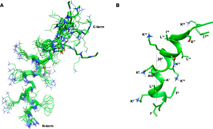
Staphylococcus aureus, a bacterium resistant to multiple drugs, is a significant cause of illness and death worldwide. Antimicrobial peptides (AMPs) provide an excellent potential strategy to cope with this threat. Recently, we characterized a derivative of the frog-skin AMP esculentin-1a, Esc(1-21) (1) that is endowed with potent activity against Gram-negative bacteria but poor efficacy against Gram-positive strains. In this study, three analogues of peptide 1 were designed by replacing Gly8 with α-aminoisobutyric acid (Aib), Pro, and dPro (2-4, respectively). The single substitution Gly8 → Aib8 in peptide 2 makes it active against the planktonic form of Gram-positive bacterial strains, especially Staphylococcus aureus, including multidrug-resistant clinical isolates, with an improved biostability without resulting in cytotoxicity to mammalian cells. Moreover, peptide 2 showed a higher antibiofilm activity than peptide 1 against both reference and clinical isolates of S. aureus. Peptide 2 was also able to induce rapid bacterial killing, suggesting a membrane-perturbing mechanism of action. Structural analysis of the most active peptide 2 evidenced that the improved biological activity of peptide 2 is the consequence of a combination of higher biostability, higher α helical content, and ability to reduce membrane fluidity and to adopt a distorted helix, bent in correspondence of Aib8. Overall, this study has shown how a strategic single amino acid substitution is sufficient to enlarge the spectrum of activity of the original peptide 1, and improve its biological properties for therapeutic purposes, thus paving the way to optimize AMPs for the development of new broad-spectrum anti-infective agents.

Keywords: Staphylococcus aureus; antimicrobial peptides; bent helical structure; biofilm; multidrug-resistant strains; α-aminoisobutyric acid.


Conflict of interest statement

The authors declare no competing financial interest.

Figures

Figure 1
Figure 1
Activity of peptides 1 and 2 against the biofilm of S. aureus ATCC 25923 and S. aureus #4, after 2 h of treatment. Biofilm viability was evaluated by measuring the reduction of MTT to its insoluble formazan (as reported in the Materials and Methods section) and expressed as percentage compared to that of untreated samples (bacterial biofilm not treated with the peptide, 100% viability). Data are the mean ± standard error of the mean (SEM) of three independent experiments performed in duplicate. Statistical analysis was conducted using two-way ANOVA to determine the significance between the two peptides. **, p < 0.01; ***, p < 0.001; ****, p < 0.0001; ns, not significant.
Figure 2
Figure 2
Effect of peptide 2 on the survival of HaCaT cells was assessed using the MTT assay after 24 h of treatment. The results are presented as percentage compared to the untreated control cells and represent the mean of three independent experiments ± SEM.
Figure 3
Figure 3
Stability of peptide 2 in 50% fresh bovine serum at different incubation times at 37 °C. (A) The panel reports the most representative RP-HPLC chromatograms of peptide 2 at 0, 5, and 16 h. (B) The panel reports the percentage of nondegraded peptide (%) after 1, 3, 4, 5, 16, and 24 h. Data represent the mean ± standard deviation (SD) of three independent experiments.
Figure 4
Figure 4
Kinetics of cytoplasmic membrane permeabilization of S. epidermidis ATCC 12228 and S. aureus ATCC 25923 induced by the addition of peptide 1 (panels A and C) and peptide 2 (panels B and D) at different concentrations. Alterations of the permeability of the cytoplasmic membrane allowed the Sytox Green probe (1 μM) to enter the cell and bind intracellular nucleic acids, resulting in an increase of fluorescence intensity. Controls (Ctrl) are microbial cells without the addition of any peptide. The reported values are from one representative experiment out of three.
Figure 5
Figure 5
Effect of peptide 2 on S. aureus ATCC 25923 cell viability. Bacteria (1 × 10⁶ CFU/mL) were incubated with peptide 2 at 2 × MIC (25 μM), MIC (12.5 μM), 1/2 × MIC (6.25 μM), and 1/4 × MIC (3.12 μM) in phosphate-buffered saline (PBS) at 37 °C. The number of surviving cells (CFU/mL) was calculated at different time points (5, 15, 30, 60, 90, and 120 min). Data represent the mean ± SD of four independent experiments.
Figure 6
Figure 6
ThT fluorescence as a function of the peptide concentration of 1 and 2 in liposomes mimicking Gram-positive bacterial membranes (POPG/POCL, 6:4, mol:mol) (100 μM). On the y-axis, F indicates the value of fluorescence after peptide addition, while F0 represents the initial fluorescence in the absence of peptide. Data represent the mean ± SD of three independent experiments.
Figure 7
Figure 7
Effect of different concentrations of peptide 2 on the leakage of CF encapsulated into POPG/POCL (6:4, mol:mol) LUVs after 5 min. LUVs were used at a final lipid concentration of 100 μM. Data points are the mean ± SD of three different experiments.
Figure 8
Figure 8
Circular dichroism spectra of peptides 1 (red line), 2 (blue line), and 3 (orange line) at 20 μM measured in (A) water and (B) in the presence of POPG/POCL (6:4 mol/mol) LUVs (500 μM).
Figure 9
Figure 9
(A) NMR-derived 10 lowest-energy structures of peptide 2. Heavy atoms are shown in atom-type coloring (carbon, green; nitrogen, blue; oxygen, red; hydrogen, white). For the sake of clarity, only polar hydrogen atoms are shown. (B) Representative structure of peptide 2. Backbone is shown as a ribbon and helical axes are shown as dotted lines.
Scheme 1
Scheme 1. Schematic Description of the Hypothetical Mechanism Involved in Peptide 2 Activity

Similar articles

Cited by

References

    1. Romanescu M.; Oprean C.; Lombrea A.; Badescu B.; Teodor A.; Constantin G. D.; Andor M.; Folescu R.; Muntean D.; Danciu C.; Dalleur O.; Batrina S. L.; Cretu O.; Buda V. O. Current State of Knowledge Regarding WHO High Priority Pathogens-Resistance Mechanisms and Proposed Solutions through Candidates Such as Essential Oils: A Systematic Review. Int. J. Mol. Sci. 2023, 24, 9727.10.3390/ijms24119727. - DOI - PMC - PubMed
    1. Giudice P. Skin Infections Caused by Staphylococcus aureus. Acta Derm. Venereol. 2020, 100, adv0011010.2340/00015555-3466. - DOI - PMC - PubMed
    1. Weiner-Lastinger L. M.; Abner S.; Edwards J. R.; Kallen A. J.; Karlsson M.; Magill S. S.; Pollock D.; See I.; Soe M. M.; Walters M. S.; Dudeck M. A. Antimicrobial-resistant pathogens associated with adult healthcare-associated infections: Summary of data reported to the National Healthcare Safety Network, 2015–2017. Infect. Control Hosp. Epidemiol. 2020, 41, 1–18. 10.1017/ice.2019.296. - DOI - PMC - PubMed
    1. Tickler I. A.; Goering R. V.; Mediavilla J. R.; Kreiswirth B. N.; Tenover F. C.; Consortium H. A. I. Continued expansion of USA300-like methicillin-resistant Staphylococcus aureus (MRSA) among hospitalized patients in the United States. Diagn. Microbiol. Infect. Dis. 2017, 88, 342–347. 10.1016/j.diagmicrobio.2017.04.016. - DOI - PubMed
    1. Datta M.; Rajeev A.; Chattopadhyay I. Application of antimicrobial peptides as next-generation therapeutics in the biomedical world. Biotechnol. Genet. Eng. Rev. 2023, 1–39. 10.1080/02648725.2023.2199572. - DOI - PubMed

MeSH terms

LinkOut - more resources