. 2024 Jun 7;10(23):eadj3194.
doi: 10.1126/sciadv.adj3194. Epub 2024 Jun 7.

Nuclear factor κB overactivation in the intervertebral disc leads to macrophage recruitment and severe disc degeneration

Affiliations

Nuclear factor κB overactivation in the intervertebral disc leads to macrophage recruitment and severe disc degeneration

Kevin G Burt et al. Sci Adv. .

Abstract

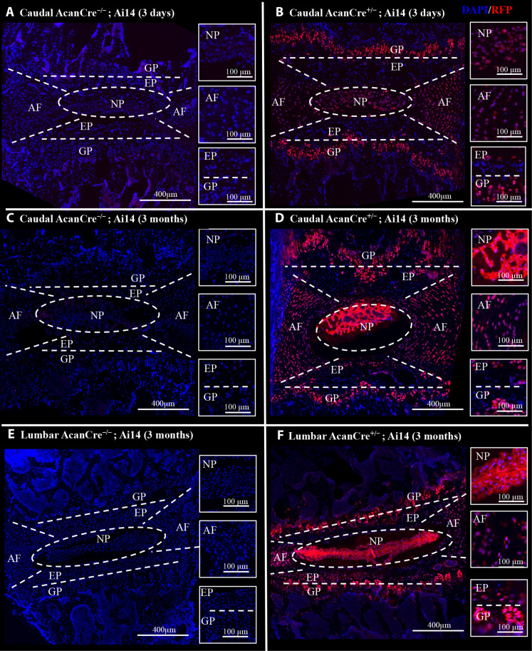
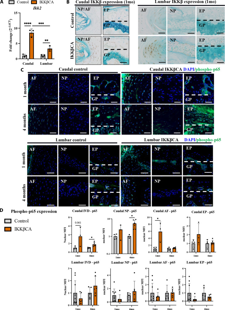
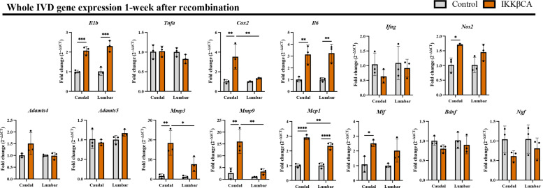
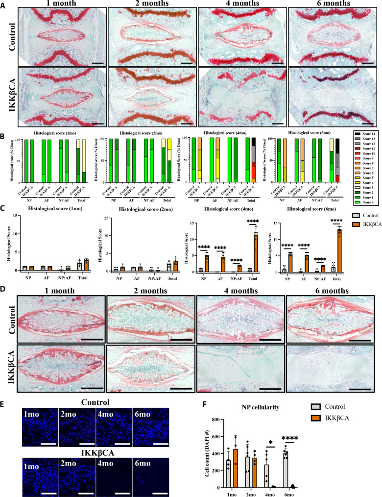
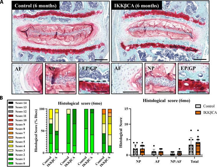
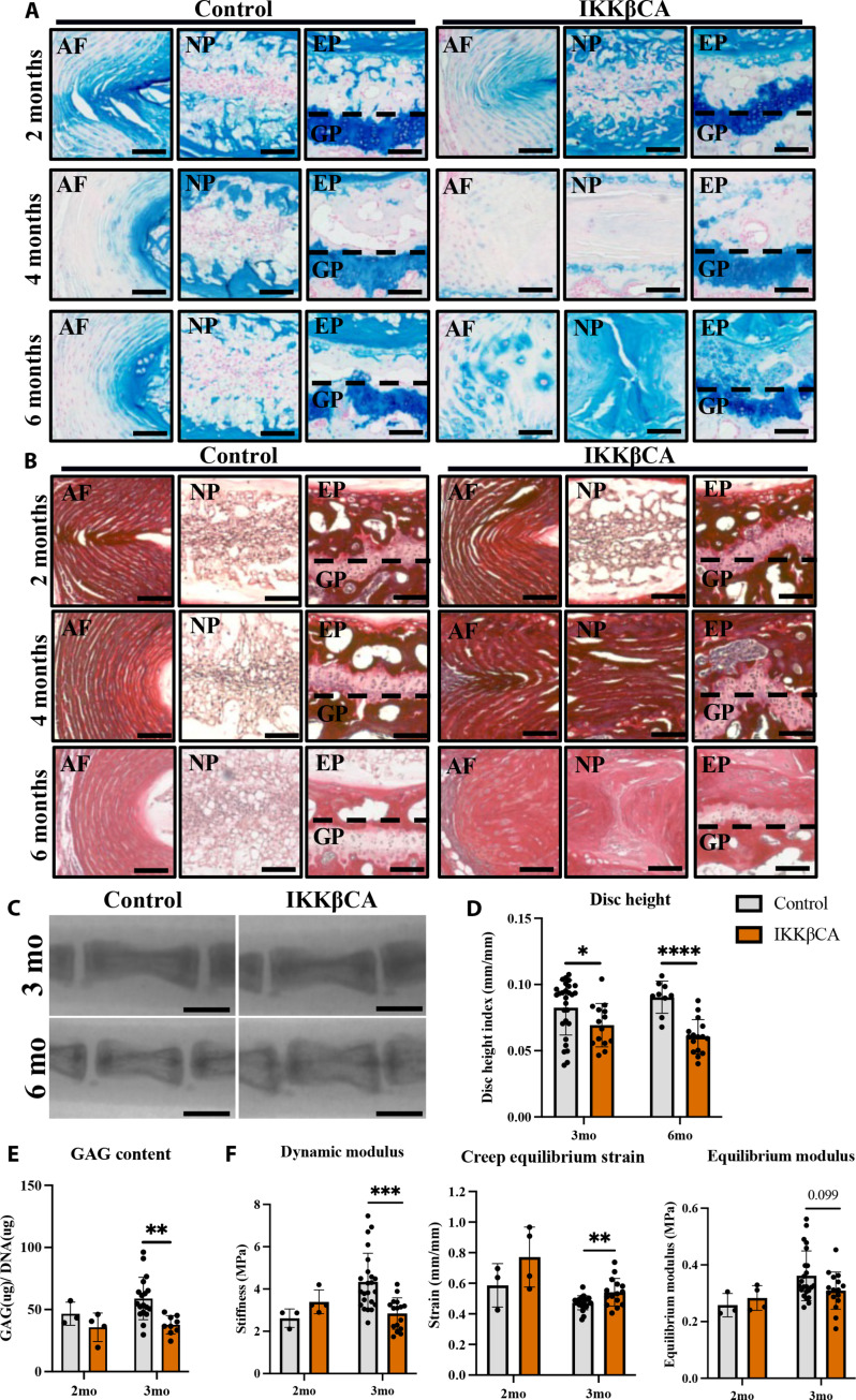
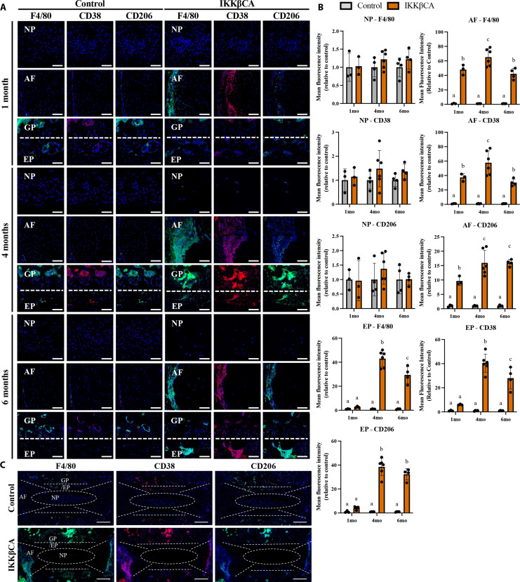
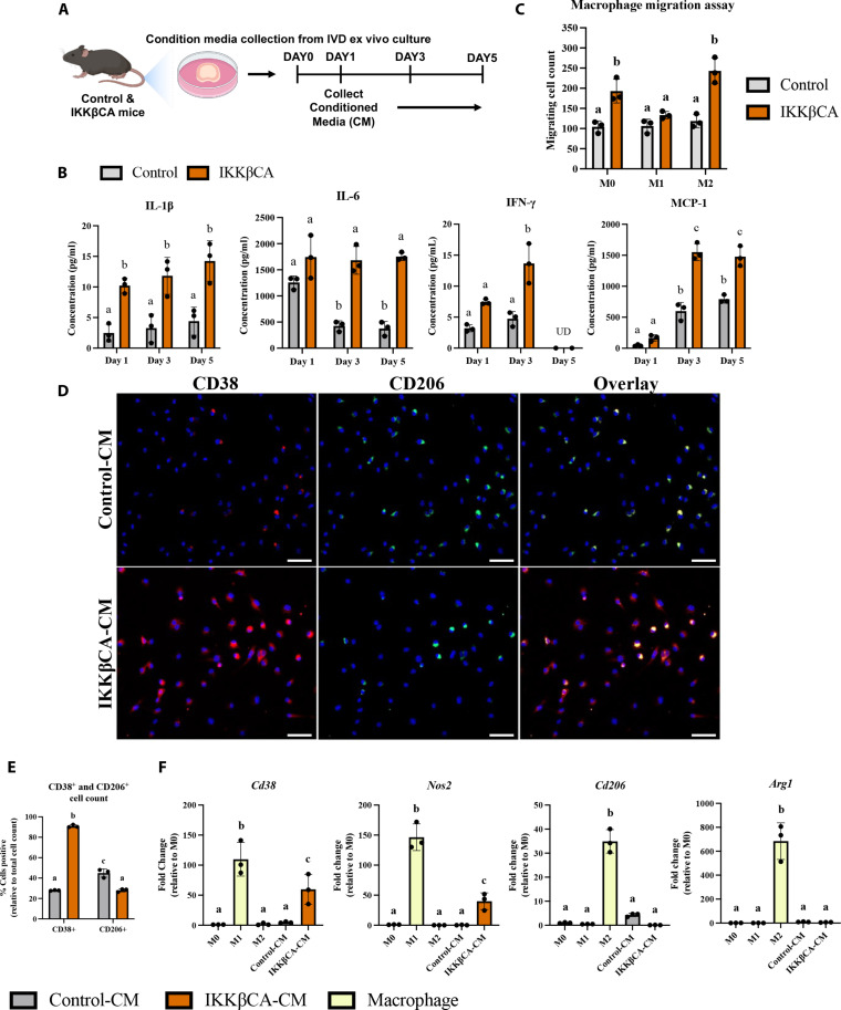
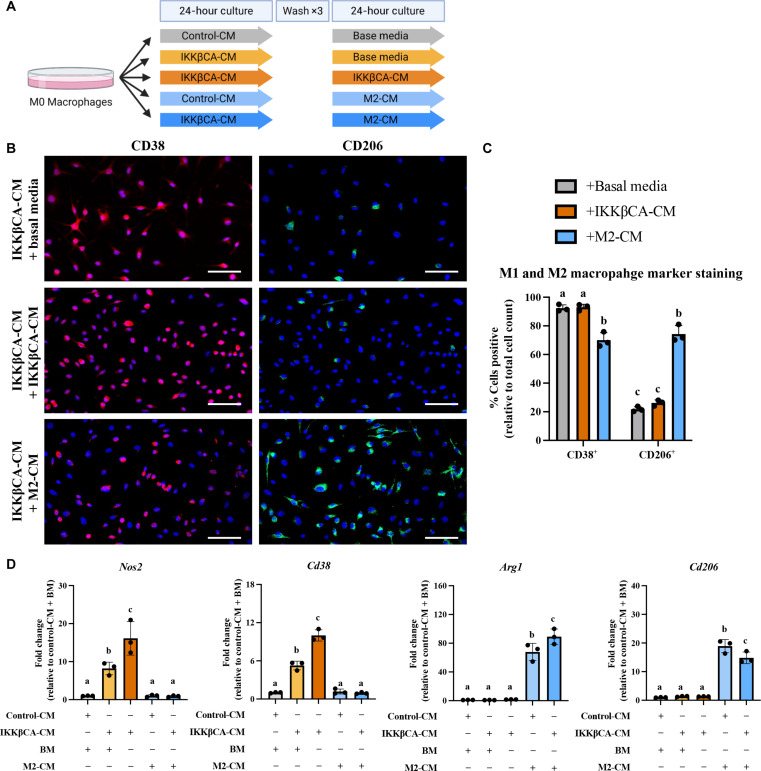
Persistent inflammation has been associated with severe disc degeneration (DD). This study investigated the effect of prolonged nuclear factor κB (NF-κB) activation in DD. Using an inducible mouse model, we genetically targeted cells expressing aggrecan, a primary component of the disc extra cellular matrix, for activation of the canonical NF-κB pathway. Prolonged NF-κB activation led to severe structural degeneration accompanied by increases in gene expression of inflammatory molecules (Il1b, Cox2, Il6, and Nos2), chemokines (Mcp1 and Mif), and catabolic enzymes (Mmp3, Mmp9, and Adamts4). Increased recruitment of proinflammatory (F4/80+,CD38+) and inflammatory resolving (F4/80+,CD206+) macrophages was observed within caudal discs. We found that the secretome of inflamed caudal disc cells increased macrophage migration and inflammatory activation. Lumbar discs did not exhibit phenotypic changes, suggestive of regional spinal differences in response to inflammatory genetic overactivation. Results suggest prolonged NF-κB activation can induce severe DD through increases in inflammatory cytokines, chemotactic proteins, catabolic enzymes, and the recruitment and activation of macrophage cell populations.


Figures

Fig. 1.
Fig. 1.. The AcanCreERT2 mouse targets all compartments of the IVD and GP.
(A to D) Representative images of immunofluorescent (IF) staining for RFP within mid-sagittal sections of caudal AcanCre−/−;Ai14 and AcanCre+/−;Ai14 reporter mice 3 days and 3 months following tamoxifen IP injections. (E and F) Images of IF staining for RFP within mid-sagittal sections of lumbar AcanCre−/−;Ai14 and AcanCre+/−;Ai14 reporter mice 3 months following tamoxifen IP injections. NP, AF, EP, and GP compartments are delineated (white dashed lines).
Fig. 2.
Fig. 2.. IKKβ overexpression and NF-κB activation within IKKβCA mice.
(A) Gene expression of Ikk2 within caudal and lumbar IVDs expressed as fold change relative to control (AcanCre−/−;Ikk2cafl/fl) 1 week after recombination (n = 3 mice, one disc per mouse). (B) IHC staining for IKKβ within mid-sagittal sections of 1 month control and IKKβCA IVDs. Scale bar, 100 μm. (C) Representative IF staining for phosphorylated p65 (green) within mid-sagittal sections of control and IKKβCA IVDs. Scale bars, 50 μm. (D) Nuclear MFI quantification (normalized to control within time point) of phosphorylated p65 (n = 3 to 6 mice per group, one to two IVDs per mouse). *P < 0.05, **P < 0.01, and ***P < 0.001.
Fig. 3.
Fig. 3.. IKKβ overexpression up-regulates inflammatory cytokine, chemokine, catabolic enzyme, and neurotrophic factor gene expression.
Gene expression changes (relative to control) from total RNA isolated from control (AcanCre−/−;Ikk2cafl/fl) and IKKβCA whole caudal and lumbar IVDs containing NP, AF, and EP, 1 week after recombination. *P < 0.05, **P < 0.01, ***P < 0.001, and ****P < 0.0001 (n = 3 mice per genotype, one IVD per mouse).
Fig. 4.
Fig. 4.. IKKβ overexpression produces severe caudal DD.
(A) Representative images of safranin-O–stained mid-sagittal sections of control (AcanCre−/−;Ikk2cafl/fl) and IKKβCA caudal IVDs 1, 2, 4, and 6 months after recombination. Scale bars, 250 μm. Histological scoring legend, ranging from 0 (healthy) to 14 (most severe). (B) Distribution of histological scores. (C) Histological scoring within NP, AF, and NP:AF border compartments, and total score. (D) Representative images of safranin-O–stained mid-sagittal sections of the NP region of control and IKKβCA caudal IVDs at 1, 2, 4, and 6 months after recombination (n = 4 to 6 mice per genotype and time point, one to three caudal IVDs per mouse). Scale bars, 250 μm. (E) Representative images of 4′,6-diamidino-2-phenylindole (DAPI; nuclear)–stained mid-sagittal caudal IVD sections. Scale bars, 100 μm. (F) Quantification of caudal NP cellularity within hand-drawn ROIs of DAPI nuclear-stained mid-sagittal sections. *P < 0.05 and ****P < 0.0001. (n = 4 to 6 mice per genotype and time point, one to three caudal IVDs per mouse).
Fig. 5.
Fig. 5.. IKKβ overexpression produces no significant changes to lumbar disc health/structure at 6 months.
(A) Representative images of safranin-O–stained mid-coronal sections of control (AcanCre−/−;Ikk2cafl/fl) and IKKβCA lumbar IVDs 6 months after recombination. Total IVD scale bars = 400 μm. Compartment image scale bars = 200 μm. (B) Distribution of histological grades within NP, AF, and NP:AF border compartments and total score. Histological scoring severity legend ranging from 0 (healthy) to 14 (most severe). No significant differences observed (n = 3 mice per genotype, three to four lumbar IVDs per mouse).
Fig. 6.
Fig. 6.. IKKβ overexpression mediates a loss of caudal ECM, disc height, and weakened mechanical properties.
(A) Representative Alcian blue (GAG) and (B) Picrosirius red (collagen)–stained images of control (AcanCre−/−;Ikk2cafl/fl) and IKKβCA caudal IVDs mid-sagittal sections at 2, 4, and 6 months after recombination. Scale bars, 100 μm. (C) Representative fluoroscopic images of control and IKKβCA C6-C8 IVDs 3 and 6 months after recombination. Scale bars, 1 mm. (D) IVD height quantified via DHI of control and IKKβCA caudal IVDs 3 and 6 months after recombination (n = 3 to 6 mice per genotype and time point, three to five IVDs per mouse). (E) GAG content (μg) normalized to total DNA content (μg) within control and IKKβCA caudal IVD digests 2 and 3 months after recombination (n = 3 to 6 mice per genotype, one to three discs per mouse). (F) Dynamic modulus (MPa), creep equilibrium strain (mm/mm), and equilibrium modulus (MPa) of control and IKKβCA caudal IVDs 2 and 3 months after recombination (n = 3 to 6 mice per genotype, one to three discs per mouse). *P < 0.05, **P < 0.01, ***P < 0.001, and ****P < 0.0001.
Fig. 7.
Fig. 7.. IKKβ overexpression increases macrophage presence within the caudal IVD.
(A) Representative images of IF staining for F4/80, CD38, and CD206 in mid-sagittal sections of control (AcanCre−/−;Ikk2cafl/fl) and IKKβCA caudal IVDs 1, 4, and 6 months after recombination. DAPI nuclear stain (blue). Scale bars, 100 μm. (B) MFI quantification of F4/80, CD38, and CD206 expression within individual NP, AF, and EP compartments. Letters (a), (b), and (c) indicate statistically significant (P < 0.05) different groupings. (C) Representative images of whole disc sagittal sections of control and IKKβCA IVDs 4 months after recombination with delineation of tissue compartments. DAPI nuclear stain (blue). Scale bars, 200 μm. (n = 3 to 6 mice per group, one to two discs per mouse).
Fig. 8.
Fig. 8.. Caudal IKKβCA-CM increases macrophage migration and polarization toward an inflammatory phenotype.
(A) Study design schematic of whole-organ in vitro culture and CM collection from caudal control (AcanCre−/−;Ikk2cafl/fl) and IKKβCA IVDs harvested 1 week after recombination. (B) Protein concentrations (pg/ml) within CM analyzed after 1, 3, and 5 days in culture. UD, undetectable reading. Letters (a), (b), and (c) indicate statistically significant (P < 0.05) different groupings (n = 3 per genotype). (C) Quantification of M0, M1, and M2 macrophage migration through a transwell membrane via DAPI nuclear count (n = 3). (D) Representative images of IF staining for CD38 and CD206 within BMDMs cultured in 2D monolayer and stimulated with control or IKKβCA CM. DAPI nuclear stain (blue). Scale bars, 100 μm. (E) Quantification of % positivity for CD38 and CD206 within bone marrow–derived macrophages treated with CM (n = 3). (F) Gene expression of M1 and M2 phenotypic markers within M0, M1, or M2 macrophages in basal media, or M0 macrophages with or without IVD CM stimulation (n = 3). Letters (a), (b), and (c) indicate statistically significant (P < 0.05) different groupings.
Fig. 9.
Fig. 9.. Caudal IKKβCA-CM mediated macrophage inflammatory responses are mitigated by costimulation with an M2 secretome.
(A) Study design schematic for CM stimulation of M0 macrophages. (B) Representative images of IF staining for CD38 and CD206 within BMDMs cultured in 2D monolayer and stimulated with IKKβCA-CM followed by basal media or M2 macrophage CM. Scale bars, 100 μm. (C) Quantification of % positivity for CD38 and CD206 within BMDMs following CM stimulation (n = 3). (D) Gene expression of M1 and M2 phenotypic markers within M0 macrophages following CM stimulation (n = 3). Letters (a), (b), and (c) indicate statistically significant (P < 0.05) different groupings.

Update of

References

    1. Murray C. J., Atkinson C., Bhalla K., Birbeck G., Burstein R., Chou D., Dellavalle R., Danaei G., Ezzati M., Fahimi A., Flaxman D., Foreman, Gabriel S., Gakidou E., Kassebaum N., Khatibzadeh S., Lim S., Lipshultz S. E., London S., Lopez, MacIntyre M., Mokdad A. H., Moran A., Moran A. E., Mozaffarian D., Murphy T., Naghavi M., Pope C., Roberts T., Salomon J., Schwebel D. C., Shahraz S., Sleet D. A., Murray, Abraham J., Ali M. K., Atkinson C., Bartels D. H., Bhalla K., Birbeck G., Burstein R., Chen H., Criqui M. H., Dahodwala, Jarlais, Ding E. L., Dorsey E. R., Ebel B. E., Ezzati M., Fahami, Flaxman S., Flaxman A. D., Gonzalez-Medina D., Grant B., Hagan H., Hoffman H., Kassebaum N., Khatibzadeh S., Leasher J. L., Lin J., Lipshultz S. E., Lozano R., Lu Y., Mallinger L., McDermott M., Micha R., Miller T. R., Mokdad A. A., Mokdad A. H., Mozaffarian D., Naghavi M., Narayan K. M., Omer S. B., Pelizzari P. M., Phillips D., Ranganathan D., Rivara F. P., Roberts T., Sampson U., Sanman E., Sapkota A., Schwebel D. C., Sharaz S., Shivakoti R., Singh G. M., Singh D., Tavakkoli M., Towbin J. A., Wilkinson J. D., Zabetian A., Murray, Abraham J., Ali M. K., Alvardo M., Atkinson C., Baddour L. M., Benjamin E. J., Bhalla K., Birbeck G., Bolliger I., Burstein R., Carnahan E., Chou D., Chugh S. S., Cohen A., Colson K. E., Cooper L. T., Couser W., Criqui M. H., Dabhadkar K. C., Dellavalle R. P., Jarlais, Dicker D., Dorsey E. R., Duber H., Ebel B. E., Engell R. E., Ezzati M., Felson D. T., Finucane M. M., Flaxman S., Flaxman A. D., Fleming T., Foreman, Forouzanfar M. H., Freedman G., Freeman M. K., Gakidou E., Gillum R. F., Gonzalez-Medina D., Gosselin R., Gutierrez H. R., Hagan H., Havmoeller R., Hoffman H., Jacobsen K. H., James S. L., Jasrasaria R., Jayarman S., Johns N., Kassebaum N., Khatibzadeh S., Lan Q., Leasher J. L., Lim S., Lipshultz S. E., London S., Lopez, Lozano R., Lu Y., Mallinger L., Meltzer M., Mensah G. A., Michaud C., Miller T. R., Mock C., Moffitt T. E., Mokdad A. A., Mokdad A. H., Moran A., Naghavi M., Narayan K. M., Nelson R. G., Olives C., Omer S. B., Ortblad K., Ostro B., Pelizzari P. M., Phillips D., Raju M., Razavi H., Ritz B., Roberts T., Sacco R. L., Salomon J., Sampson U., Schwebel D. C., Shahraz S., Shibuya K., Silberberg D., Singh J. A., Steenland K., Taylor J. A., Thurston G. D., Vavilala M. S., Vos T., Wagner G. R., Weinstock M. A., Weisskopf M. G., Wulf S., Murray, U.S. Burden of Disease Collaborators , The state of US health, 1990-2010: Burden of diseases, injuries, and risk factors. JAMA 310, 591–608 (2013). - PMC - PubMed
    1. Katz J. N., Lumbar disc disorders and low-back pain: Socioeconomic factors and consequences. J. Bone Joint Surg. Am. 88, 21–24 (2006). - PubMed
    1. Luoma K., Riihimäki H., Luukkonen R., Raininko R., Viikari-Juntura E., Lamminen A., Low back pain in relation to lumbar disc degeneration. Spine 25, 487–492 (2000). - PubMed
    1. Nerlich A. G., Bachmeier B. E., Schleicher E., Rohrbach H., Paesold G., Boos N., Immunomorphological analysis of rage receptor expression and nf-κb activation in tissue samples from normal and degenerated intervertebral discs of various ages. Ann. N. Y. Acad. Sci. 1096, 239–248 (2007). - PubMed
    1. Abraham A. C., Shah S. A., Golman M., Song L., Li X., Kurtaliaj I., Akbar M., Millar N. L., Abu-Amer Y., Galatz L. M., Thomopoulos S., Targeting the NF-κB signaling pathway in chronic tendon disease. Sci. Transl. Med. 11, eaav4319 (2019). - PMC - PubMed

MeSH terms