. 2024 Jun 7;19(6):e0291568.
doi: 10.1371/journal.pone.0291568. eCollection 2024.

Understanding the biosynthesis of human IgM SAM-6 through a combinatorial expression of mutant subunits that affect product assembly and secretion

Affiliations

Understanding the biosynthesis of human IgM SAM-6 through a combinatorial expression of mutant subunits that affect product assembly and secretion

Haruki Hasegawa et al. PLoS One. .

Abstract

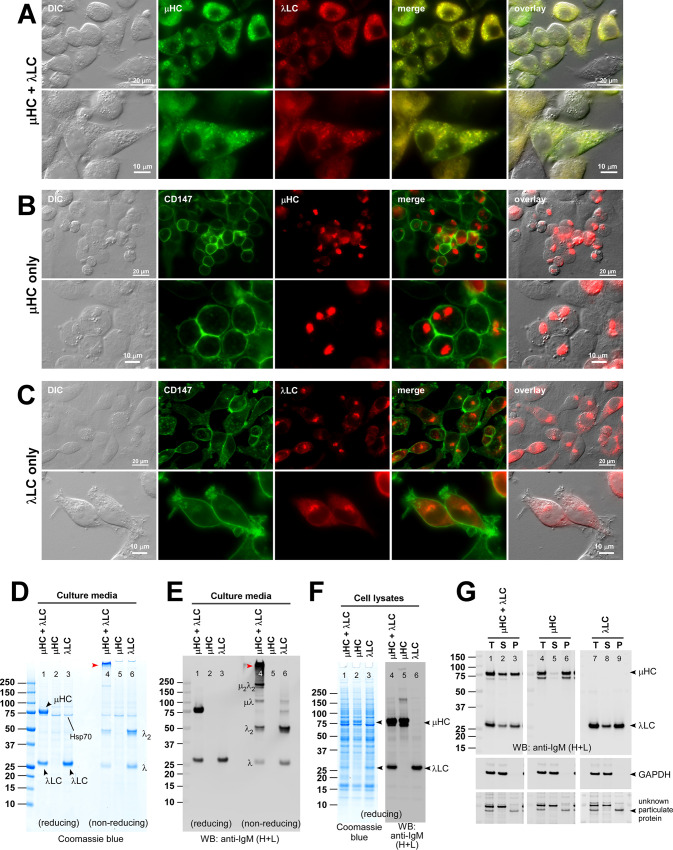
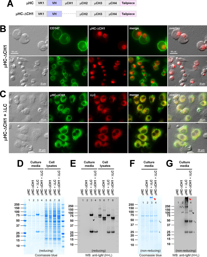
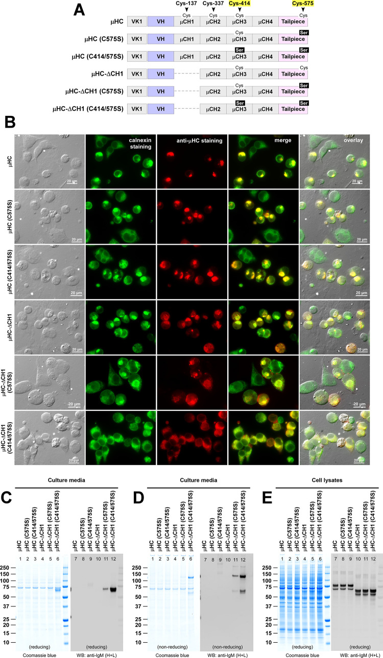
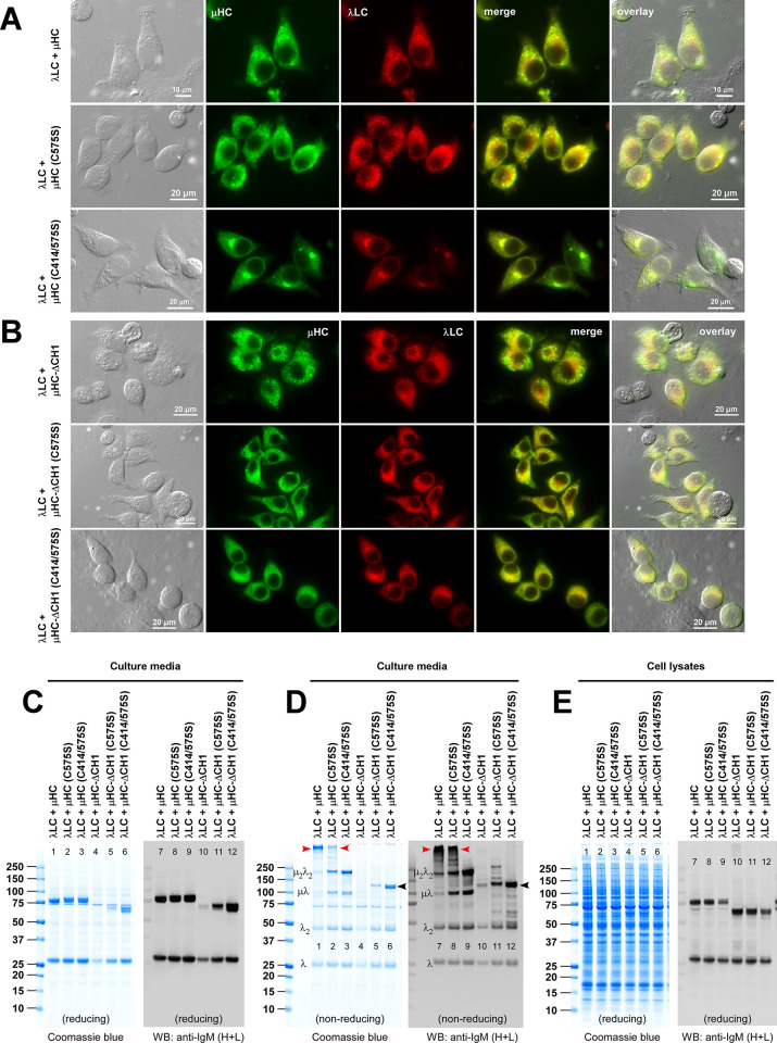
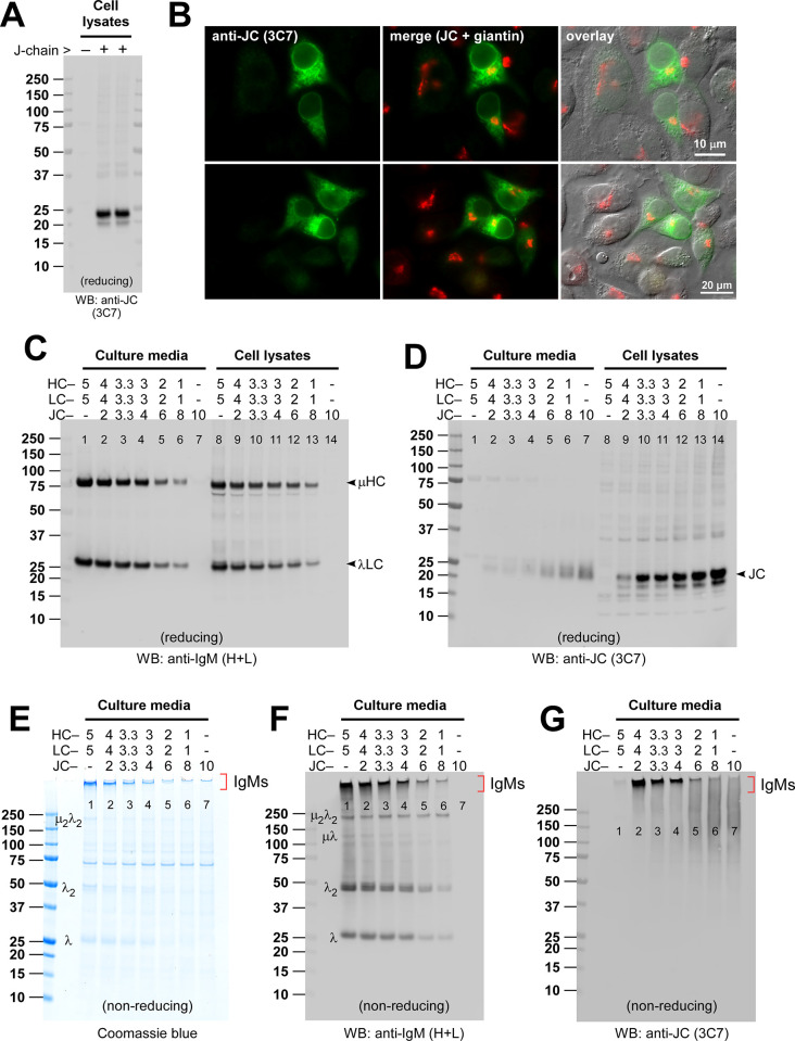
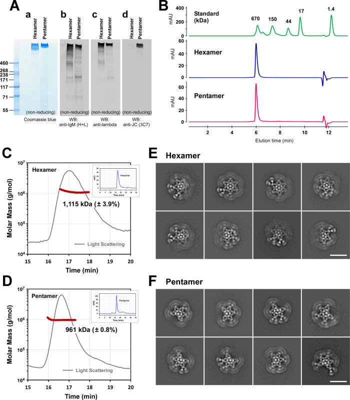
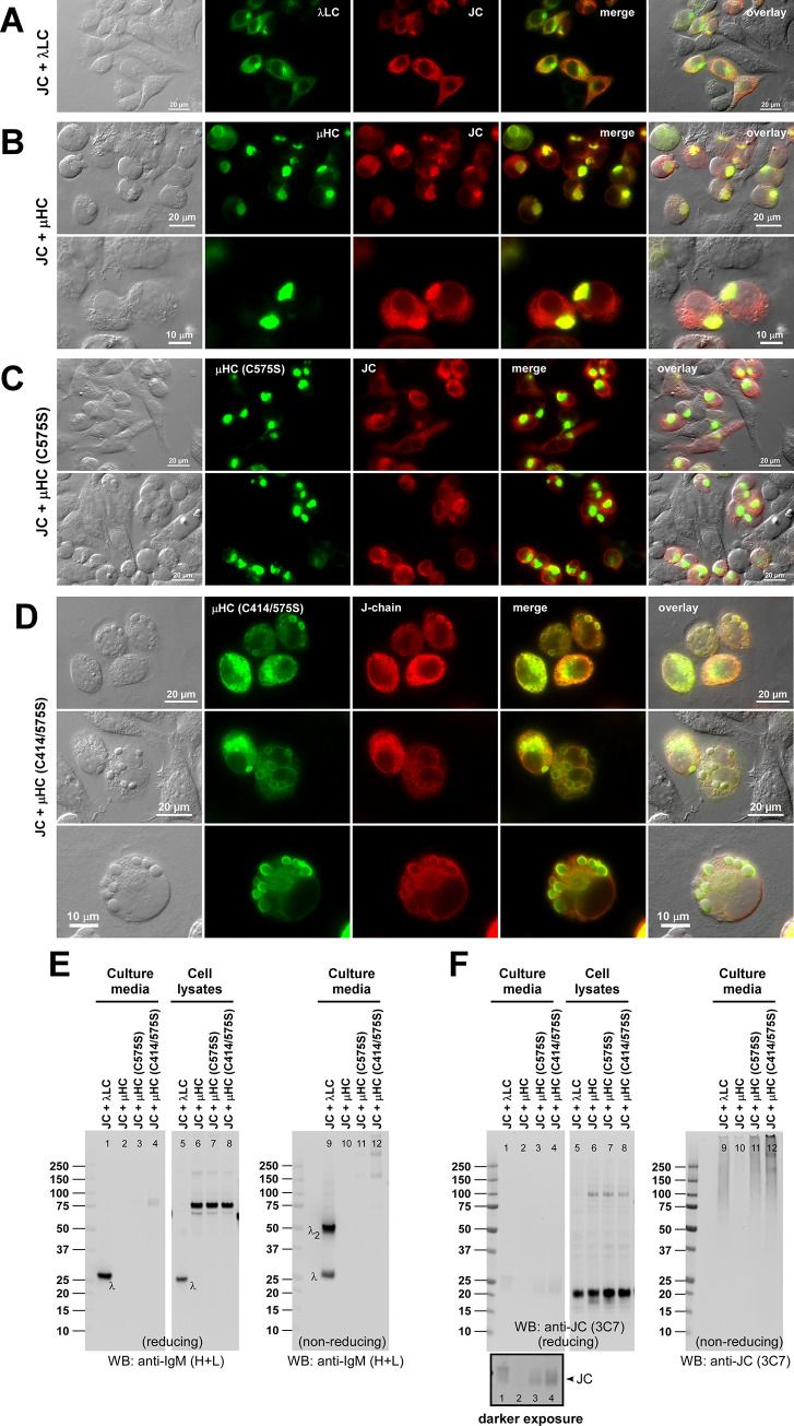
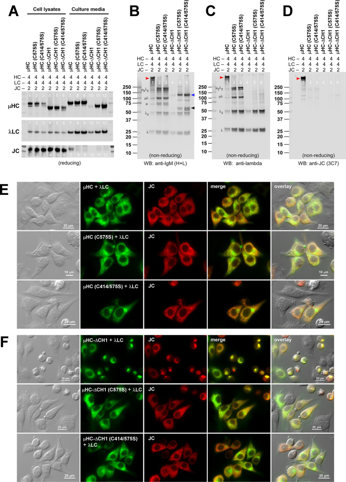
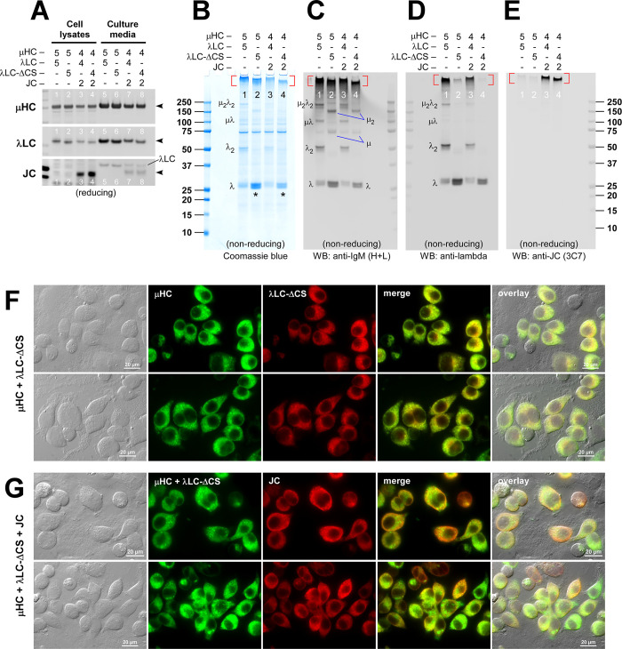
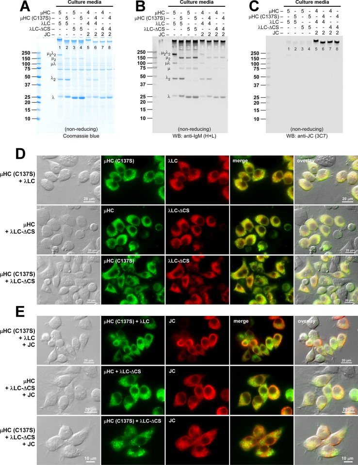
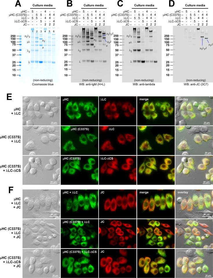
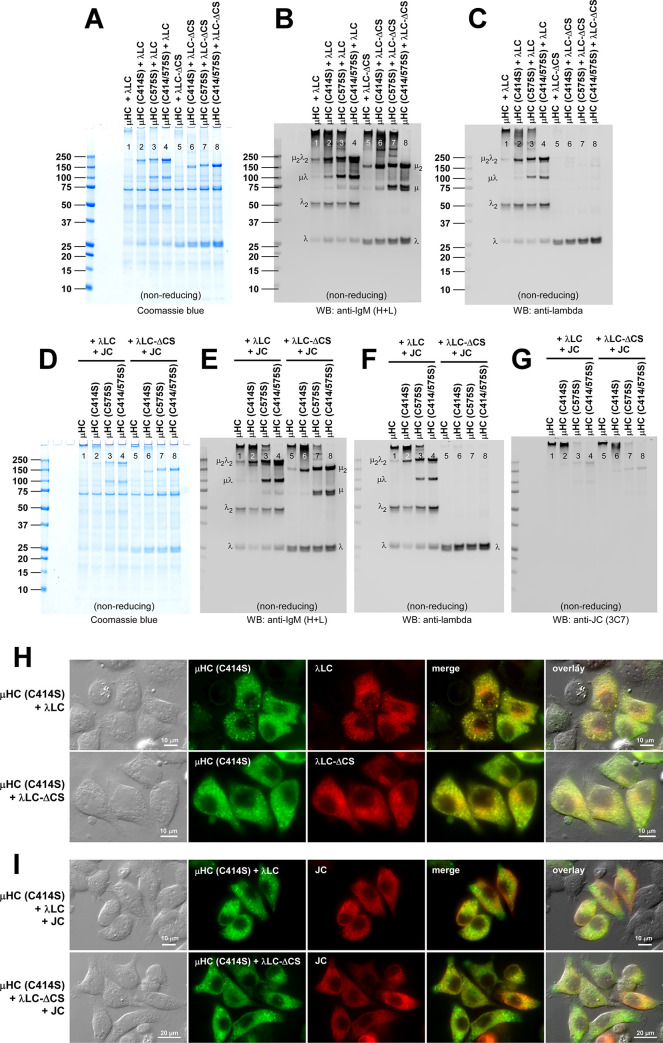
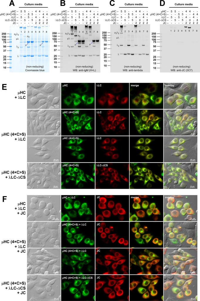
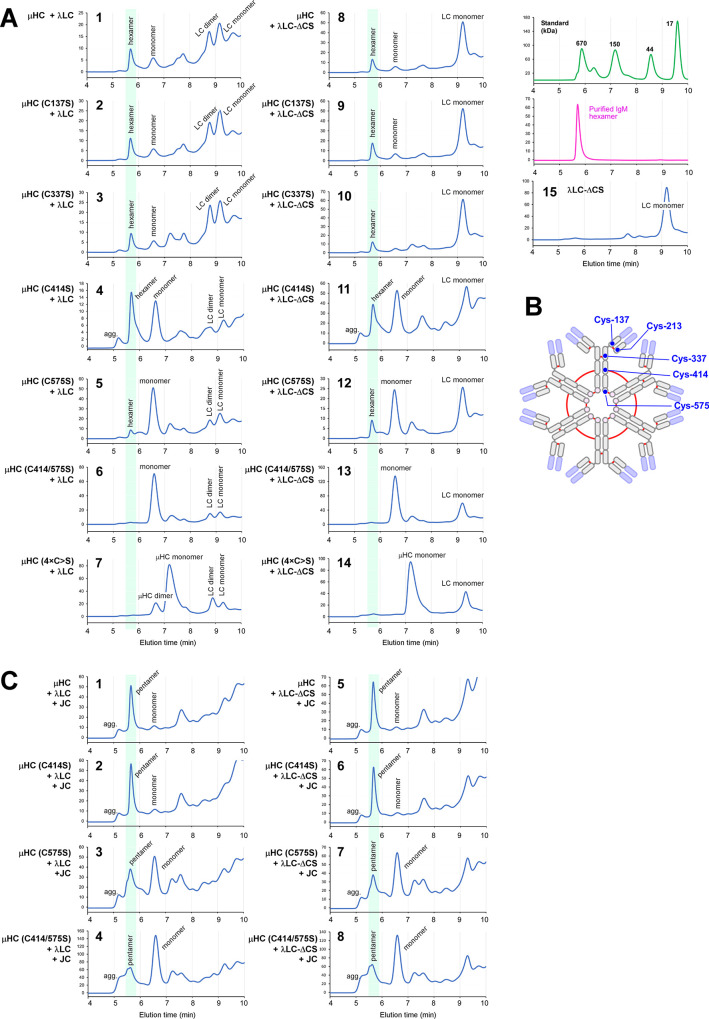
Polymeric IgMs are secreted from plasma cells abundantly despite their structural complexity and intricate multimerization steps. To gain insights into IgM's assembly mechanics that underwrite such high-level secretion, we characterized the biosynthetic process of a natural human IgM, SAM-6, using a heterologous HEK293(6E) cell platform that allowed the production of IgMs both in hexameric and pentameric forms in a controlled fashion. By creating a series of mutant subunits that differentially disrupt secretion, folding, and specific inter-chain disulfide bond formation, we assessed their effects on various aspects of IgM biosynthesis in 57 different subunit chain combinations, both in hexameric and pentameric formats. The mutations caused a spectrum of changes in steady-state subcellular subunit distribution, ER-associated inclusion body formation, intracellular subunit detergent solubility, covalent assembly, secreted IgM product quality, and secretion output. Some mutations produced differential effects on product quality depending on whether the mutation was introduced to hexameric IgM or pentameric IgM. Through this systematic combinatorial approach, we consolidate diverse overlapping knowledge on IgM biosynthesis for both hexamers and pentamers, while unexpectedly revealing that the loss of certain inter-chain disulfide bonds, including the one between μHC and λLC, is tolerated in polymeric IgM assembly and secretion. The findings highlight the differential roles of underlying non-covalent protein-protein interactions in hexamers and pentamers when orchestrating the initial subunit interactions and maintaining the polymeric IgM product integrity during ER quality control steps, secretory pathway trafficking, and secretion.


Conflict of interest statement

The authors have declared that no competing interests exist.

Figures

Fig 1
Fig 1. Subcellular distribution and secretion of co-expressed μHC and λLC subunits during hexameric IgM expression.
Fluorescent micrographs of HEK293 cells transfected with (A) [μHC + λLC] construct pair, (B) μHC subunit alone, or (C) λLC subunit alone. (A) Co-staining was performed using FITC-labeled anti-μHC and Texas Red-labeled anti-λLC. (B) Co-stained with FITC-labeled anti-CD147 and Texas Red-labeled anti-μHC. (C) Co-stained with FITC-labeled anti-CD147 and Texas Red-labeled anti-λLC. Green and red image fields were superimposed to create ‘merge’ views. DIC and ‘merge’ were superimposed to make ‘overlay’ views in A. DIC and red image fields were superimposed to create ‘overlay’ views in B and C. (D) Coomassie blue stained gel showing the secreted IgM and subunits. HEK293 cells were transfected with [μHC + λLC] pair (lanes 1 and 4), μHC only (lanes 2 and 5), or λLC (lanes 3 and 6). Cell culture media were harvested on day-7 post-transfection and analyzed by SDS-PAGE under reducing conditions (lanes 1‒3) or non-reducing conditions (lanes 4‒6). The detectable subunit chain is pointed by an arrowhead and labeled in lanes 1‒3. High molecular weight hexameric IgM is pointed by a red arrowhead in lane 4. Monomeric and dimeric λLC subunit is labeled in lane 6. A cell host-derived ~74 kDa protein visible in all culture media lanes was identified as human Hsp70 by mass spectrometry. (E) An identical sample set was analyzed by Western blotting. The membrane was probed with polyclonal anti-IgM (H+L) to detect μHC and λLC subunits simultaneously and any assembly intermediates composed of μHC or λLC or both. The protein band corresponding to the assembled hexameric IgM is pointed by a red arrowhead in lane 4. Identifiable assembly intermediates are labeled next to the corresponding protein bands in lane 4. (F) Cell lysate samples were prepared on day-7 post-transfection and analyzed by SDS-PAGE or Western blotting after the proteins were resolved under reducing conditions. Subunit chains are pointed by arrowhead and labeled. (G) The detergent solubility of intracellular subunits was assessed on day-3 post-transfection. Whole cell lysates were prepared under non-denaturing conditions and subjected to 15,000 g centrifugation. Total (T), soluble (S), and particulate (P) fractions were resolved in SDS-PAGE under reducing conditions. Membranes were probed with anti-IgM (H+C) (top panels) and anti-GAPDH (middle and bottom panels). Nonspecifically cross-reacting two proteins that are partitioned to soluble and particulate fractions are shown in the bottom panel.
Fig 2
Fig 2. Effects of CH1 domain deletion on hexameric IgM assembly and secretion.
(A) Schematic representation of SAM-6 μHC subunit and μHC-ΔCH1 mutant, which lacks the CH1 domain. Individual domain names are indicated in each box. ER targeting is driven by a heterologous signal sequence adapted from a VK1 encoding gene. (B) Fluorescent micrographs of HEK293 cells expressing μHC-ΔCH1 mutant. On day-3 post-transfection, cells were fixed, permeabilized, and immunostained with FITC-labeled anti-CD147 and Texas Red-labeled anti-μHC. (C) Fluorescent micrographs of HEK293 cells expressing [μHC-ΔCH1 + λLC] pair. Immunostaining was performed using FITC-labeled anti-μHC and Texas Red-labeled anti-λLC. (D‒G) HEK293 cells were transfected with μHC (lanes 1 and 5), μHC-ΔCH1 (lanes 2 and 6), [μHC + λLC] pair (lanes 3 and 7), or [μHC-ΔCH1 + λLC] pair (lanes 4 and 8). Cell culture media were harvested on day-7 post-transfection and analyzed by SDS-PAGE under reducing conditions (D, E; lanes 1‒4) or non-reducing conditions (F, G; lanes 1‒4). Day-7 cell lysate samples were analyzed by SDS-PAGE (D, lanes 5‒8) or Western blotting (E, lanes 5‒8). Western blotting was performed using polyclonal anti-IgM (H+L) to detect μHC and λLC subunits simultaneously. A faintly detectable μHC-ΔCH1 is pointed by a black arrowhead in panel E (lane 4) and panel G (lane 4). The assembled hexameric IgM product is pointed by a red arrowhead (F, G; lane 3). Identifiable assembly intermediates are labeled next to the corresponding bands in panels F and G.
Fig 3
Fig 3. Roles of Cys-414 and Cys-575 residues in μHC subunit synthesis and secretion.
(A) Schematic representation of parental SAM-6 μHC (top) and its CH1 deletion mutant μHC-ΔCH1 (fourth row) as well as their C575S and C414/575S mutant variants. The positions of key cysteine residue participating in the inter-chain disulfide bond are marked at the top. (B) Fluorescent micrographs of HEK293 cells transfected with the six constructs shown in panel A. The transfected construct name is shown on the left side of each row. On day-3 post-transfection, cells were fixed, permeabilized, and immunostained with polyclonal anti-calnexin (green) and Texas Red-labeled anti-μHC (red). Green and red image fields were superimposed in ‘merge’ views. DIC and ‘merge’ were superimposed in ‘overlay’ views. (C, D) Cell culture media were harvested on day-7 post-transfection and analyzed by SDS-PAGE and Western blotting after resolving the proteins under reducing (C) or non-reducing (D) conditions. (E) Day-7 cell lysate samples were analyzed by SDS-PAGE (lanes 1‒6) or Western blotting (lanes 7‒12). Western blotting was performed using polyclonal anti-IgM (H+L).
Fig 4
Fig 4. Roles of Cys-414 and Cys-575 in covalent assembly and secretion of hexameric IgM.
(A) Fluorescent micrographs of HEK293 cells co-transfected with [λLC + μHC] pair (top row), [λLC + μHC (C575S)] pair (middle row), and [λLC + μHC (C414/575S)] pair (bottom row). On day-3 post-transfection, cells were fixed, permeabilized, and co-stained with FITC-labeled anti-μHC and Texas Red-labeled anti-λLC. (B) Fluorescent micrographs of HEK293 cells co-transfected with [λLC + μHC-ΔCH1] pair (top row), [λLC + μHC-ΔCH1 (C575S)] pair (second row), and [λLC + μHC-ΔCH1 (C414/575S)] pair (third row). Cell fixation, immunostaining, and image processing were performed as described above. (C‒E) The transfected construct pair is shown at the top of each lane. Cell culture media (C, D) and cell lysates (E) were harvested on day-7 post-transfection and analyzed by SDS-PAGE and by Western blotting after resolving the proteins under reducing conditions (C and E) or non-reducing conditions (D). Western blotting was performed using polyclonal anti-IgM (H+L) to detect μHC and λLC subunits simultaneously. A protein band corresponding to the covalently assembled hexameric IgM is pointed by a red arrowhead in panel D, lanes 1, 2, 7, and 8. Identifiable assembly intermediates are labeled next to the corresponding protein bands in panel D, lanes 1 and 7. Secreted μHC-ΔCH1 (C414/575S) mutant dimers are pointed by black arrowhead in panel D, lanes 6 and 12.
Fig 5
Fig 5. Optimization of pentameric IgM secretion by titrating J-chain subunit expression.
(A, B) HEK293 cells were transfected with a human JC encoding construct, and the expression was verified by Western blotting and immunofluorescent microscopy. (A) Cell lysates were prepared on day-3 post-transfection and analyzed by Western blotting using a monoclonal anti-JC (clone 3C7). The mock-transfected cell lysate was analyzed as a control for anti-JC detection specificity. (B) On day-3 post-transfection, cells were fixed, permeabilized, and co-stained with monoclonal anti-JC (green) and anti-giantin (red). (C, D) To produce a pentameric form of IgM, three subunit chains (μHC, λLC, and JC) were co-transfected using varying plasmid DNA ratios indicated at the top of individual lanes without changing the total amount of transfected DNA (= 10 μg). The numbers at the top of each lane represent the amount of each construct in μg. On day-7 post-transfection, cell culture media were harvested (lanes 1‒7), and cell lysates were prepared (lanes 8‒14) to run SDS-PAGE under reducing conditions followed by Western blotting. Membranes were probed (C) with polyclonal anti-IgM (H+L) to detect μHC and λLC subunits simultaneously or (D) with monoclonal anti-JC. (E‒G) Cell culture media harvested on day-7 post-transfection were resolved by SDS-PAGE under non-reducing conditions, which were Coomassie blue stained (E) or analyzed by Western blotting (F, G). Membranes were probed with polyclonal anti-IgM (H+L) (panel F) or with monoclonal anti-JC to detect free JC and JC-containing protein complexes (panel G). In panels E and F, protein bands corresponding to λLC monomers, dimers, and assembly intermediates are labeled on the left side of lane 1. The protein band corresponding to hexameric or pentameric IgM is marked with a bracket on the right side of lane 7.
Fig 6
Fig 6. Characteristics of purified recombinant hexameric and pentameric IgMs.
(A, a) Two-step purified hexameric and pentameric IgMs were resolved by SDS-PAGE under non-reducing conditions using a 3‒8% Tris-Acetate gradient gel. Sample loading was 2.5 μg per lane. (A, b‒d) Identically run samples were analyzed by Western blotting using three different detection probes: (A, b) polyclonal anti-IgM (H+L), (A, c) polyclonal anti-λLC, and (A, d) monoclonal anti-JC. (B) The protein purity of hexameric or pentameric IgMs was assessed by analytical SEC. About 5 μg of purified protein was injected into the column. (C, D) Purified IgM proteins (100 μg each) were analyzed by SEC-MALS. Light scattering and estimated molecular mass are plotted by solid gray line and bold brown line, respectively. The average molecular mass for (C) hexameric IgM peak and (D) pentameric IgM peak is stated in the graphs. For calculation purposes, we arbitrarily used the mass of Man9GlcNac2 (= 1,865.64 Da) for the N-linked glycan. Simultaneously collected SEC UV trace data are shown in the inset of C and D. (E, F) Negative-stain transmission electron micrographs of purified recombinant IgM. Eight representative 2D class averages for (E) hexameric IgM and (F) pentameric IgM are shown. Scale bar, 25 nm.
Fig 7
Fig 7. Intracellular J-chain distribution is dictated by the Cys-575 of the co-expressed μHC subunit.
Fluorescent micrographs of HEK293 cells co-transfected with JC and one of the following constructs: (A) λLC, (B) μHC, (C) μHC (C575S), or (D) μHC (C414/575S). Co-transfected construct pair is also shown on the left side of each row. On day-3 post-transfection, cells were fixed, permeabilized, and co-stained with FITC-labeled anti-λLC (A) or FITC-labeled anti-μHC (B‒D) and monoclonal anti-JC followed by AlexaFluor594-conjugated secondary antibody (A‒D, shown in red). (E) On day-7 post-transfection, cell culture media (lanes 1‒4; lanes 9‒12) and cell lysates (lanes 5‒8) were analyzed by Western blotting. Membranes were probed with polyclonal anti-IgM (H+L). Co-transfected construct pairs are shown at the top of each lane. (F) The same set of culture media and cell lysate samples were analyzed by Western blotting using monoclonal anti-JC. A longer exposed Western blot result is shown underneath the corresponding lanes in a black box (panel F, lanes 1‒7).
Fig 8
Fig 8. Differential effects of ΔCH1, C575S, and C414/575S mutations on pentameric IgM assembly and secretion.
Three subunit chains were co-transfected to produce pentameric IgM species at the ratio of μHC: λLC: JC = 4: 4: 2. (A) On day-7 post-transfection, cell lysates (lanes 1‒6) and cell culture media samples (lanes 7‒12) were prepared and resolved by SDS-PAGE under reducing conditions followed by Western blotting. Membranes were probed with polyclonal anti-IgM (H+L) to detect μHC and its mutants (top panel) and the λLC subunit (middle panel) as well as with monoclonal anti-JC (bottom panel). (B‒D) Day-7 cell culture media were analyzed by Western blotting after resolving the proteins under non-reducing conditions. Membranes were probed with (B) polyclonal anti-IgM (H+L), (C) polyclonal anti-λLC, or (D) monoclonal anti-JC. Identifiable assembly intermediates are labeled next to the corresponding protein bands in panels B and C. The protein band corresponding to the assembled pentameric IgM is pointed by a red arrowhead in lane 1 of panels B‒D. Secreted μHC-ΔCH1 (C414/575S) mutant monomers and dimers are pointed by black and blue arrowheads in panel B (lanes 6), respectively. (E) Fluorescent micrographs of HEK293 cells co-transfected with the following set of 3 constructs: (top row) [JC + λLC + μHC]; (middle row) [JC + λLC + μHC (C575S)]; (bottom row) [JC + λLC + μHC (C414/575S)]. On day-3 post-transfection, cells were fixed, permeabilized, and co-stained with a 1-to-1 mix of FITC-labeled anti-μHC and FITC-labeled anti-λLC to stain both subunits simultaneously (shown in green) and monoclonal anti-JC (shown in red). (F) Fluorescent micrographs of HEK293 cells expressing the following set of 3 constructs: (top row) [JC + λLC + μHC-ΔCH1]; (middle row) [JC + λLC + μHC-ΔCH1 (C575S)]; (bottom row) [JC + λLC + μHC-ΔCH1 (C414/575S)]. Transfected cells were seeded, fixed, and immunostained as above.
Fig 9
Fig 9. Polymeric IgM-like product is assembled and secreted in the absence of inter-chain disulfide linkage between μHC and λLC.
(A) Effect of LC-ΔCS mutation on protein expression and secretion was tested in a 2-chain co-expression (lanes 1‒2 and 5‒6) and a 3-chain co-expression setting (lanes 3‒4 and 7‒8). Subunit chains were co-transfected at the DNA ratio shown at the top of each lane. On day-7 post-transfection, cell lysates (lanes 1‒4) and cell culture media (lanes 5‒8) were resolved by SDS-PAGE followed by Western blotting. Membranes were probed with polyclonal anti-IgM (H+L) to detect μHC (top panel) and λLC or λLC-ΔCS (second panel) or with monoclonal anti-JC (third panel). (B‒E) Day-7 cell culture media were resolved by SDS-PAGE under non-reducing conditions, which were Coomassie blue stained in panel B and analyzed by Western blotting in panels C‒E. Membranes were probed with (C) polyclonal anti-IgM (H+L) to simultaneously detect IgM and various assembly intermediates composed of μHC or λLC or both; (D) polyclonal anti-λLC to selectively detect IgM and subsets of assembly intermediates containing λLC or λLC-ΔCS; or (E) monoclonal anti-JC to detect pentameric IgM or any assembly intermediates containing the JC subunit. The hexameric or pentameric IgM proteins are indicated by red brackets in panels B‒E. Protein bands corresponding to λLC monomers and dimers and assembly intermediates are labeled on individual gels shown in panels B‒D. (F) Fluorescent micrographs of HEK293 cells co-transfected with [μHC + λLC-ΔCS] pair. (G) Likewise, [μHC + λLC-ΔCS + JC] combination. On day-3 post-transfection, cells were fixed, permeabilized, and co-stained with (F) FITC-labeled anti-μHC and Texas Red-labeled anti-λLC or (G) a 1-to-1 mix of FITC-labeled anti-μHC and FITC-labeled anti-λLC (shown in green) and monoclonal anti-JC (shown in red).
Fig 10
Fig 10. Polymeric IgM-like product is assembled and secreted without covalent linkage between μHC and λLC.
(A‒C) The effect of μHC (C137S) mutation on covalent polymeric IgM assembly and secretion was assessed in a 2-chain co-expression (lanes 1‒4) and a 3-chain co-expression setting (lanes 5‒8). Parental subunit chains and mutants were co-transfected at the DNA ratio shown at the top of each lane. Day-7 cell culture media were resolved by SDS-PAGE under non-reducing conditions, which were Coomassie blue stained (A) or analyzed by Western blotting (B, C). Membranes were probed with (B) polyclonal anti-IgM (H+L) and (C) monoclonal anti-JC. Protein bands corresponding to λLC monomers and dimers, as well as assembly intermediates, are labeled in panels A and B. (D, E) Fluorescent micrographs of HEK293 cells co-transfected with a combination of parental or mutant subunit chains that abolish inter-chain disulfide bond between μHC and λLC. Cells were transfected with 2-chain (D) and 3-chain (E) construct sets. The transfected subunit chain combination is shown on the left side of each row. On day-3 post-transfection, cells were fixed, permeabilized, and co-stained with (D) FITC-labeled anti-μHC and Texas Red-labeled anti-λLC or (E) a 1-to-1 mix of FITC-labeled anti-μHC and FITC-labeled anti-λLC (shown in green) and monoclonal anti-JC (shown in red).
Fig 11
Fig 11. Effects of C337S point mutation on hexameric and pentameric IgM product assembly and secretion.
(A‒D) The role of Cys-337 in polymeric IgM formation was tested by replacing the parental μHC subunit with μHC (C337S) mutant in a 2-chain co-expression (lanes 1‒3) and a 3-chain co-expression (lanes 4‒6). Subunit chains were co-transfected at the DNA ratio shown at the top of each lane. Day-7 cell culture media were resolved by SDS-PAGE under non-reducing conditions, which were Coomassie blue stained in panel A or analyzed by Western blotting in panels B‒D. Membranes were probed with (B) polyclonal anti-IgM (H+L), (C) polyclonal anti-λLC, or (D) monoclonal anti-JC. Protein bands corresponding to λLC monomers and dimers, as well as other identifiable assembly intermediates, are labeled on individual gels. (E, F) Fluorescent micrographs of HEK293 cells co-transfected with a combination of parental or mutant subunit chains. Cells were transfected with 2-chain (E) and 3-chain (F) construct sets. The transfected subunit chain combination is shown on the left side of each row. On day-3 post-transfection, cells were fixed, permeabilized, and co-stained with (E) FITC-labeled anti-μHC and Texas Red-labeled anti-λLC or (F) a 1-to-1 mix of FITC-labeled anti-μHC and FITC-labeled anti-λLC (shown in green) and monoclonal anti-JC (shown in red).
Fig 12
Fig 12. Effects of C414S point mutation on hexameric and pentameric IgM product assembly and secretion.
(A‒C) The role of Cys-414 in polymeric IgM formation was tested by replacing the parental μHC subunit with μHC (C414S) mutant in a 2-chain co-expression setting and compared with the effects of μHC (C575S) and μHC (C414/575S). Subunit chains were co-transfected as shown at the top of each lane. Day-7 cell culture media were resolved by SDS-PAGE under non-reducing conditions, which were Coomassie blue stained in panel A or analyzed by Western blotting in panels B and C. Membranes were probed with (B) polyclonal anti-IgM (H+L) and (C) polyclonal anti-λLC. Protein bands corresponding to λLC monomers and dimers, as well as other identifiable assembly intermediates, are labeled on individual gels. (D‒G) Effects of μHC (C414S) mutation were compared with those of μHC (C575S) and μHC (C414/575S) in a 3-chain co-expression setting. As above, membranes were probed with (E) polyclonal anti-IgM (H+L), (F) polyclonal anti-λLC, and (G) monoclonal anti-JC. (H, I) Fluorescent micrographs of HEK293 cells co-transfected with a combination of parental or mutant subunit chains. Cells were transfected with a 2-chain (H) and 3-chain (I) scheme. The transfected subunit combination is shown on the left side of each row. On day-3 post-transfection, cells were fixed, permeabilized, and co-stained with (H) FITC-labeled anti-μHC and Texas Red-labeled anti-λLC or (I) a 1-to-1 mix of FITC-labeled anti-μHC and FITC-labeled anti-λLC (shown in green) and monoclonal anti-JC (shown in red).
Fig 13
Fig 13. μHC subunit cannot produce covalent intermediates when all four cysteine residues are mutated.
(A‒D) The effect of simultaneous inter-chain disulfide bridge ablation was tested in a 2-chain co-expression (lanes 1‒4) and a 3-chain co-expression (lanes 5‒8) using μHC (4×C>S) mutant subunit. Day-7 cell culture media were resolved by SDS-PAGE under non-reducing conditions, which were Coomassie blue stained in panel A and analyzed by Western blotting in panels B‒D. Membranes were probed with (B) polyclonal anti-IgM (H+L), (C) polyclonal anti-λLC, or (D) monoclonal anti-JC. Protein bands corresponding to λLC monomers and dimers, as well as identifiable assembly intermediates, are labeled on individual gels. (E, F) Cells were transfected with (E) 2-chain and (F) 3-chain construct sets. The transfected subunit combination is shown on the left side of each row. Cells were fixed, permeabilized, and co-stained with (E) FITC-labeled anti-μHC and Texas Red-labeled anti-λLC or (F) a 1-to-1 mix of FITC-labeled anti-μHC and FITC-labeled anti-λLC (shown in green) and monoclonal anti-JC (shown in red).
Fig 14
Fig 14. The integrity of polymeric IgMs is maintained partially through non-covalent protein-protein interactions.
(A) The secreted product quality was assessed by analytical SEC under physiological conditions. The harvested day-7 cell culture media obtained from 14 different [μHC + λLC] subunit chain combinations were directly analyzed by analytical SEC. The transfected subunit chains are shown on the left side of each chromatogram. Purified hexameric IgM was analyzed as a reference for polymeric IgM elution (right-most column, second panel). In panel 15, the elution profile of λLC-ΔCS mutant is shown as a monomeric λLC protein. The elution peak corresponding to hexameric IgM is shaded in light green in individual chromatograms. (B) The position of Cys residues involved in inter-chain disulfide bonds is shown in the context of hexameric IgM structure. Solid red lines represent the inter-chain disulfide bond connectivity. (C) SEC elution chromatograms for day-7 culture media containing the parental pentameric IgM (panel 1) and its mutant series (panels 2–8). The transfected subunit chain combination is also shown on the left side of each chromatogram. The elution peak corresponding to the designated pentameric IgM is shaded in light green in individual chromatograms. An aggregated protein peak fraction eluted immediately after 5 min is indicated as “agg.”.

Similar articles

References

    1. Gong S. and Ruprecht R.M., Immunoglobulin M: An Ancient Antiviral Weapon—Rediscovered. Front Immunol, 2020. 11: p. 1943. doi: 10.3389/fimmu.2020.01943 - DOI - PMC - PubMed
    1. Pomarici N.D., et al.., Evolution of the Immunoglobulin Isotypes—Variations of Biophysical Properties among Animal Classes. Biomolecules, 2023. 13(5): p. 801. doi: 10.3390/biom13050801 - DOI - PMC - PubMed
    1. Matsumoto M.L., Molecular Mechanisms of Multimeric Assembly of IgM and IgA. Annu Rev Immunol, 2022. 40: p. 221–247. doi: 10.1146/annurev-immunol-101320-123742 - DOI - PubMed
    1. Boes M., Role of natural and immune IgM antibodies in immune responses. Mol Immunol, 2000. 37(18): p. 1141–9. doi: 10.1016/s0161-5890(01)00025-6 - DOI - PubMed
    1. Kaveri S.V., Silverman G.J., and Bayry J., Natural IgM in immune equilibrium and harnessing their therapeutic potential. J Immunol, 2012. 188(3): p. 939–45. doi: 10.4049/jimmunol.1102107 - DOI - PMC - PubMed