

Coumarin/β-Cyclodextrin Inclusion Complexes Promote Acceleration and Improvement of Wound Healing
- PMID: 38848495
- PMCID: PMC11194811
- DOI: 10.1021/acsami.4c05069
Coumarin/β-Cyclodextrin Inclusion Complexes Promote Acceleration and Improvement of Wound Healing
Abstract
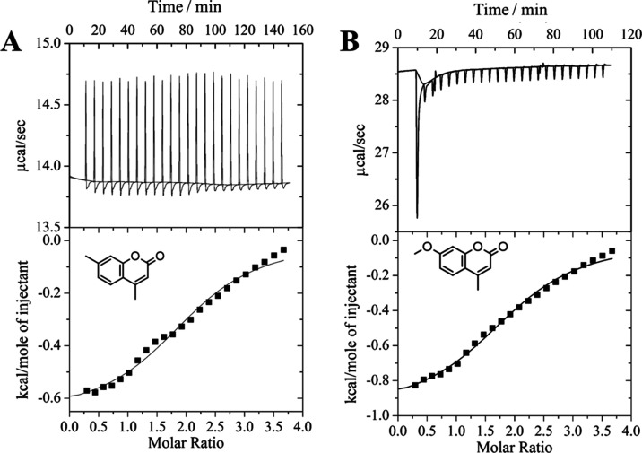
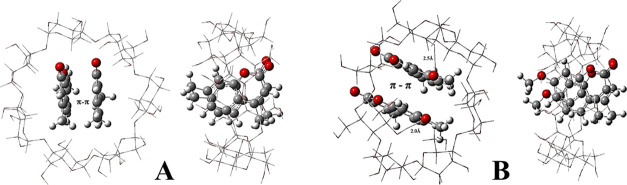
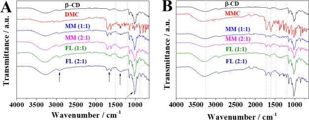
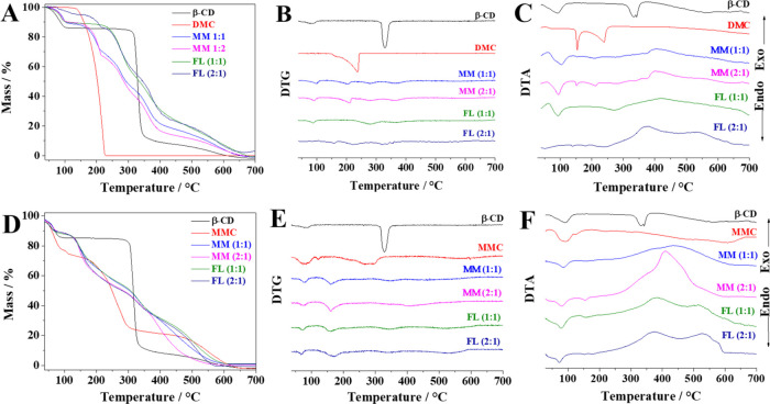
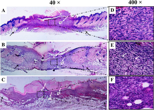
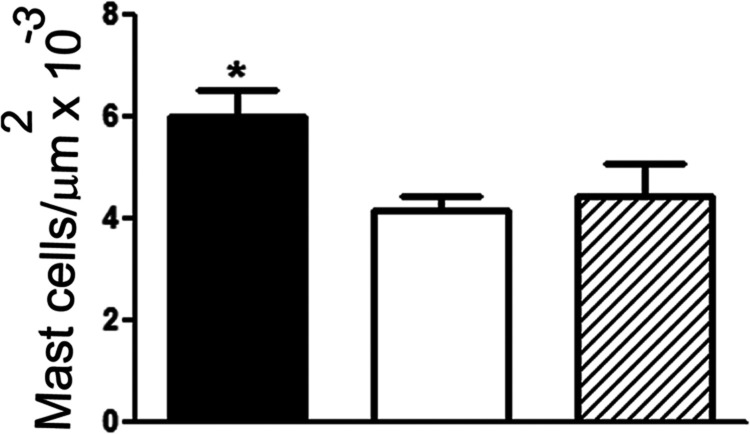
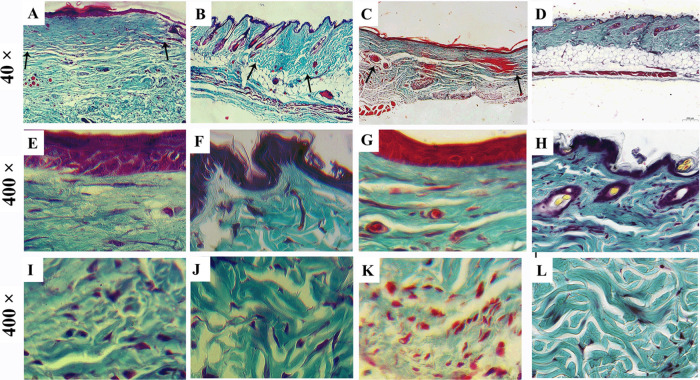
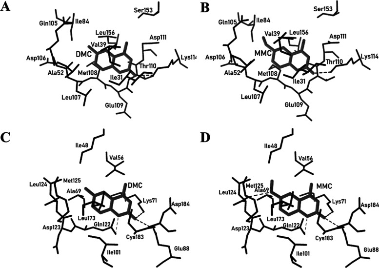
Coumarins have great pharmacotherapeutic potential, presenting several biological and pharmaceutical applications, like antibiotic, fungicidal, anti-inflammatory, anticancer, anti-HIV, and healing activities, among others. These molecules are practically insoluble in water, and for biological applications, it became necessary to complex them with cyclodextrins (CDs), which influence their bioavailability in the target organism. In this work, we studied two coumarins, and it was possible to conclude that there were structural differences between 4,7-dimethyl-2H-chromen-2-one (DMC) and 7-methoxy-4-methyl-2H-chromen-2-one (MMC)/β-CD that were solubilized in ethanol, frozen, and lyophilized (FL) and the mechanical mixtures (MM). In addition, the inclusion complex formation improved the solubility of DMC and MMC in an aqueous medium. According to the data, the inclusion complexes were formed and are more stable at a molar ratio of 2:1 coumarin/β-CD, and hydrogen bonds along with π-π stacking interactions are responsible for the better stability, especially for (MMC)2@β-CD. In vivo wound healing studies in mice showed faster re-epithelialization and the best deposition of collagen with the (DMC)2@β-CD (FL) and (MMC)2@β-CD (FL) inclusion complexes, demonstrating clearly that they have potential in wound repair. Therefore, (DMC)2@β-CD (FL) deserves great attention because it presented excellent results, reducing the granulation tissue and mast cell density and improving collagen remodeling. Finally, the protein binding studies suggested that the anti-inflammatory activities might exert their biological function through the inhibition of MEK, providing the possibility of development of new MEK inhibitors.
Keywords: coumarin; cyclodextrin; healing wounds; inclusion complex; skin lesions.
Conflict of interest statement
The authors declare no competing financial interest.
Figures
Similar articles
-
Bioadhesive hydrogel comprising bilirubin/β-cyclodextrin inclusion complexes promote diabetic wound healing.Pharm Biol. 2021 Dec;59(1):1139-1149. doi: 10.1080/13880209.2021.1964543. Pharm Biol. 2021. PMID: 34425063 Free PMC article.
-
Inclusion complexes of tadalafil with natural and chemically modified beta-cyclodextrins. I: preparation and in-vitro evaluation.Eur J Pharm Biopharm. 2008 Nov;70(3):819-27. doi: 10.1016/j.ejpb.2008.06.024. Epub 2008 Jul 4. Eur J Pharm Biopharm. 2008. PMID: 18655829
-
Preparation and Biophysical Characterization of Quercetin Inclusion Complexes with β-Cyclodextrin Derivatives to be Formulated as Possible Nose-to-Brain Quercetin Delivery Systems.Mol Pharm. 2020 Nov 2;17(11):4241-4255. doi: 10.1021/acs.molpharmaceut.0c00672. Epub 2020 Oct 19. Mol Pharm. 2020. PMID: 32986435
-
Enhancement of oral bioavailability of cilostazol by forming its inclusion complexes.AAPS PharmSciTech. 2009;10(2):660-9. doi: 10.1208/s12249-009-9249-7. Epub 2009 May 21. AAPS PharmSciTech. 2009. PMID: 19459053 Free PMC article.
-
Short Review on the Biological Activity of Cyclodextrin-Drug Inclusion Complexes Applicable in Veterinary Therapy.Molecules. 2023 Jul 21;28(14):5565. doi: 10.3390/molecules28145565. Molecules. 2023. PMID: 37513437 Free PMC article. Review.
Cited by
-
A computational study on the enantioselective separation of cyhalothrin enantiomers by β-cyclodextrins.J Mol Model. 2025 Aug 11;31(9):238. doi: 10.1007/s00894-025-06469-7. J Mol Model. 2025. PMID: 40788417
-
Bacterial cellulose-based scaffold with in-situ cationic micelle modification for urethral stricture disease: Sustained drug components release, cytokines recruitment, and bacterial microenvironment regulation.Bioact Mater. 2025 May 19;51:306-317. doi: 10.1016/j.bioactmat.2025.04.031. eCollection 2025 Sep. Bioact Mater. 2025. PMID: 40491686 Free PMC article.
-
Cyclodextrins as multifunctional tools for advanced biomaterials in tissue repair and regeneration.Bioact Mater. 2025 Mar 27;49:627-651. doi: 10.1016/j.bioactmat.2025.03.018. eCollection 2025 Jul. Bioact Mater. 2025. PMID: 40212783 Free PMC article. Review.
-
Characterization and In Vitro Prebiotic Activity of Pterostilbene/β-Cyclodextrin Inclusion Complexes.Molecules. 2025 Mar 18;30(6):1363. doi: 10.3390/molecules30061363. Molecules. 2025. PMID: 40142136 Free PMC article.
References
MeSH terms
Substances
LinkOut - more resources
Full Text Sources
Research Materials
Miscellaneous
