

Power Density Titration of Reversible Photoisomerization of a Fluorescent Protein Chromophore in the Presence of Thermally Driven Barrier Crossing Shown by Quantitative Millisecond Serial Synchrotron X-ray Crystallography
- PMID: 38848551
- PMCID: PMC11191680
- DOI: 10.1021/jacs.3c12883
Power Density Titration of Reversible Photoisomerization of a Fluorescent Protein Chromophore in the Presence of Thermally Driven Barrier Crossing Shown by Quantitative Millisecond Serial Synchrotron X-ray Crystallography
Abstract
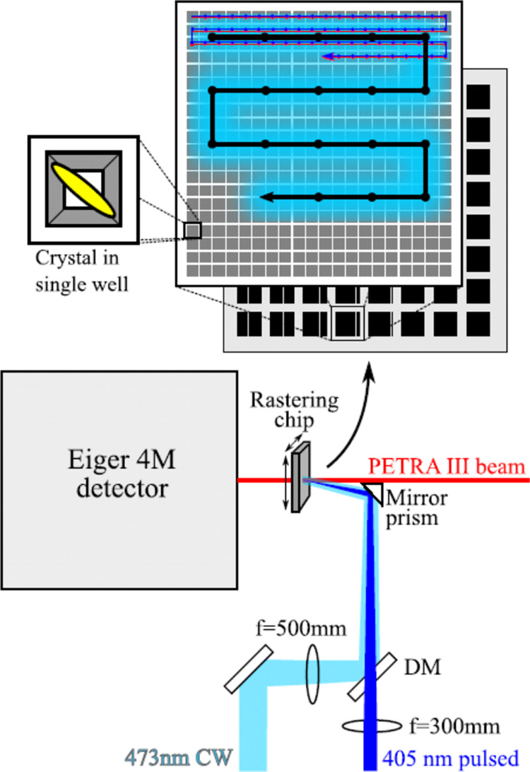
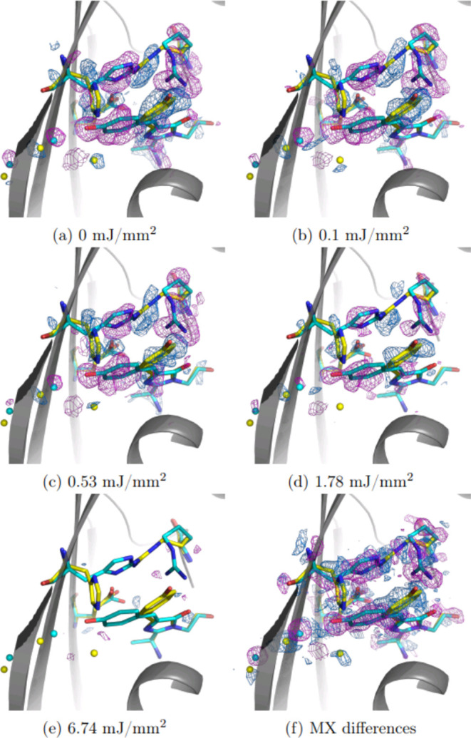
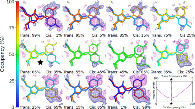
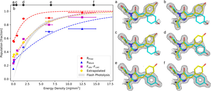
We present millisecond quantitative serial X-ray crystallography at 1.7 Å resolution demonstrating precise optical control of reversible population transfer from Trans-Cis and Cis-Trans photoisomerization of a reversibly switchable fluorescent protein, rsKiiro. Quantitative results from the analysis of electron density differences, extrapolated structure factors, and occupancy refinements are shown to correspond to optical measurements of photoinduced population transfer and have sensitivity to a few percent in concentration differences. Millisecond time-resolved concentration differences are precisely and reversibly controlled through intense continuous wave laser illuminations at 405 and 473 nm for the Trans-to-Cis and Cis-to-Trans reactions, respectively, while the X-ray crystallographic measurement and laser illumination of the metastable Trans chromophore conformation causes partial thermally driven reconversion across a 91.5 kJ/mol thermal barrier from which a temperature jump between 112 and 128 K is extracted.
Conflict of interest statement
The authors declare no competing financial interest.
Figures
Similar articles
-
Observation of Cation Chromophore Photoisomerization of a Fluorescent Protein Using Millisecond Synchrotron Serial Crystallography and Infrared Vibrational and Visible Spectroscopy.J Phys Chem B. 2022 Nov 17;126(45):9288-9296. doi: 10.1021/acs.jpcb.2c06780. Epub 2022 Nov 3. J Phys Chem B. 2022. PMID: 36326150 Free PMC article.
-
Kindling fluorescent protein from Anemonia sulcata: dark-state structure at 1.38 A resolution.Biochemistry. 2005 Apr 19;44(15):5774-87. doi: 10.1021/bi047644u. Biochemistry. 2005. PMID: 15823036
-
The nature of the primary photochemical events in rhodopsin and isorhodopsin.Biophys J. 1988 Mar;53(3):367-85. doi: 10.1016/S0006-3495(88)83114-X. Biophys J. 1988. PMID: 2964878 Free PMC article.
-
Bacteriorhodopsin: Structural Insights Revealed Using X-Ray Lasers and Synchrotron Radiation.Annu Rev Biochem. 2019 Jun 20;88:59-83. doi: 10.1146/annurev-biochem-013118-111327. Epub 2019 Apr 3. Annu Rev Biochem. 2019. PMID: 30830799 Review.
-
Photoswitching of E222Q GFP mutants: "concerted" mechanism of chromophore isomerization and protonation.Photochem Photobiol Sci. 2010 Oct 28;9(10):1307-19. doi: 10.1039/c0pp00189a. Epub 2010 Sep 21. Photochem Photobiol Sci. 2010. PMID: 20859582 Review.
References
-
- Seddon E. A.; Clarke J. A.; Dunning D. J.; Masciovecchio C.; Milne C. J.; Parmigiani F.; Rugg D.; Spence J. C. H.; Thompson N. R.; Ueda K.; Vinko S. M.; Wark J. S.; Wurth W. Short-Wavelength Free-Electron Laser Sources and Science: A Review. Rep. Prog. Phys. 2017, 80 (11), 11590110.1088/1361-6633/aa7cca. - DOI - PubMed
-
- Pande K.; Hutchison C. D. M.; Groenhof G.; Aquila A.; Robinson J. S.; Tenboer J.; Basu S.; Boutet S.; DePonte D. P.; Liang M.; White T. A.; Zatsepin N. A.; Yefanov O.; Morozov D.; Oberthuer D.; Gati C.; Subramanian G.; James D.; Zhao Y.; Koralek J.; Brayshaw J.; Kupitz C.; Conrad C.; Roy-Chowdhury S.; Coe J. D.; Metz M.; Xavier P. L.; Grant T. D.; Koglin J. E.; Ketawala G.; Fromme R.; Šrajer V.; Henning R.; Spence J. C. H.; Ourmazd A.; Schwander P.; Weierstall U.; Frank M.; Fromme P.; Barty A.; Chapman H. N.; Moffat K.; van Thor J. J.; Schmidt M. Femtosecond Structural Dynamics Drives the Trans/Cis Isomerization in Photoactive Yellow Protein. Science 2016, 352 (6286), 725–729. 10.1126/science.aad5081. - DOI - PMC - PubMed
-
- Coquelle N.; Sliwa M.; Woodhouse J.; Schirò G.; Adam V.; Aquila A.; Barends T. R. M.; Boutet S.; Byrdin M.; Carbajo S.; De la Mora E.; Doak R. B.; Feliks M.; Fieschi F.; Foucar L.; Guillon V.; Hilpert M.; Hunter M. S.; Jakobs S.; Koglin J. E.; Kovacsova G.; Lane T. J.; Lévy B.; Liang M.; Nass K.; Ridard J.; Robinson J. S.; Roome C. M.; Ruckebusch C.; Seaberg M.; Thepaut M.; Cammarata M.; Demachy I.; Field M.; Shoeman R. L.; Bourgeois D.; Colletier J.-P.; Schlichting I.; Weik M. Chromophore Twisting in the Excited State of a Photoswitchable Fluorescent Protein Captured by Time-Resolved Serial Femtosecond Crystallography. Nat. Chem. 2018, 10 (1), 31–37. 10.1038/nchem.2853. - DOI - PubMed
LinkOut - more resources
Full Text Sources
