. 2024 Jul:105:105185.
doi: 10.1016/j.ebiom.2024.105185. Epub 2024 Jun 7.

Intranasal SARS-CoV-2 Omicron variant vaccines elicit humoral and cellular mucosal immunity in female mice

Affiliations

Intranasal SARS-CoV-2 Omicron variant vaccines elicit humoral and cellular mucosal immunity in female mice

Stefan Slamanig et al. EBioMedicine. 2024 Jul.

Abstract

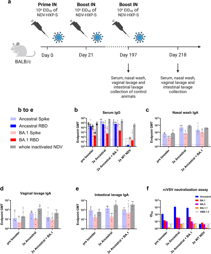
Background: In order to prevent the emergence and spread of future variants of concern of severe acute respiratory syndrome coronavirus 2 (SARS-CoV-2), developing vaccines capable of stopping transmission is crucial. The SARS-CoV-2 vaccine NDV-HXP-S can be administered live intranasally (IN) and thus induce protective immunity in the upper respiratory tract. The vaccine is based on Newcastle disease virus (NDV) expressing a stabilised SARS-CoV-2 spike protein. NDV-HXP-S can be produced as influenza virus vaccine at low cost in embryonated chicken eggs.

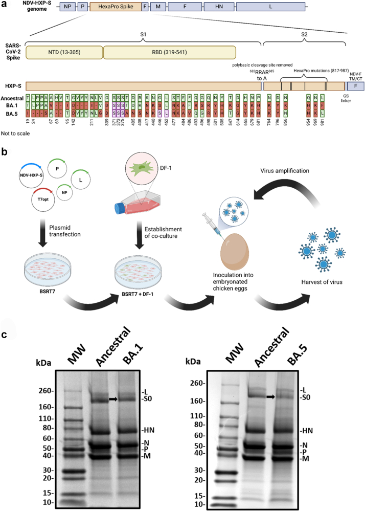
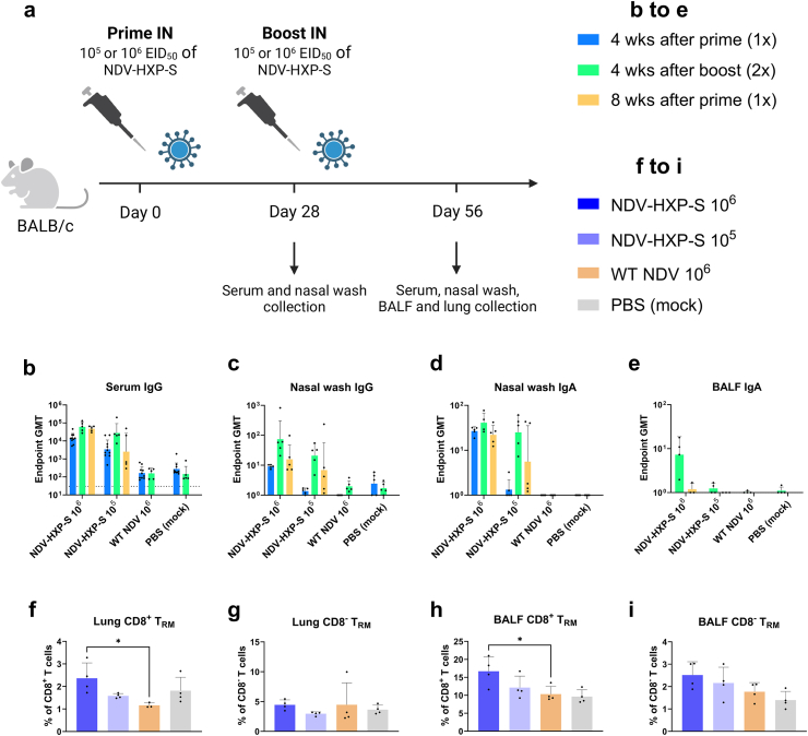
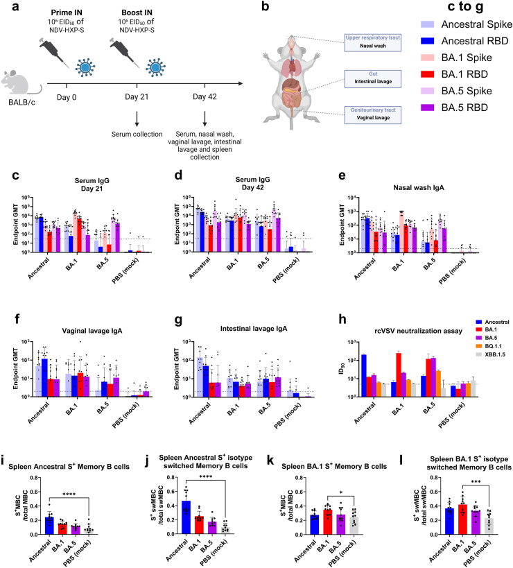
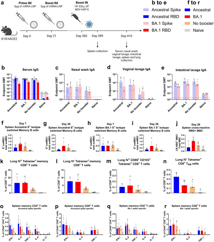
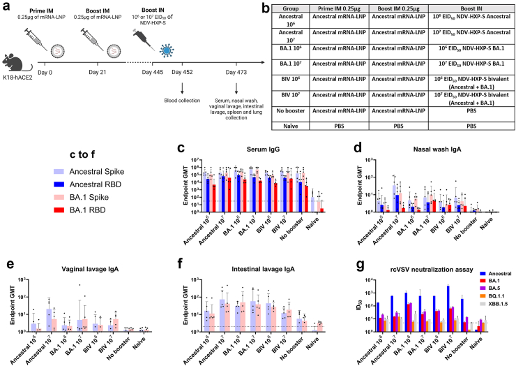
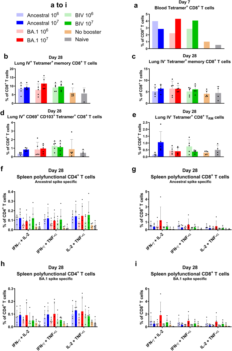
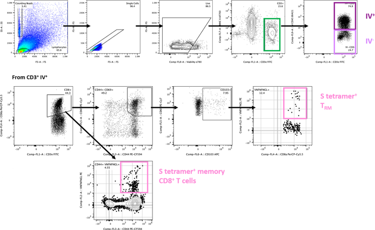
Methods: The NDV-HXP-S vaccine was genetically engineered to match the Omicron variants of concern (VOC) BA.1 and BA.5 and tested as an IN two or three dose vaccination regimen in female mice. Furthermore, female mice intramuscularly (IM) vaccinated with mRNA-lipid nanoparticles (LNPs) were IN boosted with NDV-HXP-S. Systemic humoral immunity, memory T cell responses in the lungs and spleens as well as immunoglobulin A (IgA) responses in distinct mucosal tissues were characterised.

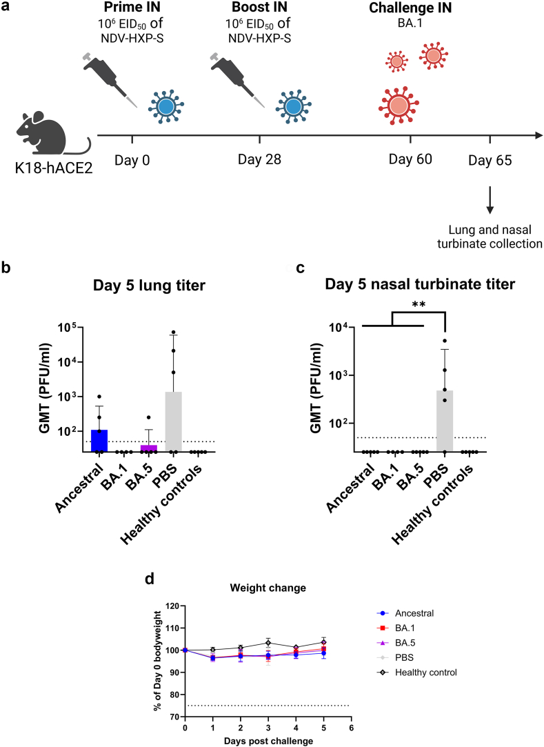
Findings: NDV-HXP-S Omicron variant vaccines elicited high mucosal IgA and serum IgG titers against respective SARS-CoV-2 VOC in female mice following IN administration and protected against challenge from matched variants. Additionally, antigen-specific memory B cells and local T cell responses in the lungs were induced. Host immunity against the NDV vector did not interfere with boosting. Intramuscular vaccination with mRNA-LNPs was enhanced by IN NDV-HXP-S boosting resulting in improvement of serum neutralization titers and induction of mucosal immunity.

Interpretation: We demonstrate that NDV-HXP-S Omicron variant vaccines utilised for primary immunizations or boosting efficiently elicit humoral and cellular immunity. The described induction of systemic and mucosal immunity has the potential to reduce infection and transmission.

Funding: This work was partially funded by the NIAIDCenters of Excellence for Influenza Research and Response (CEIRR) and by the NIAID Collaborative Vaccine Innovation Centers and by institutional funding from the Icahn School of Medicine at Mount Sinai. See under Acknowledgements for details.

Keywords: COVID-19; Low cost vaccine platform; Mucosal immune response; NDV vector; Prime-pull vaccination; Variant vaccine; mRNA vaccine.


Conflict of interest statement

Declaration of interests The Icahn School of Medicine at Mount Sinai has filed patent applications entitled “RECOMBINANT NEWCASTLE DISEASE VIRUS EXPRESSING SARS-COV-2 SPIKE PROTEIN AND USES THEREOF” which names P.P., A.G.S, F.K. and W.S. as inventors. Mount Sinai is seeking to commercialise this vaccine; therefore, the institution and its faculty inventors could benefit financially. I.G.D. has co-chaired at the ninth ESWI Influenza conference, which has no competing interest with this work. The M.S. laboratory has received unrelated research funding in sponsored research agreements from 7Hills Pharma, ArgenX N.V., Moderna and Phio Pharmaceuticals, which has no competing interest with this work. F.K. has consulted for Merck, Seqirus, Curevac and Pfizer, and is currently consulting for Pfizer, Third Rock Ventures, GSK and Avimex. The FK laboratory is also collaborating with Pfizer on animal models of SARS-CoV-2. The A.G.-S. laboratory has received research support from GSK, Pfizer, Senhwa Biosciences, Kenall Manufacturing, Blade Therapeutics, Avimex, Johnson & Johnson, Dynavax, 7Hills Pharma, Pharmamar, ImmunityBio, Accurius, Nanocomposix, Hexamer, N-fold LLC, Model Medicines, Atea Pharma, Applied Biological Laboratories and Merck. A.G.S. has consulting agreements for the following companies involving cash and/or stock: Castlevax, Amovir, Vivaldi Biosciences, Contrafect, 7Hills Pharma, Avimex, Pagoda, Accurius, Esperovax, Farmak, Applied Biological Laboratories, Pharmamar, CureLab Oncology, CureLab Veterinary, Synairgen, Paratus and Pfizer. A.G.S. has been an invited speaker in meeting events organised by Seqirus, Janssen, Abbott and Astrazeneca. A.G.S. is inventor on patents and patent applications on the use of antivirals and vaccines for the treatment and prevention of virus infections and cancer, owned by the Icahn School of Medicine at Mount Sinai, New York. Specifically, A.G.S., a member of the faculty of the Icahn School of Medicine at Mount Sinai (Mount Sinai) is an inventor of a novel COVID-19 vaccine currently being investigated in clinical trials. Mount Sinai is advancing the development of this vaccine and related technologies for potential commercial use. Mount Sinai has created CastleVax Inc., a Mount Sinai company, and has licensed the applicable IP to it. Mount Sinai will receive financial compensation from CastleVax Inc. pursuant to that license if vaccine development proceeds and as an owner of the company subject to the sale of its ownership interest in the future. Subject to Mount Sinai receiving such financial consideration, A.G.S. will receive a portion of that consideration pursuant to the terms of the Mount Sinai Intellectual Property Policy. All other authors declare no competing interests.

Figures

Fig. 1
Fig. 1
Design and production of NDV-HXP-S Omicron BA.1 and BA.5 variant vaccines. (a) Structure and design of the NDV-HXP-S genome. The SARS-CoV-2 HexaPro spike sequences were inserted between the P and the M genes of the LaSota NDV strain. The ectodomain of the spike was connected to the transmembrane domain and the cytoplasmic tail (TM/CT) of the F protein of NDV (ectodomain of the spike in beige; NDV components in blue). The original polybasic cleavage site was deleted by mutating 682RRAR685 to 682--A-685. The spike-stabilizing HexaPro (F817P, A892P, A899P, A942P, K986P, and V987P) mutations were introduced. The sequence was codon-optimised for mammalian host expression. The ancestral HXP-S sequence aligned with the new Omicron variants BA.1 and BA.5 is depicted. Amino acids of the ancestral original HXP-S sequence are shown in green while changes for BA.1 or BA.5 are depicted in red. Amino acids that have been genetically engineered are shown in purple. (b) Experimental scheme of NDV-HXP-S variant rescue. BSRT7 cells were transfected with pNDV-HXP-S and the helper plasmids pTM1-NP, pTM1-P, pTM1-L, and pCI-T7opt. The next day, transfected BSRT7 cells were co-cultured with DF-1 cells. After two or three days of incubation, co-cultures were inoculated into 8- or 9-day old specific pathogen-free (SPF) embryonated chicken eggs. Following three days of incubation at 37 °C, eggs were cooled at 4 °C overnight and subsequently virus containing allantoic fluid was harvested. The recombinant virus was amplified and passaged multiple times to confirm genetic stability. (c) Protein analysis of NDV-HXP-S BA.1 and BA.5. NDV-HXP-S BA.1, BA.5 and ancestral were purified from allantoic fluid via ultra-centrifugation through a sucrose cushion and resuspended in PBS. The purified viruses were resolved on 4–20% SDS page and the viral proteins visualised by Coomassie blue staining (L, S0, HN, N, P, and M). The uncleaved SARS-CoV-2 S0 spike protein is highlighted with an arrow.
Fig. 2
Fig. 2
Intranasal administration of NDV-HXP-S vaccine induces mucosal humoral and T cell memory. (a) Design of the study. Six to eight week-old female BALB/c mice were intranasally immunised with 105 or 106 fifty percent of egg embryo infectious dose (EID50) of NDV-HXP-S expressing ancestral spike. Four weeks later, half of the mice were boosted. The empty WT LaSota NDV vector (106 EID50) and PBS were used for vector-only and negative control groups, respectively. Twenty-eight days after prime, sera (n = 10) and nasal washes (n = 5 or 4) were collected and 56 days after prime (28 days after boost), serum (n = 5), nasal washes (n = 5 or 4), and bronchoalveolar lavage fluid (BALF) (n = 4 or 3) were collected to assess binding antibody responses. Lung tissues and BALF were collected for T cell analysis (n = 5 or 4). (b–e) Measurement of ancestral spike-specific (b) serum IgG, (c) nasal wash IgG, (d) nasal wash IgA, and (e) BALF IgA in naïve mice, mice immunised with empty vector, mice immunised with 105 EID50 or mice immunised with 106 EID50 of NDV-HXP-S ancestral; at four weeks after prime, four weeks after boost and eight weeks after prime. The geometric mean titer (GMT) endpoint titers of the ELISA were graphed (an endpoint titer of one was assigned to negative samples). The dashed line indicates the limit of detection (for nasal wash and BALF x-axis equals limit of detection). The error bars represent geometric standard deviation (SD). (f–i) Staining for CD3, CD8, CD44, CD69, and CD103 was used to assess tissue resident memory cells (TRM) (f) in the lungs and positive for CD8, (g) in the lungs and negative for CD8, (h) in the BALF and positive for CD8 and (i) in the BALF and negative for CD8. Mean with SD was graphed. Statistical significance was calculated by means of [(b)(e)] log transforming the data set to normalise followed by a two-way analysis of variance (ANOVA) or [(f)(i)] one-way ANOVA followed by Tukey’s multiple comparisons test. The experiment was conducted twice, pooled data are shown ∗p < 0.05.
Fig. 3
Fig. 3
NDV-HXP-S BA.1 and BA.5 variant vaccinations increase strain specific humoral immunity. (a) Design of the study. Six to eight week-old female BALB/c mice were intranasally immunised twice 21 days apart with 106 EID50 of NDV-HXP-S ancestral, BA.1 or BA.5. PBS was used for a negative control group. Twenty-one days after prime, serum was collected and 42 days after prime (21 days after boost), serum, nasal washes, vaginal lavages, and intestinal lavages were collected to assess binding and neutralizing antibody response. Spleens were collected for memory B cell analysis. (n = 10) (b) Anatomical scheme of harvesting nasal wash, intestinal lavage, and vaginal lavage. From each animal, nasal washes from the upper respiratory tract, intestinal lavages from the gut and vaginal lavages from the genitourinary tract were harvested for IgA measurement. (c–e) Measurement of ancestral spike, ancestral RBD, BA.1 spike, BA.1 RBD, BA.5 spike, and BA.5 RBD-specific (c) serum IgG at the 1st time point (21 days post prime), (d) serum IgG at the 2nd time point (42 days post prime, 21 days post booster) and (e) nasal wash IgA in mice (mock) immunised with PBS or 106 EID50 of NDV-HXP-S ancestral, BA.1 or BA.5. (f and g) Measurement of ancestral spike, ancestral RBD, BA.1 RBD, and BA.5 RBD-specific (f) vaginal lavage IgA and (g) intestinal lavage IgA in mice (mock) immunised with PBS or 106 EID50 of NDV-HXP-S ancestral, BA.1 or BA.5. The GMT endpoint titers of the ELISA assay were graphed (an endpoint titer of one was assigned to negative samples). The dashed line indicates the limit of detection. The error bars represent geometric SD. (h) Neutralization titers against SARS-CoV-2 ancestral, BA.1, BA.5, BQ.1.1, or XBB.1.5 spike-pseudotyped vesicular stomatitis virus expressing GFP (rcVSVeGFP-CoV-2-S) were measured in technical duplicates from pooled sera. Geometric mean with geometric SD of the inhibitory dilution at which 50% neutralization is achieved (ID50) was graphed (ID50 of 1.5 was assigned to negative samples). The dashed line indicates the limit of detection. (i–l) Using two separate tetrameric B cell probes for ancestral or BA.1 spike respectively, B cell subsets in the spleens were measured. This included (i) ancestral spike specific memory B cells (CD3- Live+ B220+ CD19+ IgD GL7- CD38+), (j) isotype switched memory B cells specific for ancestral spike (CD3- Live+ B220+ CD19+ IgM IgD GL7- CD38+), (k) BA.1 spike specific memory B cells and (l) isotype switched memory B cells specific for BA.1. Mean with SD was graphed. Statistical significance was calculated by means of [(c)(g)] log transforming the data set to normalise followed by a two-way ANOVA or [(i)(l)] one-way ANOVA followed by Tukey’s multiple comparisons test. ∗p < 0.05; ∗∗p < 0.01; ∗∗∗p < 0.001; ∗∗∗∗p < 0.0001.
Fig. 4
Fig. 4
Intranasal immunization with NDV-HXP-S variant vaccines protects mice from homologous infection. (a) Design of the study. Six to eight week-old female K18-hACE2 mice were intranasally immunised twice 28 days apart with 106 EID50 of NDV-HXP-S ancestral, BA.1 or BA.5. PBS was used for a negative control group. Thirty-two days after the boost, the mice were challenged with 3 × 104 plaque-forming units (PFU) of SARS-CoV-2 BA.1 (n = 5 or 4). One animal of the BA.1 vaccination group died during the challenge process unrelated to the vaccination or virus. A healthy control group was kept uninfected (n = 5). (b and c) Measurement of viral titers in the (b) lungs and (c) nasal turbinates. Tissues of the animals were collected on day 5 post challenge. The whole lungs or nasal turbinates were homogenised in 1 mL of PBS. The viral load was measured by plaque assay on VERO E6 TMPRSS2 T2A ACE2 cells and graphed as GMT of PFU/ml (a titer of 25 PFU/mL was assigned to negative samples). The dashed line indicates the limit of detection. The error bars represent geometric SD. (d) Body weight of mice infected with SARS-CoV-2 BA.1. Data are mean with SD. Statistical significance was calculated by means of [(b) and (c)] Kruskal–Wallis test followed by Dunn’s multiple comparisons test. ∗p < 0.05; ∗∗p < 0.01.
Fig. 5
Fig. 5
A third vaccination increases mucosal as well as serum antibody titers with no interference due to vector immunity. (a) Design of the study. Six to eight week-old female BALB/c mice were intranasally immunised twice 21 days apart with 106 EID50 of NDV-HXP-S ancestral or WT LaSota NDV vector. After 197 days, serum of the WT NDV control group as well as serum, nasal washes, vaginal lavages, and intestinal lavages of the animals of the control group pre booster (vaccinated twice with NDV-HXP-S expressing ancestral spike) were collected to assess binding and neutralizing antibody response. The other mice were boosted IN with 106 EID50 of NDV-HXP-S ancestral or BA.1. Twenty-one days later, on day 218, serum, nasal washes, vaginal lavages, and intestinal lavages of booster groups 3x ancestral and 2x ancestral + BA.1 were collected to assess binding and neutralizing antibody response. (n = 5 or 15) (b) Measurement of ancestral spike, ancestral RBD, BA.1 spike, BA.1 RBD, and whole inactivated NDV-specific serum IgG. (c–e) Measurement of ancestral spike, BA.1 spike, and whole inactivated NDV-specific (c) nasal wash IgA, (d) vaginal lavage IgA and (e) intestinal lavage IgA in mice immunised twice with 106 EID50 of NDV-HXP-S ancestral, three times with NDV-HXP-S ancestral or immunised twice with NDV-HXP-S ancestral and boosted once with BA.1. The GMT endpoint titers of the ELISA assay were graphed (an endpoint titer of one was assigned to negative samples). The dashed line indicates the limit of detection (for vaginal lavage x-axis equals limit of detection). The error bars represent geometric SD. (f) Neutralization titers against SARS-CoV-2 ancestral, BA.1, BA.5, BQ.1.1, or XBB.1.5 spike-pseudotyped vesicular stomatitis virus expressing GFP (rcVSVeGFP-CoV-2-S) were measured in technical duplicates from pooled sera. Geometric mean with geometric SD of the ID50 was graphed (ID50 of 1.5 was assigned to negative samples). The dashed line indicates the limit of detection. Statistical significance was calculated by means of [(b)(e)] log transforming the data set to normalise followed by a two-way ANOVA followed by Tukey’s multiple comparisons test.
Fig. 6
Fig. 6
A mucosal boosting with NDV-HXP-S in mice with ceiling serum antibodies induced by high-dose mRNA vaccinations improves cellular and mucosal immunity. (a) Design of the study. Six to eight week-old female K18-hACE2 mice were intramuscularly immunised twice 21 days apart with 5 μg of mRNA-lipid nanoparticles (LNPs, Pfizer/BioNTech BNT162b2). After 382 days, the mice were boosted IN with 106 EID50 of NDV-HXP-S ancestral or BA.1. A no booster group received PBS. A naïve control group received PBS throughout the whole vaccination schedule. After 389 days (seven days after boosting), spleens were collected. After 410 days (28 days after boosting), serum, nasal washes, vaginal lavages, and intestinal lavages were collected to assess binding antibody response. Spleens were collected to analyse memory B and T cells. Lungs were collected to analyse CD8+ T cells. (n = 5 or 4) (b) Measurement of ancestral spike, ancestral RBD, BA.1 spike and BA.1 RBD-specific serum IgG harvested 28 days after the booster. (c–e) Measurement of ancestral spike and BA.1 spike-specific (c) nasal wash IgA, (d) vaginal lavage IgA and (e) intestinal lavage IgA harvested 28 days after boosting mice with 106 EID50 of NDV-HXP-S ancestral or BA.1 or PBS (no booster and naïve control groups). The GMT endpoint titers of the ELISA assay were graphed (an endpoint titer of one was assigned to negative samples). The dashed line indicates the limit of detection (for vaginal lavage x-axis equals limit of detection). The error bars represent geometric SD. (f–j) Using two separate tetrameric B cell probes for ancestral or BA.1 spike respectively as well as an ancestral RBD probe, B cell subsets in the spleens were measured. This included (f) isotype switched memory B cells specific for ancestral spike seven days after boosting, (g) isotype switched memory B cells specific for ancestral spike 28 days after boosting, (h) isotype switched memory B cells specific for BA.1 spike seven days after boosting, (i) isotype switched memory B cells specific for BA.1 spike 28 days after boosting (CD3- Live+ B220+ CD19+ IgM IgD GL7- CD38+) and (j) cross-reactive memory B cells (gated ancestral RBD vs BA.1 spike, CD3- Live+ B220+ CD19+ IgD GL7- CD38+). Mean with SD was graphed. (k–n) Using CD45 intravascular (IV) labelling as well as tetramer staining, intravascular (IV+) and extravascular (IV) CD8+ T cells specific for SARS-CoV-2 spike were measured in the lungs 28 days after boosting. This included (k) IV+ memory CD8+ T cells, (l) IV memory CD8+ T cells (CD3+ MHC II CD44+), (m) IV+ CD69+ CD103+ CD8+ T cells (CD3+ MHC II CD44+ CD69+ CD103+), and (n) IV CD8+ TRM cells (CD3+ MHC II CD44+ CD69+ CD103+). Mean with SD was graphed. (o–r) Twenty-eight days after boosting, splenocytes were isolated and restimulated with an ancestral or BA.1 spike specific peptide pool. Intracellular cytokine staining was used to measure antigen-specific production of IFN-γ, TNF-α, IL-2, IL-4, and IL-17 by CD44+ CD4+ or CD44+ CD8+ T cells out of total CD44+ CD4+ or CD44+ CD8+ T cells. Mean with SD was graphed. Statistical significance was calculated by means of [(b)(e)] log transforming the data set to normalise followed by a two-way ANOVA, or [(f)(n)] one-way ANOVA, or [(o)(r)] two-way ANOVA followed by Tukey’s multiple comparisons test.
Fig. 7
Fig. 7
A mucosal boost with NDV-HXP-S in mice after low-dose mRNA vaccinations improves serum antibody binding and neutralization titers and induces mucosal IgA responses. (a and b) Design of the study and groups. Six to eight week-old female K18-hACE2 mice were intramuscularly immunised twice 21 days apart with 0.25 μg of mRNA-LNPs (Pfizer/BioNTech BNT162b2). After 445 days, the mice were boosted IN with 106 or 107 EID50 of NDV-HXP-S ancestral, BA.1 or bivalent (BIV, equal amounts of ancestral and BA.1 to a total of 106 or 107 EID50). A no booster group received PBS. A naïve control group received PBS throughout the whole vaccination schedule. After 452 days (seven days after boosting), serum was collected to assess blood circulating CD8+ T cells. After 473 days (28 days after boosting) serum, nasal washes, vaginal lavages, and intestinal lavages were collected to assess binding and neutralizing antibody response. Spleens were collected to analyse T cells. Lungs were collected to analyse CD8+ T cells. (n = 5) (c and d) Measurement of ancestral spike, ancestral RBD, BA.1 spike, and BA.1 RBD-specific (b) serum IgG and (c) nasal wash IgA. (e and f) Measurement of ancestral spike and BA.1 spike-specific (d) vaginal lavage IgA and (e) intestinal lavage IgA harvested 28 days after boosting mice with 106 or 107 EID50 of NDV-HXP-S ancestral, BA.1 or BIV or PBS (no booster and naïve control groups). The GMT endpoint titers of the ELISA assay were graphed (an endpoint titer of one was assigned to negative samples). The dashed line indicates the limit of detection (for nasal wash and vaginal lavage x-axis equals limit of detection). The error bars represent geometric SD. (g) Neutralization titers against SARS-CoV-2 ancestral, BA.1, BA.5, BQ.1.1, or XBB.1.5 spike-pseudotyped vesicular stomatitis virus expressing GFP (rcVSVeGFP-CoV-2-S) were measured in technical duplicates from pooled sera. Geometric mean with geometric SD of the ID50 was graphed (ID50 of 1.5 was assigned to negative samples). The dashed line indicates the limit of detection. Statistical significance was calculated by means of [(c)(f)] log transforming the data set to normalise followed by a two-way ANOVA followed by Tukey’s multiple comparisons test.
Fig. 8
Fig. 8
A mucosal boost with NDV-HXP-S in mice after low-dose mRNA vaccinations improves T cell responses in blood, lungs and spleens. (a) Using tetramer staining, CD8+ T cells specific for SARS-CoV-2 spike were measured in the blood seven days after boosting (CD3+ MHC II). Blood of individual animals was pooled for each group. Mean was graphed. (b–e) Using CD45 IV labelling as well as tetramer staining, intravascular (IV+) and extravascular (IV) CD8+ T cells specific for SARS-CoV-2 spike were measured in the lungs 28 days after boosting (n = 5 or 4). This included (b) IV+ memory CD8+ T cells, (c) IV memory CD8+ T cells (CD3+ MHC II CD44+), (d) IV+ CD69+ CD103+ CD8+ T cells (CD3+ MHC II CD44+ CD69+ CD103+), and (e) IV CD8+ TRM cells (CD3+ MHC II CD44+ CD69+ CD103+). Mean with SD was graphed. (f–i) Twenty-eight days after boosting, splenocytes were isolated and restimulated with an ancestral or BA.1 spike specific peptide pool (n = 5 or 4). Intracellular cytokine staining was used to measure antigen-specific production of IFN-γ, TNF-α, and IL-2 by CD4+ or CD8+ T cells out of total CD4+ or CD8+ T cells. Polyfunctional T cell populations positive for two markers are shown. Mean with SD was graphed. Statistical significance was calculated by means of [(b)(e)] one-way ANOVA or [(f)(i)] two-way ANOVA followed by Tukey’s multiple comparisons test.
Supplementary Fig. S1
Supplementary Figure S1. Serum passive transfer decreases viral load in the lungs. (a) Design of the study and groups. Sera from sequentially intranasally vaccinated BALB/c mice were pooled and administered intraperitoneally (IP) to six to eight week-old female K18-hACE2 mice. After two hours, the mice were challenged with 3 x 10⁴ plaque-forming units (PFU) of SARS-CoV-2 BA.1 (n = 5). A healthy control group was kept uninfected (n = 5). (b and c) Measurement of viral titers in the (b) lungs and (c) nasal turbinates. Tissues of the animals were collected on day 5 post challenge. The whole lungs or nasal turbinates were homogenised in 1 mL of PBS. The viral load was measured by plaque assay on VERO E6 TMPRSS2 T2A ACE2 cells and graphed as GMT of PFU/ml (a titer of 25 PFU/ml was assigned to negative samples). The dashed line indicates the limit of detection. The error bars represent geometric SD. Statistical significance was calculated by means of Kruskal-Wallis test followed by Dunn’s multiple comparisons test.
Supplementary Fig. S2
Supplementary Figure S2. Memory B cells gating strategy and S protein probe staining. (a) Representative gating strategy for the identification of memory B cells (MBC) and isotype switched memory B cells (swMBC) in the spleen. (b) Representative flow cytometry plots of ancestral S protein probe staining (PE and APC) for MBC and swMBC. (c) Representative flow cytometry plots of BA.1 S protein probe staining (PE and APC) for memory B cells (MBC) and isotype switched memory B cells (swMBC).
Supplementary Fig. S3
Supplementary Figure S3. Gating strategy for S tetramer specific memory CD8+T cells in the lung. Representative gating strategy for the identification of lung IV+/- tetramer+ memory CD8+ T cells or TRM (CD69+ CD103+) cells. In the specific example shown, the IV+ population is further analysed, but the same gating strategy was applied for IV- populations.
Supplementary Fig. S4
Supplementary Figure S4. Gating strategy for intracellular cytokine staining of T cells. Representative gating strategy of splenocytes for the identification of CD4+ CD44+ T cells producing IFN-γ, TNF-α, IL-2, IL-4, or IL-17 and CD8+ CD44+ T cells producing IFN-γ, TNF-α or IL-2 upon restimulation with an ancestral or BA.1 spike specific peptide pool. The specific examples shown are positive control samples.
Supplementary Fig. S5
Supplementary Figure S5. A mucosal boost with NDV-HXP-S in mice after low-dose mRNA vaccinations increases the frequency of IFN-γ+CD8+T cells. (a–d) Twenty-eight days after boosting, splenocytes were isolated and restimulated with an ancestral or BA.1 spike specific peptide pool. Intracellular cytokine staining was used to measure antigen-specific production of IFN-γ, TNF-α, and IL-2 by CD44+ CD4+ or CD44+ CD8+ T cells out of total CD44+ CD4+ or CD44+ CD8+ T cells. Mean with SD was graphed. Statistical significance was calculated by means of two-way ANOVA followed by Tukey’s multiple comparisons test.

Similar articles

Cited by

References

    1. Carvalho T., Krammer F., Iwasaki A. The first 12 months of COVID-19: a timeline of immunological insights. Nat Rev Immunol. 2021;21:245–256. doi: 10.1038/s41577-021-00522-1. - DOI - PMC - PubMed
    1. Gupta R.K., Topol E.J. COVID-19 vaccine breakthrough infections. Science. 2021;374:1561–1562. doi: 10.1126/science.abl8487. - DOI - PubMed
    1. Lucas C., Vogels C.B.F., Yildirim I., et al. Impact of circulating SARS-CoV-2 variants on mRNA vaccine-induced immunity. Nature. 2021;600:523–529. doi: 10.1038/s41586-021-04085-y. - DOI - PMC - PubMed
    1. Tartof S.Y., Slezak J.M., Fischer H., et al. Effectiveness of mRNA BNT162b2 COVID-19 vaccine up to 6 months in a large integrated health system in the USA: a retrospective cohort study. Lancet. 2021;398:1407–1416. doi: 10.1016/S0140-6736(21)02183-8. - DOI - PMC - PubMed
    1. Chalkias S., Harper C., Vrbicky K., et al. A bivalent omicron-containing booster vaccine against covid-19. N Engl J Med. 2022;387:1279–1291. doi: 10.1056/NEJMoa2208343. - DOI - PMC - PubMed

MeSH terms

Supplementary concepts