

Fly into tranquility: GABA's role in Drosophila sleep
- PMID: 38848811
- PMCID: PMC11290982
- DOI: 10.1016/j.cois.2024.101219
Fly into tranquility: GABA's role in Drosophila sleep
Abstract
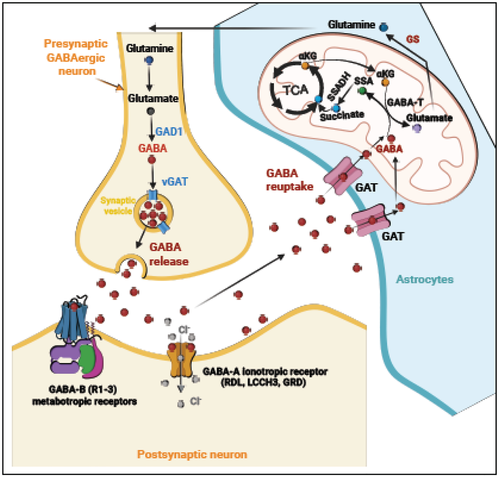
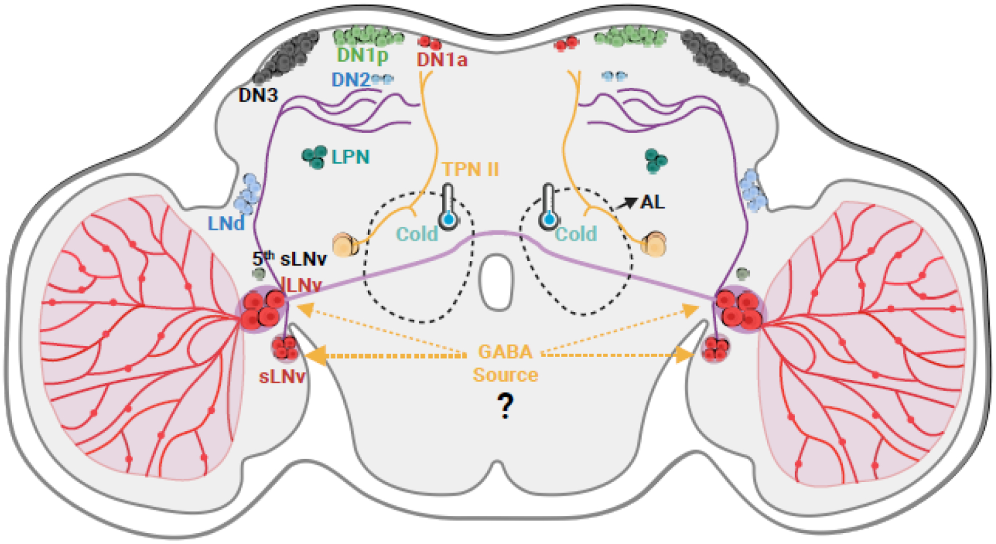
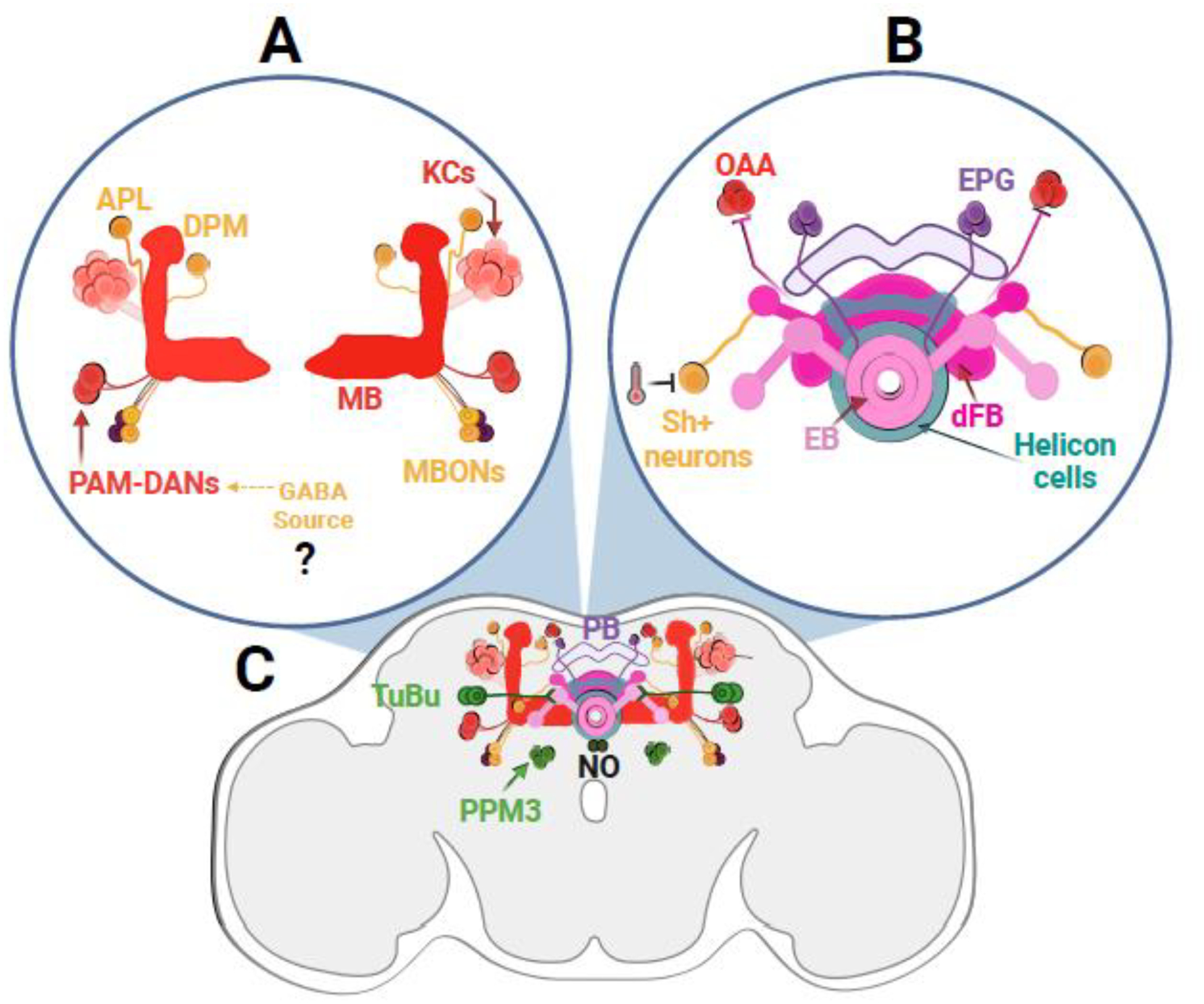
Sleep is conserved across the animal kingdom, and Drosophila melanogaster is a prime model to understand its intricate circadian and homeostatic control. GABA (gamma-aminobutyric acid), the brain's main inhibitory neurotransmitter, plays a central role in sleep. This review delves into GABA's complex mechanisms of actions within Drosophila's sleep-regulating neural networks. We discuss how GABA promotes sleep, both by inhibiting circadian arousal neurons and by being a key neurotransmitter in sleep homeostatic circuits. GABA's impact on sleep is modulated by glia through astrocytic GABA recapture and metabolism. Interestingly, GABA can be coexpressed with other neurotransmitters in sleep-regulating neurons, which likely contributes to context-based sleep plasticity.
Copyright © 2024 Elsevier Inc. All rights reserved.
Conflict of interest statement
Declaration of Competing Interest The authors declare that they have no known competing financial interests or personal relationships that could have appeared to influence the work reported in this paper.
Figures
Similar articles
-
Neuropeptide-Dependent Spike Time Precision and Plasticity in Circadian Output Neurons.Eur J Neurosci. 2025 Mar;61(5):e70037. doi: 10.1111/ejn.70037. Eur J Neurosci. 2025. PMID: 40080910 Free PMC article.
-
Wakefulness can be distinguished from general anesthesia and sleep in flies using a massive library of univariate time series analyses.PLoS Biol. 2025 Jul 10;23(7):e3003217. doi: 10.1371/journal.pbio.3003217. eCollection 2025 Jul. PLoS Biol. 2025. PMID: 40638578 Free PMC article.
-
Stress-induced Cdk5 activity enhances cytoprotective basal autophagy in Drosophila melanogaster by phosphorylating acinus at serine437.Elife. 2017 Dec 11;6:e30760. doi: 10.7554/eLife.30760. Elife. 2017. PMID: 29227247 Free PMC article.
-
The Lived Experience of Autistic Adults in Employment: A Systematic Search and Synthesis.Autism Adulthood. 2024 Dec 2;6(4):495-509. doi: 10.1089/aut.2022.0114. eCollection 2024 Dec. Autism Adulthood. 2024. PMID: 40018061 Review.
-
Developing a role for patients and the public in the implementation of health and social care research evidence into practice: the PIPER study (Pathways to Implementation for Public Engagement in Research) realist evaluation protocol.Res Involv Engagem. 2025 Jul 14;11(1):80. doi: 10.1186/s40900-025-00728-w. Res Involv Engagem. 2025. PMID: 40660365 Free PMC article. Review.
Cited by
-
The Extract of Piper nigrum Improves the Cognitive Impairment and Mood in Sleep-Deprived Mice Through the JAK1/STAT3 Signalling Pathway.Int J Mol Sci. 2025 Feb 21;26(5):1842. doi: 10.3390/ijms26051842. Int J Mol Sci. 2025. PMID: 40076470 Free PMC article.
-
Foods for Sleep Improvement: A Review of the Potential and Mechanisms Involved.Foods. 2025 Mar 21;14(7):1080. doi: 10.3390/foods14071080. Foods. 2025. PMID: 40238208 Free PMC article. Review.
References
-
- Anafi RC, Kayser MS, and Raizen DM, Exploring phylogeny to find the function of sleep. Nat Rev Neurosci, 2019. 20(2): p. 109–116. - PubMed
Publication types
MeSH terms
Substances
Grants and funding
LinkOut - more resources
Full Text Sources
Molecular Biology Databases
