Review
. 2024 Sep:199:106550.
doi: 10.1016/j.nbd.2024.106550. Epub 2024 Jun 6.

Brain development and bioenergetic changes

Affiliations
Review

Brain development and bioenergetic changes

Arjun Rajan et al. Neurobiol Dis. 2024 Sep.

Abstract

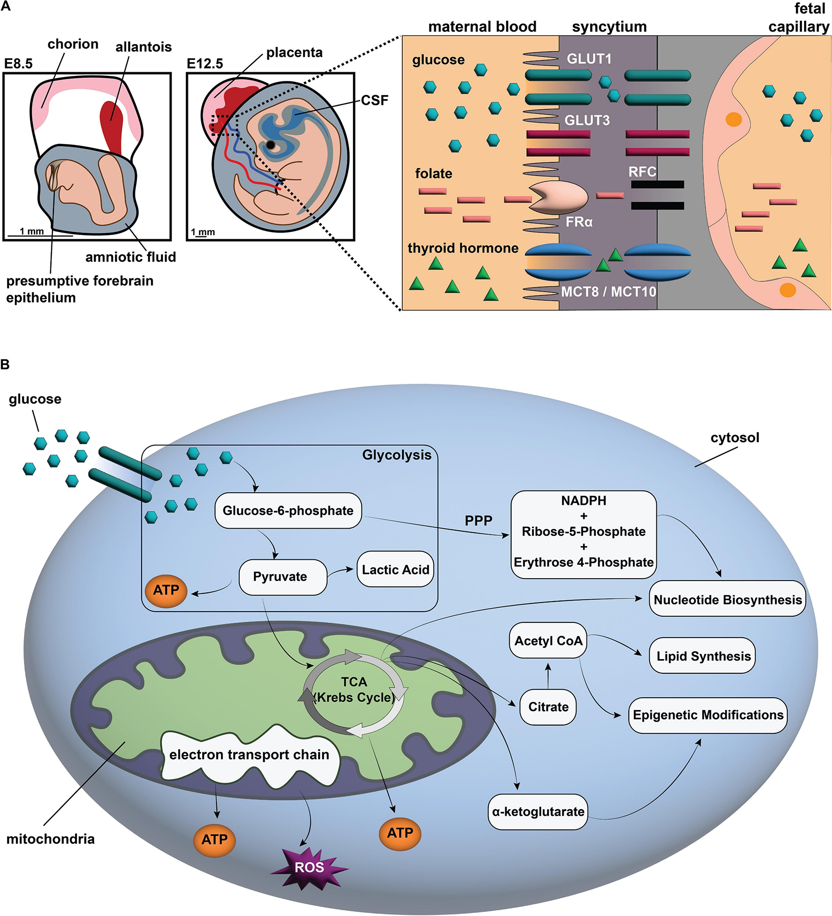
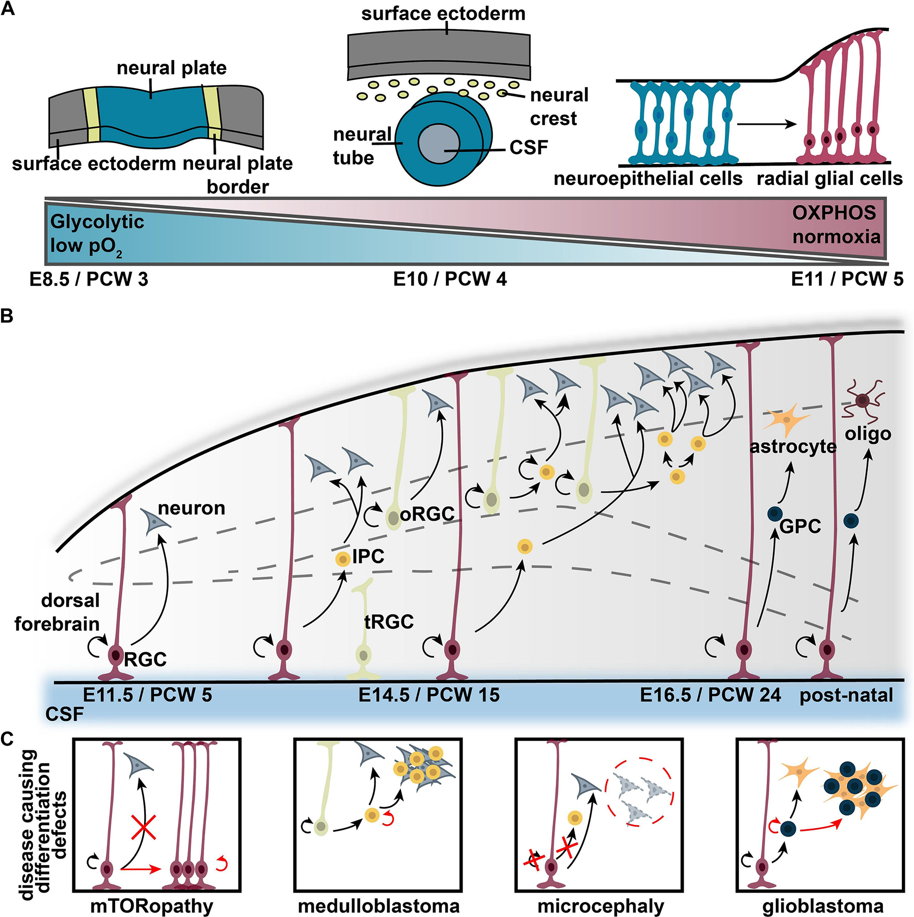
Bioenergetics describe the biochemical processes responsible for energy supply in organisms. When these changes become dysregulated in brain development, multiple neurodevelopmental diseases can occur, implicating bioenergetics as key regulators of neural development. Historically, the discovery of disease processes affecting individual stages of brain development has revealed critical roles that bioenergetics play in generating the nervous system. Bioenergetic-dependent neurodevelopmental disorders include neural tube closure defects, microcephaly, intellectual disability, autism spectrum disorders, epilepsy, mTORopathies, and oncogenic processes. Developmental timing and cell-type specificity of these changes determine the long-term effects of bioenergetic disease mechanisms on brain form and function. Here, we discuss key metabolic regulators of neural progenitor specification, neuronal differentiation (neurogenesis), and gliogenesis. In general, transitions between glycolysis and oxidative phosphorylation are regulated in early brain development and in oncogenesis, and reactive oxygen species (ROS) and mitochondrial maturity play key roles later in differentiation. We also discuss how bioenergetics interface with the developmental regulation of other key neural elements, including the cerebrospinal fluid brain environment. While questions remain about the interplay between bioenergetics and brain development, this review integrates the current state of known key intersections between these processes in health and disease.

Keywords: Bioenergetic pathways; Neural progenitors; Neurodevelopmental diseases.


Conflict of interest statement

Declaration of competing interest The authors declare no conflicts of interest.

Figures

Fig. 1.
Fig. 1.
Overview of metabolic processing in development. (A) Schematic of the developing embryo pre- and post-neural tube closure (NTC). Embryos become directly connected to the maternal blood supply at E11.5. The transport of maternal metabolic products occurs across the syncytium between maternal blood and the fetal capillaries. Maternal glucose is taken up into embryonic cells and converted into ATP and pyruvate in the cytoplasm. Embryonic neural cells increasingly use oxidative phosphorylation post-neural tube closure. The products of glycolysis are processed through the citric acid (TCA) cycle and the electron transport chain (ETC) to produce large amounts of ATP and other metabolic products critical for cellular processes. Glucose-6-phosphate can also be converted via the pentose phosphate pathway (PPP) to create nicotinamide adenine dinucleotide phosphate (NADPH) and other critical cellular products. (B) Glucose transporter 1 (GLUT1), Glucose transporter 3 (GLUT3), Folate Receptor Alpha (FRα), Reduced Folate Carrier (RFC), Thyroid Hormone Transporters MCT8/MCT10 (MCT8/MCT10).
Fig. 2.
Fig. 2.
Overview of neural development and disease. (A) Major structures during neurulation and generation of radial glia. During neurulation, neural progenitors switch from glycolytic to oxidative phosphorylation. (B) After radial glia are generated, neurogenesis begins with radial glial cells (RGCs) asymmetrically dividing to produce either neurons or intermediate progenitors (IPCs), which go on to produce neurons. Outer radial glial cells (oRGCs) that lack apical endfeet and truncated radial glial cells (tRGCs) that lack pial processes also generate neurons. Later, gliogenesis begins, and radial glia switch to producing glial progenitor cells (GPCs), which go on to generate astrocytes or glia. (C) Different neurodevelopmental diseases originate due to defects in self-renewal and differentiation during neurogenesis and gliogenesis. Post-conception week (PCW), embryonic day (E), ventricular zone (VZ), subventricular zone (SVZ), inner SVZ (iSVZ), outer SVZ (oSVZ), cortical plate (CP).

Similar articles

Cited by

References

    1. Adelman DM, Gertsenstein M, Nagy A, Simon MC, Maltepe E, 2000. Placental cell fates are regulated in vivo by HIF-mediated hypoxia responses. Genes Dev. 14, 3191–3203. - PMC - PubMed
    1. Adzick NS, Thom EA, Spong CY, Brock JW 3rd, Burrows PK, Johnson MP, Howell LJ, Farrell JA, Dabrowiak ME, Sutton LN, et al., 2011. A randomized trial of prenatal versus postnatal repair of myelomeningocele. N. Engl. J. Med 364, 993–1004. - PMC - PubMed
    1. Aleck KA, Kaplan AM, Sherwood WG, Robinson BH, 1988. In utero central nervous system damage in pyruvate dehydrogenase deficiency. Arch. Neurol 45, 987–989. - PubMed
    1. Alexander JF, Seua AV, Arroyo LD, Ray PR, Wangzhou A, Heibeta-Luckemann L, Schedlowski M, Price TJ, Kavelaars A, Heijnen CJ, 2021. Nasal administration of mitochondria reverses chemotherapy-induced cognitive deficits. Theranostics 11, 3109–3130. - PMC - PubMed
    1. Almeida A, Gonzalez-Buitrago JM, Bolanos JP, Medina JM, 1996. Fuel utilization by early newborn brain is preserved under congenital hypothyroidism in the rat. Pediatr. Res 40, 410–414. - PubMed

Publication types

LinkOut - more resources