. 2024 Jun 7;14(1):13134.
doi: 10.1038/s41598-024-63804-3.

Impact of in vitro SARS-CoV-2 infection on breast cancer cells

Affiliations

Impact of in vitro SARS-CoV-2 infection on breast cancer cells

Michele Sommariva et al. Sci Rep. .

Abstract

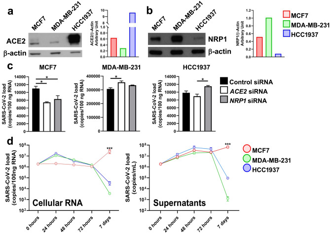
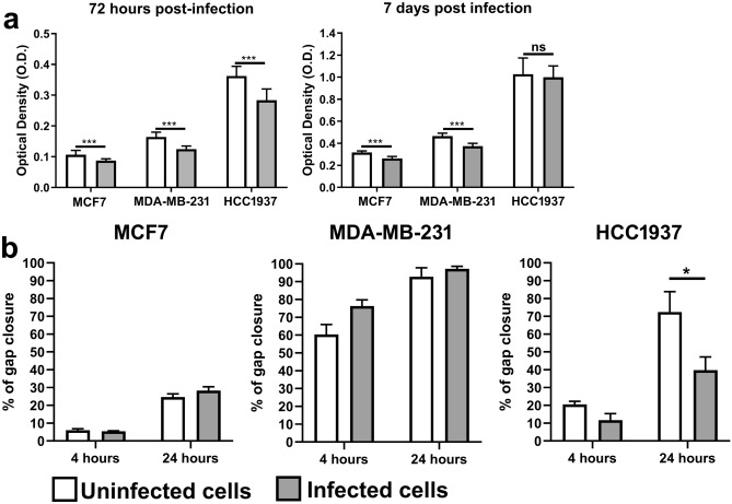
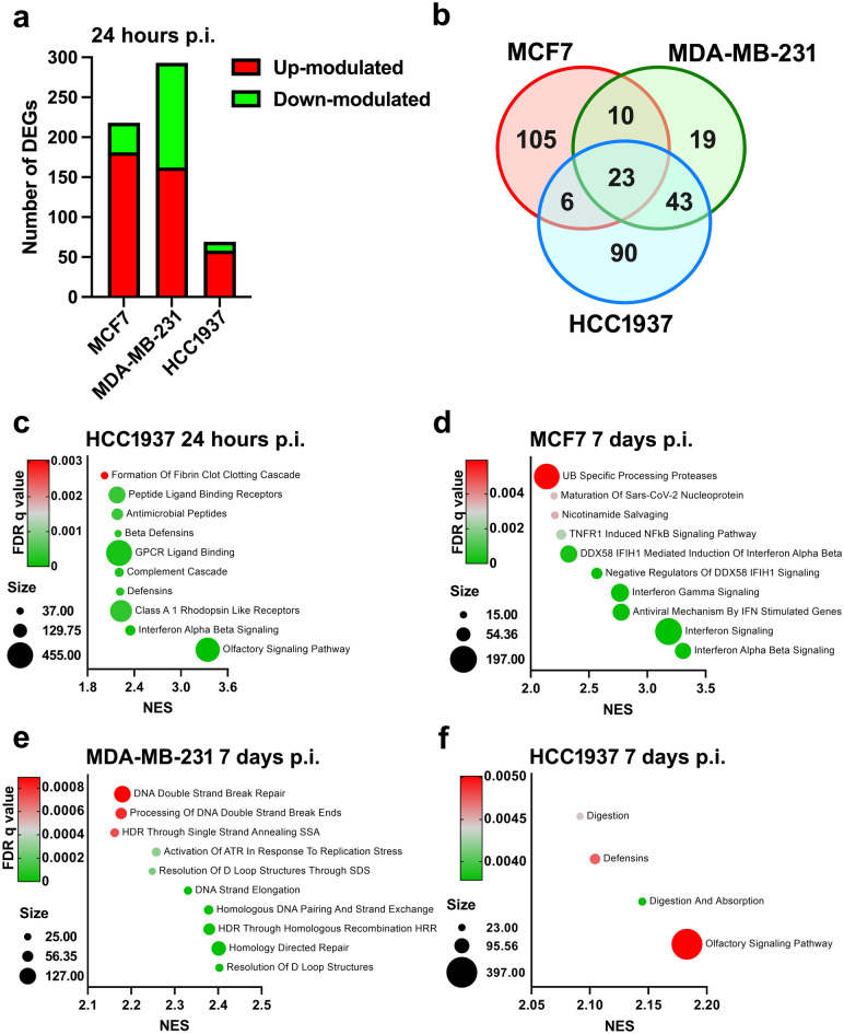
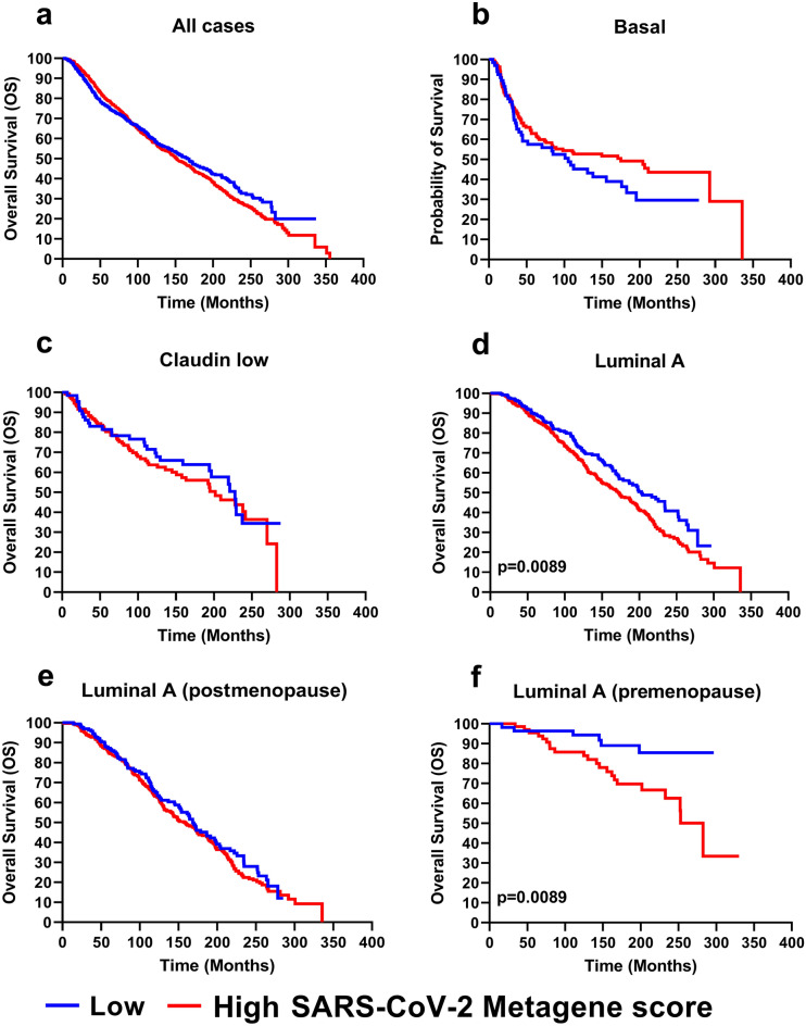
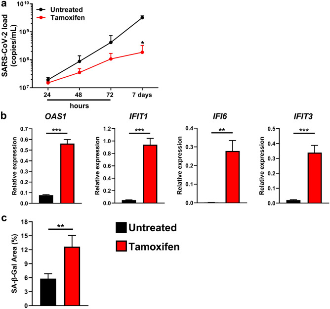
The pandemic of coronavirus disease 19 (COVID-19), caused by severe respiratory syndrome coronavirus 2 (SARS-CoV-2), had severe repercussions for breast cancer patients. Increasing evidence indicates that SARS-CoV-2 infection may directly impact breast cancer biology, but the effects of SARS-CoV-2 on breast tumor cells are still unknown. Here, we analyzed the molecular events occurring in the MCF7, MDA-MB-231 and HCC1937 breast cancer cell lines, representative of the luminal A, basal B/claudin-low and basal A subtypes, respectively, upon SARS-CoV-2 infection. Viral replication was monitored over time, and gene expression profiling was conducted. We found that MCF7 cells were the most permissive to viral replication. Treatment of MCF7 cells with Tamoxifen reduced the SARS-CoV-2 replication rate, suggesting an involvement of the estrogen receptor in sustaining virus replication in malignant cells. Interestingly, a metagene signature based on genes upregulated by SARS-CoV-2 infection in all three cell lines distinguished a subgroup of premenopausal luminal A breast cancer patients with a poor prognosis. As SARS-CoV-2 still spreads among the population, it is essential to understand the impact of SARS-CoV-2 infection on breast cancer, particularly in premenopausal patients diagnosed with the luminal A subtype, and to assess the long-term impact of COVID-19 on breast cancer outcomes.

Keywords: Breast cancer; Estrogen receptor; Luminal A breast cancer; SARS-CoV-2.


Conflict of interest statement

The authors declare no competing interests.

Figures

Figure 1
Figure 1
SARS-CoV-2 replication in breast cancer cell lines. Western blot and densitometric analysis of (a) ACE2 and (b) NRP1 protein expression in the MCF7, MDA-MB-231 and HCC1937 breast cancer cell lines. (c) ACE2 and NRP1 were silenced by siRNAs in the MCF7, MDA-MB-231 and HCC1937 breast cancer cell lines, which were subsequently infected with SARS-CoV-2 lineage B1. Viral RNA, expressed as copies/100 ng RNA, was quantified in the cytoplasm 6 h p.i. by real-time PCR with primers targeting the viral N1 gene. The data are presented as the means ± SEMs and are representative of one of three independent experiments with similar results. * p < 0.05 by one-way ANOVA followed by Tukey’s multiple comparison test with a single pooled variance. (d) MCF7, MDA-MB-231 and HCC1937 breast cancer cells were infected with SARS-CoV-2, and viral RNA was quantified at different time points in the cytoplasm and in the supernatants by real-time PCR. The viral load is expressed as copies/100 ng RNA or copies/mL for the cellular or supernatant RNA, respectively. The data are presented as the means ± SEMs and are representative of one of three independent experiments with similar results. *** p < 0.001 according to two-way ANOVA with multiple comparisons; refers to the comparison between MCF7 cells and MDA-MB-231 cells and the comparison between MCF7 cells and HCC1937 cells.
Figure 2
Figure 2
Effect of SARS-CoV-2 infection on breast cancer cell proliferation and motility. (a) MTT proliferation assay of MCF7, MDA-MB-231 and HCC1937 breast cancer cells after 72 h or 7 days of SARS-CoV-2 infection. Uninfected cells served as controls. The data are presented as the means ± SEMs and are representative of one of three independent experiments with similar results. ***p < 0.001 by unpaired two-tailed t test. (b) Confluent monolayers of MCF7, MDA-MB-231 and HCC1937 cells were infected with SARS-CoV-2. After 2 h of infection, the viral inoculum was removed, and gap wounds were generated by mechanical scratching. Cell migration into the wounded area was observed at 0, 4 and 24 h p.i. Histograms showing the extent of wound closure at the indicated time points are presented, with data shown as the percentages of wound closure at a specific time point compared to 0 h. The data are presented as the means ± SEMs and are representative of one of three independent experiments with similar results. *p < 0.05 by one-way ANOVA followed by Tukey’s multiple comparison test with a single pooled variance.
Figure 3
Figure 3
Effect of SARS-CoV-2 infection on breast cancer cell gene expression profiles. MCF7, MDA-MB-231 and HCC1937 breast cancer cells were infected with SARS-CoV-2 lineage B1, and gene expression profile analysis was performed at 24 h and 7 days p.i. (a) Number of DEGs (FDR < 0.05) identified by the comparison of infected and control cells of each cell line 24 h p.i. (b) Overlapping upregulated DEGs (FDR < 0.05) identified by the comparison of infected and control cells of each cellular model 24 h p.i. (c-e) Bubble plots of the top 10 Reactome pathways with an FDR q-value < 0.05 found to be enriched in infected cells versus uninfected cells as determined by preranked GSEA. (f) Bubble plot of the only 4 Reactome pathways with an FDR q-value < 0.05 found to be enriched in infected cells versus uninfected HCC1937 cells at 7 days p.i., as determined by preranked GSEA. The X-axis shows the normalized enrichment score (NES). The bubble area is proportional to the size of the gene set, and the bubble color represents the FDR q value.
Figure 4
Figure 4
Impact of the SARS-CoV-2 Metagene on breast cancer patient survival. Breast cancer patients represented in the METABRIC dataset were stratified as having a high (red line) or a low (blue line) SARS-CoV-2 Metagene score, with the lower tertile used as the cutoff. Kaplan–Meier curves of overall survival (OS) for all patients (a), the basal subgroup (b), the claudin-low subgroup (c), the luminal A subgroup (d) and the postmenopausal (e) and premenopausal (f) luminal A breast cancer subgroups in the METABRIC dataset. Differences between groups were estimated by the log-rank test.
Figure 5
Figure 5
Effect of Tamoxifen treatment on SARS-CoV-2 replication in MCF7 cells. SARS-CoV-2-infected MCF7 cells were exposed to 10 µM Tamoxifen 24 h before infection, at the time of infection, immediately post infection and throughout the experiment. (a) The viral load in the supernatants was evaluated at 24, 48, and 72 h and at 7 days p.i. by real-time PCR. The data, expressed as copies/mL, are presented as the means ± SEMs and are representative of one of three independent experiments with similar results. *p < 0.05 by two-way analysis of variance with multiple comparisons. (b) Real-time PCR analysis of OAS1, IFIT1, IFI6, and IFIT3 gene expression was performed on mRNA extracted from the samples collected at 7 days p.i. described in (a). The results are presented as 2−ΔCt values. ** p < 0.01, *** p < 0.001 by two-tailed unpaired Student’s t test. (c) MCF7 cells were infected and exposed to 10 µM Tamoxifen throughout the experiment. Seven days p.i., the cells were stained for determination of senescence-associated β-galactosidase (SA-β-Gal) activity. The histograms show the percentage of the SA-β-Gal-positive area evaluated in four randomly selected fields for each sample acquired with constant parameters. The data are presented as the means ± SEMs. **p < 0.01 by the Mann–Whitney U test.

Similar articles

References

    1. Nascimento BD, et al. Coronavirus disease (Covid-19) pandemic: an overview of systematic reviews. Bmc Infect. Dis. 2021;21:525. doi: 10.1186/s12879-021-06214-4. - DOI - PMC - PubMed
    1. Hu B, Guo H, Zhou P, Shi Z-L. Characteristics of Sars-Cov-2 and Covid-19. Nat. Rev. Microbiol. 2021;19:141–154. doi: 10.1038/s41579-020-00459-7. - DOI - PMC - PubMed
    1. Lamers MM, Haagmans BL. Sars-Cov-2 pathogenesis. Nat. Rev. Microbiol. 2022;20:270–284. doi: 10.1038/s41579-022-00713-0. - DOI - PubMed
    1. Amador M, et al. Impact of the Covid-19 pandemic on the care of cancer patients in Spain. Esmo Open. 2021;6:100157. doi: 10.1016/j.esmoop.2021.100157. - DOI - PMC - PubMed
    1. Sheng JY, et al. Management of breast cancer during the Covid-19 pandemic: a stage- and subtype-specific approach. Jco Oncol. Pract. 2020;16:665–674. doi: 10.1200/OP.20.00364. - DOI - PMC - PubMed