

Double-modified, thio and methylene ATP analogue facilitates wound healing in vitro and in vivo
- PMID: 38849425
- PMCID: PMC11161507
- DOI: 10.1038/s41598-024-63759-5
Double-modified, thio and methylene ATP analogue facilitates wound healing in vitro and in vivo
Abstract
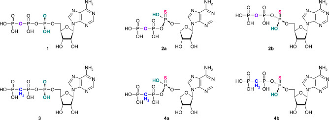
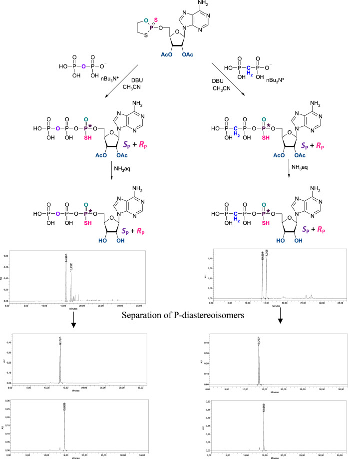
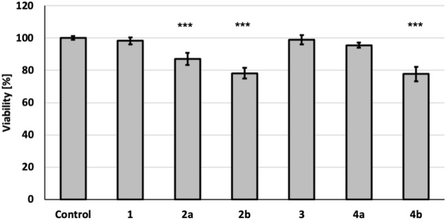
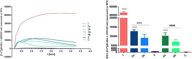
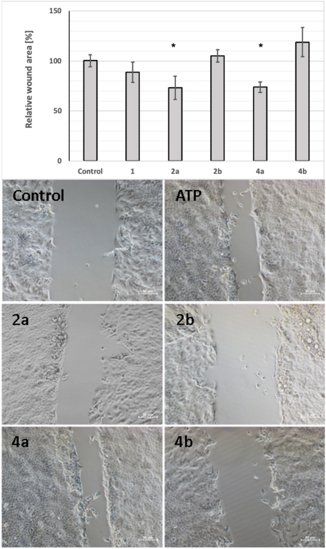
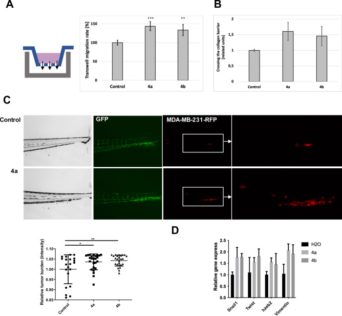
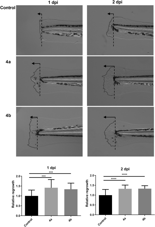
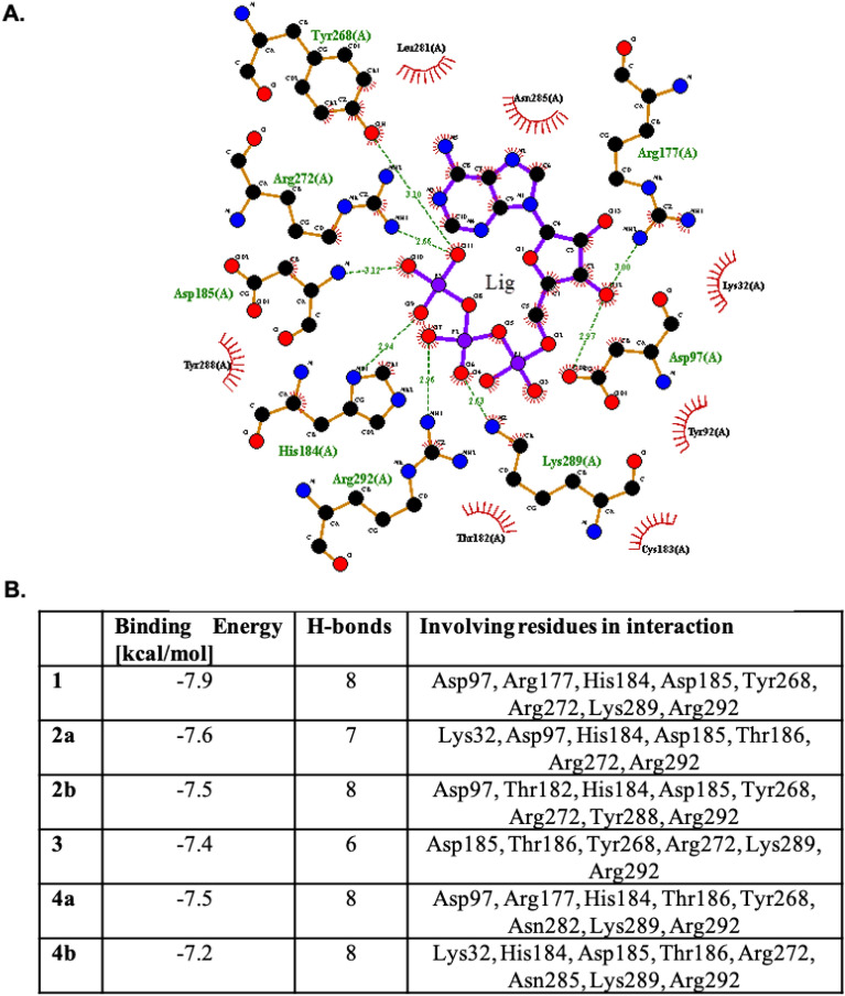
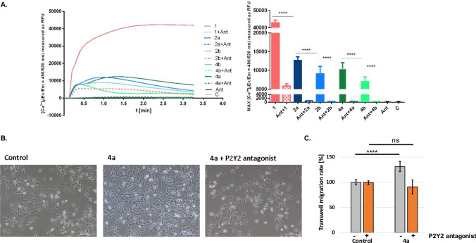
Recent data indicate that extracellular ATP affects wound healing efficacy via P2Y2-dependent signaling pathway. In the current work, we propose double-modified ATP analogue-alpha-thio-beta,gamma-methylene-ATP as a potential therapeutic agent for a skin regeneration. For the better understanding of structure-activity relationship, beside tested ATP analogues, the appropriate single-modified derivatives of target compound, such as alpha-thio-ATP and beta,gamma-methylene-ATP, were also tested in the context of their involvement in the activation of ATP-dependent purinergic signaling pathway via the P2Y2 receptor. The diastereomerically pure alpha-thio-modified-ATP derivatives were obtained using the oxathiaphospholane method as separate SP and RP diastereomers. Both the single- and double- modified ATP analogues were then tested for their impact on the viability and migration of human keratinocytes. The involvement of P2Y2-dependent purinergic signaling was analyzed in silico by molecular docking of the tested compounds to the P2Y2 receptor and experimentally by studying intracellular calcium mobilization in the human keratinocytes HaCaT. The effects obtained for ATP analogues were compared with the results for ATP as a natural P2Y2 agonist. To confirm the contribution of the P2Y2 receptor to the observed effects, the tests were also performed in the presence of the selective P2Y2 antagonist-AR-C118925XX. The ability of the alpha-thio-beta,gamma-methylene-ATP to influence cell migration was analyzed in vitro on the model HaCaT and MDA-MB-231 cells by wound healing assay and transwell migration test as well as in vivo using zebrafish system. The impact on tissue regeneration was estimated based on the regrowth rate of cut zebrafish tails. The in vitro and in vivo studies have shown that the SP-alpha-thio-beta,gamma-methylene-ATP analogue promotes regeneration-related processes, making it a suitable agent for enhance wound healing. Performed studies indicated its impact on the cell migration, induction of epithelial-mesenchymal transition and intracellular calcium mobilization. The enhanced regeneration of cut zebrafish tails confirmed the pro-regenerative activity of this ATP analogue. Based on the performed studies, the SP-alpha-thio-beta,gamma-methylene-ATP is proposed as a potential therapeutic agent for wound healing and skin regeneration treatment.
Keywords: ATP analogue; P2Y2 receptor; Purinergic signaling; Skin regeneration; Wound healing.
© 2024. The Author(s).
Conflict of interest statement
The authors declare no competing interests.
Figures
Similar articles
-
Autocrine regulation of wound healing by ATP release and P2Y2 receptor activation.Life Sci. 2021 Oct 15;283:119850. doi: 10.1016/j.lfs.2021.119850. Epub 2021 Jul 24. Life Sci. 2021. PMID: 34314735
-
P2Y2 R activation by nucleotides promotes skin wound-healing process.Exp Dermatol. 2014 Jul;23(7):480-5. doi: 10.1111/exd.12440. Exp Dermatol. 2014. PMID: 24816122
-
Diquafosol promotes corneal epithelial healing via intracellular calcium-mediated ERK activation.Exp Eye Res. 2016 Feb;143:89-97. doi: 10.1016/j.exer.2015.10.013. Epub 2015 Oct 24. Exp Eye Res. 2016. PMID: 26505315
-
Purinergic Signaling in Corneal Wound Healing: A Tale of 2 Receptors.J Ocul Pharmacol Ther. 2016 Oct;32(8):498-503. doi: 10.1089/jop.2016.0009. Epub 2016 Sep 19. J Ocul Pharmacol Ther. 2016. PMID: 27643999 Free PMC article. Review.
-
G protein coupled P2Y2 receptor as a regulatory molecule in cancer progression.Arch Biochem Biophys. 2024 Dec;762:110194. doi: 10.1016/j.abb.2024.110194. Epub 2024 Oct 30. Arch Biochem Biophys. 2024. PMID: 39486566 Review.
Cited by
-
Anticancer Activity of Enantiomeric Neplanocins A: Exploring the Role of Chirality in Tumor Suppression.Int J Mol Sci. 2025 Feb 4;26(3):1308. doi: 10.3390/ijms26031308. Int J Mol Sci. 2025. PMID: 39941076 Free PMC article.
References
MeSH terms
Substances
Grants and funding
LinkOut - more resources
Full Text Sources
Research Materials
Miscellaneous
