. 2024 Jun 7;14(1):13174.
doi: 10.1038/s41598-024-64022-7.

Fabrication of heart tubes from iPSC derived cardiomyocytes and human fibrinogen by rotating mold technology

Affiliations

Fabrication of heart tubes from iPSC derived cardiomyocytes and human fibrinogen by rotating mold technology

Birgit Andrée et al. Sci Rep. .

Abstract

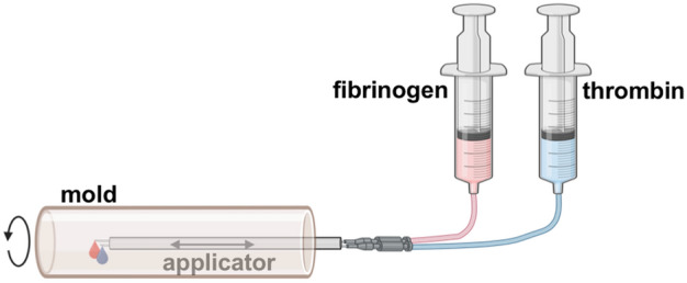
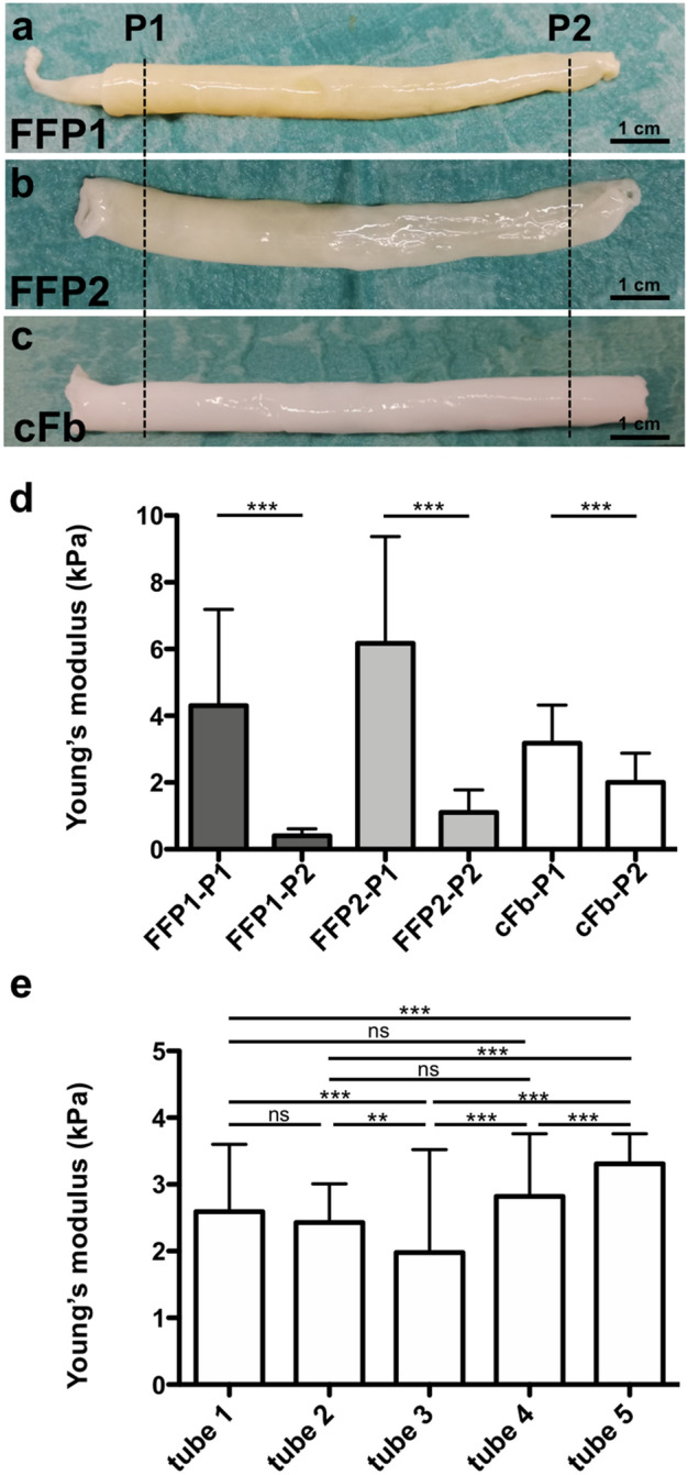
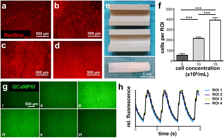
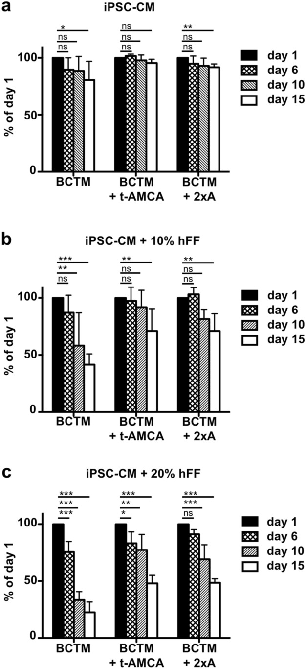
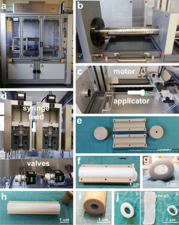
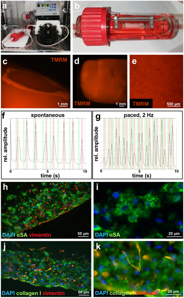
Due to its structural and functional complexity the heart imposes immense physical, physiological and electromechanical challenges on the engineering of a biological replacement. Therefore, to come closer to clinical translation, the development of a simpler biological assist device is requested. Here, we demonstrate the fabrication of tubular cardiac constructs with substantial dimensions of 6 cm in length and 11 mm in diameter by combining human induced pluripotent stem cell-derived cardiomyocytes (iPSC-CMs) and human foreskin fibroblast (hFFs) in human fibrin employing a rotating mold technology. By centrifugal forces employed in the process a cell-dense layer was generated enabling a timely functional coupling of iPSC-CMs demonstrated by a transgenic calcium sensor, rhythmic tissue contractions, and responsiveness to electrical pacing. Adjusting the degree of remodeling as a function of hFF-content and inhibition of fibrinolysis resulted in stable tissue integrity for up to 5 weeks. The rotating mold device developed in frame of this work enabled the production of tubes with clinically relevant dimensions of up to 10 cm in length and 22 mm in diameter which-in combination with advanced bioreactor technology for controlled production of functional iPSC-derivatives-paves the way towards the clinical translation of a biological cardiac assist device.


Conflict of interest statement

The authors declare no competing interests.

Figures

Figure 1
Figure 1
Schematic of the rotating mold technology. Two syringes containing fibrinogen solution and thrombin solution, respectively, are each connected to a drip line and a cannula. The cannulas are inserted into an applicator, which is reaching into a rotating mold and can move backward and forward. While moving, the two solutions are dispersed from the tip of the cannulas into the rotating mold. Upon contact of the two solutions the formation of fibrin fibers is initated. (Figure created with BioRender.com)
Figure 2
Figure 2
Commercially available fibrinogen improves uniformity of fibrin tubes. (a,b) Fibrin tubes fabricated with fibrinogen isolated from two independent batches of fresh frozen plasma (FFP), termed FFP1 and FFP2, respectively. (c) Tube produced from commercially available fibrinogen (cFb). (d) Young’s modulus of tubes shown in (ac). Young’s modulus of pieces from position 1 and 2 (P1, P2) was determined by nanoindentation (3 pieces per tube were assessed by a matrix scan of 100 indentations per piece). Mean value with SD is displayed. (e) Young’s modulus determined by nanoindentation of five independently fabricated tubes made from cFb. 2 pieces per tube were analyzed with > 100 measurement points per piece. Mean value with SD is displayed. ns = p > 0.05, ** = p < 0.01, *** = p < 0.001. FFP: fresh frozen plasma, cFb: commercially available fibrinogen. Scale bar: 1 cm.
Figure 3
Figure 3
High density of iPSC-CMs leads to synchronized calcium transients. (a–d) Fibrin tubes fabricated with increasing cell concentrations of Ruby-derived CMs and hFFs ((a) 5 × 106/mL, (b) 10 × 106/mL, (c) 15 × 106/mL, (d) 25 × 106/mL of total cell concentration with 90% CMs and 10% hFFs) after 3 days of cultivation in serum-free media (SFM). The indicated cell concentrations refer to the cell–matrix solution applied for the tissue generation process. (a–d) View onto the outer tube surface revealing nuclear RedStar fluorescence. (e) Mold for generating cell containing fibrin tubes. Length was reduced to 3 cm. Tube from (d) is shown below the mold directly after the fabrication. (f) Quantification of cells per region of interest (ROI) in dependence of initially applied cell concentration. Four ROIs with the same dimensions were analyzed per tube. Mean and SD is depicted.*** = p < 0.001 (g) Still images of GCaMP6f signal recording of the tube shown in (d) displaying a time interval of 0.25 s between pictures I to VI demonstrating GCaMP6f activity by GFP signal intensity at day 7 of cultivation. (h) Calcium fluctuations represented by GFP signal intensity were analyzed over time with ImageJ employing the recording shown in supplemental movie 1. Four regions of interest (ROI) were randomly selected. Scale bar: (ad,g) 500 µm, (e) 1 cm.
Figure 4
Figure 4
Rotation mold technology (RMT) facilitates CM coupling in contrast to static casting (SC) resulting in isolated CMs. (a,c,e,g,i) SC-tube. (b,d,f,h,j) RMT-tube. Tubes were fabricated employing the same cell concentration. (a) Macroscopic appearance of SC-tube. Left inset: 3D printed mold for static casting. Right inset: Lumen of SC-tube. (b) Macroscopic appearance of RMT-tube. Left inset: Machine for fabrication (copyright: Karin Kaiser/MHH). Right inset: Lumen of RMT-tube. (c, d) View onto the outer tube surface revealing nuclear RedStar fluorescence after 8 days of cultivation in SFM. (e, f) GCaMP6f peak fluorescence of Ruby CMs after 8 days of cultivation in SFM. Still images from movie. (g, h) Calcium fluctuations represented by GFP signal intensity were analyzed over time with ImageJ employing the recording shown in supplemental movie 2 and 3, respectively. Four regions of interest (ROI) were randomly selected. (i, j) Immunofluorescence of cross-sections stained for sarcomeric α-actinin (αSA, green), vimentin (red), and DAPI (blue). The dashed line represents the outer surface of the fibrin tube. (k) Quantification of nuclei per area for cross-sections from SC-tubes and RMT-tubes, respectively. Four regions of interest (ROI) were selected. For SC-tubes ROI were selected randomly, for RMT-tubes areas in the cell dense layers were selected. Mean and SD is depicted. *** = p < 0.001. Scale bar: (a,b) 1 cm for inset and main picture, (cf) 200 µm, (i,j) 100 µm.
Figure 5
Figure 5
Compaction of SC-tubes is medium and horse serum content dependent. SC-tubes were cut in rings and subjected to different medium compositions for 7 days. (a–c) BCT medium. (d–f) SF medium. (a,d) No horse serum (HS). (b,e) 6% HS. (c,f) 12% HS. (g–i) Immunofluorescence of cross-sections of rings in (d), (f), (c), respectively, stained for sarcomeric α-actinin (αSA, green), vimentin (red), and DAPI (blue). (j) Quantification of nuclei per area for cross-sections from of rings in (d), (f), (c), respectively. Four regions of interest (ROI) were selected in the cell containing regions. Mean and SD is depicted. * = p <0.05, *** = p < 0.001.Scale bar: (af) 1 mm, (gi) 100 µm.
Figure 6
Figure 6
Compaction of SC-tubes is influenced by hFF content and inhibitor addition. SC-tubes containing different amounts of hFFs, (a) no hFFss, (b) 10% hFFs, (c) 20% hFFs, were cut into rings and subjected to different inhibitors for 15 days (BCTM: 200 KIU/mL aprotinin; BCTM + t-AMCA: 200 KIU/mL aprotinin, 200 µM tranexamic acid (t-AMCA); BCTM + 2xA: 400 KIU/mL aprotinin). Brightfield pictures were taken and the cross-sectional area of the rings was measured in AxioVision SE64 Rel. 4.9 and expressed as percentage of the day 1 value. N = 5, Mean and SD is depicted. ns = p > 0.05, * = p < 0.05, ** = p < 0.01, *** = p < 0.001.
Figure 7
Figure 7
Automated machine for the production of fibrin tubes. (a) View of the machine. (b) Stainless steel pipe fitted on the motor for rotation. (c) Applicator equipped with 2 cannulas connected to drip lines. The motor for moving the applicator is located in the back. (d) Automated syringe feed with valves for closing and opening the lines. (e) PEEK mold for tube production. (f) Fibrin tube with a length of 10 cm and a diameter of 22 mm produced with the machine. Top view with one half shell removed. (g) Side view into the lumen of the tube in (f). (h) Fibrin tube produced in a mold with a length of 6 cm and an outer diameter of 11 mm. Top view with one half shell removed. (i) View into the lumen of a tube in the mold. (j) (I) Ring cut off from a tube produced with an outer sheath. (II) Same Ring as in (I) after the removal of the sheath which was cut open and placed on the left side of the ring. Scale bar: (fj) 1 cm.
Figure 8
Figure 8
Analysis of fibrin tubes fabricated with the new device and cultivated under pulsatile flow. (a) Bioreactor set-up for pulsatile flow. (b) Fibrin tube mounted into a bioreactor. (c) View into the lumen of the tube stained with TMRM. (d) Side view of a tube stained with TMRM. Four images were stitched together. (e) Picture with high magnification of the tube in (d). (f,g) Videos from contracting fibrin tubes stained with TMRM were recorded, imported into ImageJ and analyzed with the MYOCYTER macro to reveal the beating frequency. The amplitude is depicted in black, the maxima in red and the minima in green. (f) Analysis of spontaneous beating fibrin tube. (g) Analysis of tube from (f) paced at 2 Hz. (h–k) Immunofluorescence of cross-sections. (h,i) Staining for sarcomeric α-actinin (αSA, green), vimentin (red), and DAPI (blue). (j,k) Staining for collagen I (green), vimentin (red), and DAPI (blue). Scale bar: (c,d) 1 mm, (e) 500 µm, (h,j) 50 µm, (i,k) 20 µm.

Similar articles

Cited by

References

    1. Shadrin IY, et al. Cardiopatch platform enables maturation and scale-up of human pluripotent stem cell-derived engineered heart tissues. Nat. Commun. 2017;8:1825. doi: 10.1038/s41467-017-01946-x. - DOI - PMC - PubMed
    1. Tiburcy M, et al. Defined engineered human myocardium with advanced maturation for applications in heart failure modeling and repair. Circulation. 2017;135:1832–1847. doi: 10.1161/CIRCULATIONAHA.116.024145. - DOI - PMC - PubMed
    1. Jackman CP, Carlson AL, Bursac N. Dynamic culture yields engineered myocardium with near-adult functional output. Biomaterials. 2016;111:66–79. doi: 10.1016/j.biomaterials.2016.09.024. - DOI - PMC - PubMed
    1. Miyagawa S, et al. Case report: Transplantation of human induced pluripotent stem cell-derived cardiomyocyte patches for ischemic cardiomyopathy. Front. Cardiovasc. Med. 2022;9:950829. doi: 10.3389/fcvm.2022.950829. - DOI - PMC - PubMed
    1. Kawamura T, et al. Safety confirmation of induced pluripotent stem cell-derived cardiomyocyte patch transplantation for ischemic cardiomyopathy: First three case reports. Front. Cardiovasc. Med. 2023;10:1182209. doi: 10.3389/fcvm.2023.1182209. - DOI - PMC - PubMed