. 2024 Jul;131(2):372-386.
doi: 10.1038/s41416-024-02734-3. Epub 2024 Jun 7.

Synthetic retinoid-mediated preconditioning of cancer-associated fibroblasts and macrophages improves cancer response to immune checkpoint blockade

Affiliations

Synthetic retinoid-mediated preconditioning of cancer-associated fibroblasts and macrophages improves cancer response to immune checkpoint blockade

Takayuki Owaki et al. Br J Cancer. 2024 Jul.

Abstract

Background: The proliferation of cancer-associated fibroblasts (CAFs) hampers drug delivery and anti-tumor immunity, inducing tumor resistance to immune checkpoint blockade (ICB) therapy. However, it has remained a challenge to develop therapeutics that specifically target or modulate CAFs.

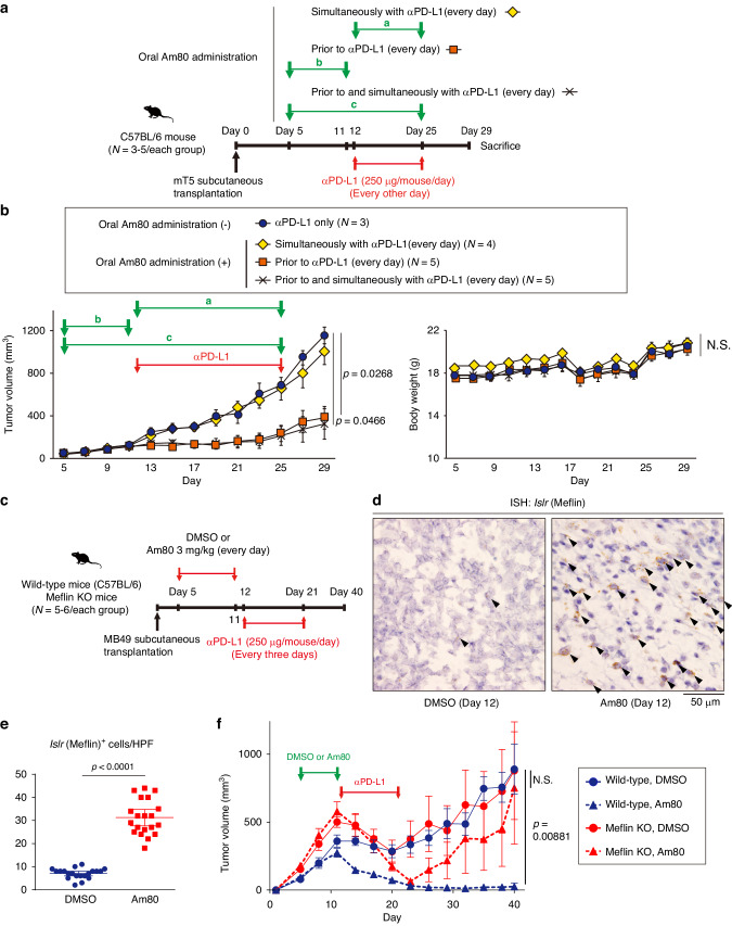
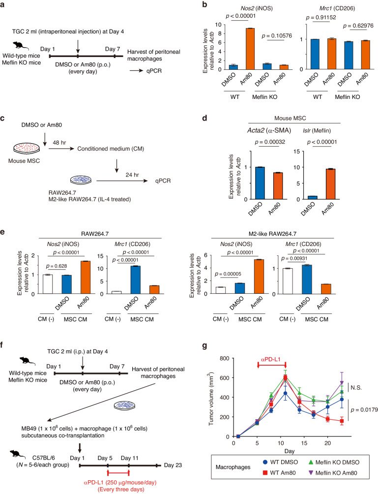
Methods: We investigated the involvement of Meflin+ cancer-restraining CAFs (rCAFs) in ICB efficacy in patients with clear cell renal cell carcinoma (ccRCC) and urothelial carcinoma (UC). We examined the effects of Am80 (a synthetic retinoid) administration on CAF phenotype, the tumor immune microenvironment, and ICB efficacy in cancer mouse models.

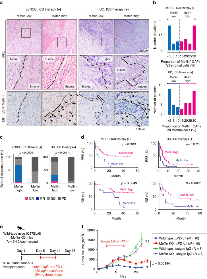
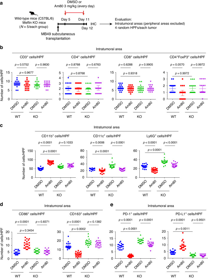
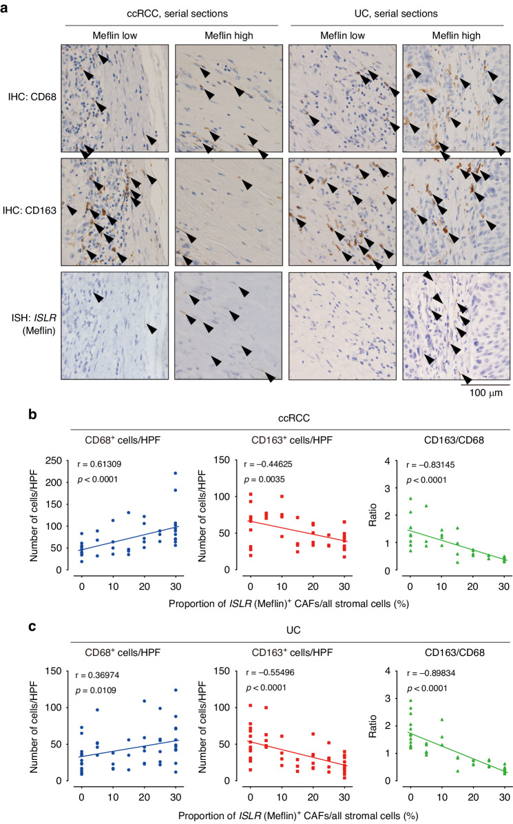
Results: High infiltration of Meflin+ CAFs correlated with ICB efficacy in patients with ccRCC and UC. Meflin+ CAF induction by Am80 administration improved ICB efficacy in the mouse models of cancer. Am80 exerted this effect when administered prior to, but not concomitant with, ICB therapy in wild-type but not Meflin-deficient mice. Am80-mediated induction of Meflin+ CAFs was associated with increases in antibody delivery and M1-like tumor-associated macrophage (TAM) infiltration. Finally, we showed the role of Chemerin produced from CAFs after Am80 administration in the induction of M1-like TAMs.

Conclusion: Our data suggested that Am80 administration prior to ICB therapy increases the number of Meflin+ rCAFs and ICB efficacy by inducing changes in TAM phenotype.


Conflict of interest statement

TM has financial interests in Denka Co., Ltd. The other authors declare no conflicts of interest.

Figures

Fig. 1
Fig. 1. Expression of Meflin in CAFs in ccRCC and UC and its significance to ICB therapy.
a Tissue sections obtained from human ccRCC and UC cases who underwent ICB therapy were hematoxylin and eosin stained (top panels), and stained for Meflin mRNA by ISH (lower panels). Boxed areas are magnified in the middle panels. Dotted lines indicate the boundaries between the tumor and the stroma. Arrowheads indicate Meflin+ CAFs. b Patients with ccRCC (top) or UC (bottom) treated with ICB therapy were stratified by the numbers of Meflin+ CAFs in the stromal tissue. c Objective response rate (ORR) of Meflin-high and -low ccRCC and UC cases who received ICB therapy. d PFS (top) and OS (bottom) of Meflin-high and -low ccRCC or UC patients treated with ICB therapy. e, f C57BL/6 wild-type and Meflin KO female mice were subcutaneously transplanted with MB49 cells (1 × 10⁶ cells/mouse) on Day 1, followed by intraperitoneal injection of anti-PD-L1 (αPD-L1) or isotype control IgG every three days from Day 5 to Day 14 (e). Tumor volume (f). The statistical methods used are a two-tailed unpaired t-test (c, f) and log-rank (Mantel–Cox) test (d). CAFs cancer-associated fibroblasts, ccRCC clear cell renal cell carcinoma, CR complete response, ICB immune checkpoint blockade, Ig immunoglobulin, ISH in situ hybridization, KO knockout, N.S. not significant, OS overall survival, PD progressive disease, PD-L1 programmed cell death ligand 1, PFS progression-free survival, PR partial response, SD stable disease, UC: urothelial carcinoma.
Fig. 2
Fig. 2. A priori oral Am80 administration enhances the efficacy of anti-PD-L1 antibody treatment in PDAC and UC mouse models.
a C57BL/6 wild-type female mice were subcutaneously transplanted with mT5 cells (1 × 10⁶ cells/mouse), followed by oral Am80 administration at the indicated periods and intraperitoneal injection of anti-PD-L1 antibodies. a, b, and c indicate the periods for oral Am80 administration. b Time courses of the volume of tumors developed in mice treated by the indicated regimens (left) and their body weights (right). c C57BL/6 wild-type or Meflin KO female mice were subcutaneously transplanted with MB49 cells (1 × 10⁶ cells/mouse), followed by oral administration of DMSO or Am80 prior to intraperitoneal injection of anti-PD-L1 antibodies. d, e Tissue sections obtained from the MB49 tumors on Day 12 were stained for the Islr gene by ISH (d), followed by quantification of Islr+ cells (e). Arrowheads in (d) indicate Islr+ cells. f Time courses of the volumes of tumors of the indicated groups. Differences between groups were analyzed using 1-way ANOVA with the Tukey test (b), and Student t-test (e, f). HPF high-power field (×40 objective lens), ICB immune checkpoint blockade, ISH in situ hybridization, KO knockout, NS not significant, PDAC pancreatic ductal adenocarcinoma, PD-L1 programmed cell death ligand 1, UC urothelial carcinoma.
Fig. 3
Fig. 3. Effects of Am80 administration on the TIME in the MB49 model.
a C57BL/6 wild-type or Meflin KO female mice were subcutaneously transplanted with MB49 cells (1 × 10⁶ cells/mouse), followed by oral administration of DMSO or Am80 and IHC for several immune cell markers. Four random HPFs from the intratumoral area for each tissue section were evaluated for quantification. be Cells positive for the indicated immune cell markers in each HPF were counted, followed by quantification. Twelve HPFs were evaluated for each group. The statistical methods used were 1-way ANOVA with the Tukey test (be). HPF high-power field (×40 objective lens), IHC immunohistochemistry, KO knockout, TIME tumor immune microenvironment.
Fig. 4
Fig. 4. Correlation between the number of Meflin+ CAFs and CD68+ macrophages in human ccRCC and UC.
a Serial sections prepared from surgically resected human ccRCC and UC tumors were stained for CD68 and CD163 by IHC and ISLR by ISH. Representative images of Meflin-low and -high cases are shown. Arrowheads denote cells positive for the indicated markers. b, c Number of cells positive for CD68 and CD163 in human ccRCC (b) and UC (c) tumor samples were counted and plotted against the proportion of Meflin+ CAFs in the stroma. The right panels show the correlation between the CD163/CD68 ratio and the number of Meflin+ CAFs. Quantitative interrelationships were determined using the Spearman’s rank-order correlation coefficient (b, c). CAFs cancer-associated fibroblasts, ccRCC clear cell renal cell carcinoma, IHC immunohistochemistry, ISH in situ hybridization, UC urothelial carcinoma.
Fig. 5
Fig. 5. Meflin expression in fibroblasts is required for Am80-mediated macrophage polarization and increases in efficacy of anti-PD-L1 antibody treatment.
a C57BL/6 wild-type or Meflin KO female mice were orally administered DMSO or Am80 every day from Day 1 to Day 7. The mice were subjected to intraperitoneal injection of 2 ml TGC (3%) on Day 4, followed by isolation of peritoneal macrophages on Day 7, and quantitative PCR (qPCR) for macrophage markers. b Expression levels of Nos2 and Mrc1 relative to Actb in the indicated macrophages as examined by qPCR. c Conditioned media (CM) from primary cultured mouse MSCs treated with either DMSO or Am80 (1 µM) for 48 h were added to control RAW264.7 cells or RAW264.7 cells that had been differentiated into M2-type macrophages using recombinant IL-4 (40 ng/ml, 48 h), followed by culture for 24 h and qPCR. d Total RNA extracted from MSCs treated with DMSO or Am80 (1 µM) for 48 h was examined for the expressions of Acta2 (left) and Islr (right) relative to Actb by qPCR. e Expression levels of Nos2 and Mrc1 relative to Actb in the indicated RAW264.7 cells as examined by qPCR. f, g Peritoneal macrophages (1 × 10⁶ cells/mouse), isolated from C57BL/6 wild-type or Meflin KO female mice that were administered DMSO or Am80 after intraperitoneal injection of 2 ml 3% TGC, were subcutaneously co-transplanted with MB49 cells (1 × 10⁶ cells/mouse) into wild-type mice on Day 1, followed by anti-PD-L1 therapy from Day 5 to Day 11 (f), and measurement of the volume of the developed tumors (g). Statistical differences were assessed with 1-way ANOVA with the Tukey test (b), Student t-test (d, g), and 1-way ANOVA with the Dunnett test (e). KO knockout, MSCs mesenchymal stem cells, NS not significant, qPCR quantitative polymerase chain reaction, PD-L1 programmed cell death ligand 1, TGC thioglycollate.
Fig. 6
Fig. 6. Expression of chemerin in Meflin+ CAFs and its role in response to anti-PD-L1 therapy in the MB49 model.
a Publicly available datasets of single-cell RNA transcriptomic analysis of human bladder UC (left) and PDAC (right) were analyzed for the expression of ISLR and RARRES2, which encode Meflin and chemerin, respectively. Shown are uniform manifold approximation and projection visualization of transcriptomes of all cells depicted by BBrowser. Arrows denote the fibroblast clusters that co-express ISLR and RARRES2. b, c Selective expression of Rarres2 in Meflin+ CAFs. Tumor sections from the MB49 model were double stained for Islr and Rarres2 by ISH (b). Boxed areas (a, b) are magnified in adjacent panels. Magenta and cyan arrowheads denote Islr and Rarres2 signals, respectively. The numbers of cells single- or double-positive for Islr and Rarres2 were counted and quantified (c). d Number of Rarres2+ cells detected in tissue sections prepared from MB49 tumors developed in wild-type and Meflin KO mice that were administered DMSO or Am80 (the experiment shown in Fig. 3a) were counted, followed by quantification. e, f C57BL/6 wild-type or Meflin KO female mice were subcutaneously transplanted with MB49 cells (1 × 10⁶ cells/mouse), followed by intratumoral injection of PBS or recombinant chemerin-9 (0.2 mg/kg) during the indicated periods, and intraperitoneal injection of anti-PD-L1 antibody (e). Tumor volumes of the developed tumors were measured over time (f). g Schematic diagram showing a working hypothesis for the mechanism of Am80-mediated tumor sensitization to ICB therapy. The mechanisms by which Meflin induces the expression of chemerin and how it is involved in the induction of M1-like macrophages remain unaddressed in the present study. Statistically significance was assessed using 1-way ANOVA with the Tukey test (d, f). HPF high-power field (×40 objective lens), ICB immune checkpoint blockade, ISH in situ hybridization, KO knockout, PDAC pancreatic ductal adenocarcinoma, PD-L1 programmed cell death ligand 1, UC urothelial carcinoma.

Similar articles

Cited by

References

    1. Biffi G, Tuveson DA. Diversity and biology of cancer-associated fibroblasts. Physiol Rev. 2021;101:147–76. 10.1152/physrev.00048.2019. 10.1152/physrev.00048.2019 - DOI - PMC - PubMed
    1. Kalluri R. The biology and function of fibroblasts in cancer. Nat Rev Cancer. 2016;16:582–98. 10.1038/nrc.2016.73. 10.1038/nrc.2016.73 - DOI - PubMed
    1. Chen Y, McAndrews KM, Kalluri R. Clinical and therapeutic relevance of cancer-associated fibroblasts. Nat Rev Clin Oncol. 2021;18:792–804. 10.1038/s41571-021-00546-5. 10.1038/s41571-021-00546-5 - DOI - PMC - PubMed
    1. Helms E, Onate MK, Sherman MH. Fibroblast heterogeneity in the pancreatic tumor microenvironment. Cancer Discov. 2020;10:648–56. 10.1158/2159-8290.CD-19-1353. 10.1158/2159-8290.CD-19-1353 - DOI - PMC - PubMed
    1. Barrett RL, Puré E. Cancer-associated fibroblasts and their influence on tumor immunity and immunotherapy. Elife. 2020;9:e57243 10.7554/eLife.57243. 10.7554/eLife.57243 - DOI - PMC - PubMed

MeSH terms

LinkOut - more resources