. 2024 Aug;27(8):1545-1554.
doi: 10.1038/s41593-024-01666-8. Epub 2024 Jun 7.

Developmental origin of oligodendrocytes determines their function in the adult brain

Affiliations

Developmental origin of oligodendrocytes determines their function in the adult brain

Sarah Foerster et al. Nat Neurosci. 2024 Aug.

Abstract

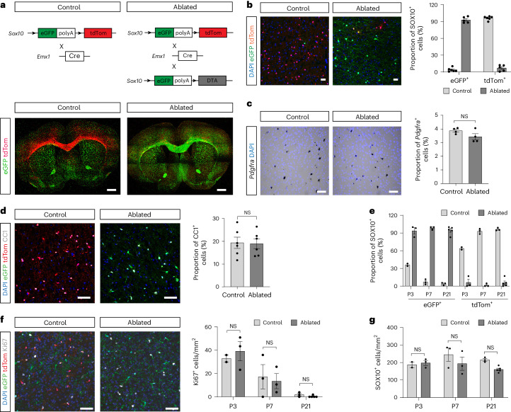
In the mouse embryonic forebrain, developmentally distinct oligodendrocyte progenitor cell populations and their progeny, oligodendrocytes, emerge from three distinct regions in a spatiotemporal gradient from ventral to dorsal. However, the functional importance of this oligodendrocyte developmental heterogeneity is unknown. Using a genetic strategy to ablate dorsally derived oligodendrocyte lineage cells (OLCs), we show here that the areas in which dorsally derived OLCs normally reside in the adult central nervous system become populated and myelinated by OLCs of ventral origin. These ectopic oligodendrocytes (eOLs) have a distinctive gene expression profile as well as subtle myelination abnormalities. The failure of eOLs to fully assume the role of the original dorsally derived cells results in locomotor and cognitive deficits in the adult animal. This study reveals the importance of developmental heterogeneity within the oligodendrocyte lineage and its importance for homeostatic brain function.


Conflict of interest statement

E.M.F. is currently employed as a Senior Editor for Nature Neuroscience. She was not involved in the decision-making process for this paper and did not have access to confidential information pertaining to the peer review and editorial process. The other authors declare no competing interests.

Figures

Fig. 1
Fig. 1. Genetic ablation of dOPCs can be fully restored by cells from ventral origin.
a, Schematic strategy for genetic fate mapping and ablation of dOLCs. Low-power images showing distribution of dorsally derived tdTom+ and ventrally derived eGFP+ cells in control (left) and ablated (right) adult mouse forebrain (images were tiled). b, Images and quantification of SOX10+ OLCs of both dorsal and ventral origin in cerebral cortex from control and ablated adult animals showing the extent of ablation of dOLCs and replacement by vOLCs. P90: control: n = 6; ablated: n = 5. c,d, OLC ablation does not alter numbers of Pdgfra+ OPCs (c) and mature CC1+ OLs (d) in adult cortex (all layers). P90: n = 4, P = 0.904 (c); n = 6, P = 0.126 (d), unpaired t-test (two-sided). e, Relative proportions of SOX10+ OLCs that are tdTom+ or eGFP+ in control and ablated animals. P3, P7 and P21: P3: control: n = 2, ablated: n = 3; P7: n = 3; P21: control: n = 3, ablated: n = 5. f, Images and quantification of Ki67+ proliferating cells in cerebral cortex. P3, P7 and P21: n = 3, except P3 control: n = 2 and P21 ablated: n = 5, P > 0.05, two-way ANOVA. g, Ablation does not result in changes to the overall densities of SOX10+ OLCs during developmental myelination. P3, P7 and P21: n = 3, except P3 control: n = 2, P > 0.05, two-way ANOVA. All data are shown as mean ± s.e.m. Scale bars, 50 μm. NS, not significant.
Fig. 2
Fig. 2. Myelination by eOLC-derived OLs exhibits a minor spatially and temporally restricted reduction in sheath thickness.
a, Quantitative analysis of G-ratio of myelinated axons in the CC of control and ablated mice. Electron microscopy images are of similar areas of CC at P13. P13, P21 and P40, n = biological replicates, N = number of axons. P13: control: n = 5, N = 498, ablated: n = 3, N = 330, P = 0.0419; P21: control: n = 4, N = 560, ablated, n = 3, N = 342, P < 0.0001; P40: control: n = 4, N = 553, ablated: n = 3, N = 421, P = 0.1315. Mann–Whitney test. b, G-ratios of axons in motor cortex (MC) layers 5–6. P21 and P40. P21: control: n = 3, N = 91, ablated: n = 3, N = 92, P = 0.195; P40: control: n = 4, N = 152, ablated: n = 3, N = 139, P < 0.0001. Mann–Whitney test. c, G-ratios in MC layers 5–6 in mice. P90: control: n = 4, N = 385, ablated: n = 3, N = 195, P < 0.0001. Mann–Whitney test. d, Immunogold electron microscopy on cryosections for eGFP and tdTom in control and ablated mice. Arrowheads, myelinated axon; it, inner tongue; ot, outer tongue; orange, OL. P90: n = 1. e, MBP and NF staining in the MC layers 5–6 in control and ablated animals. P90: n = 6, P = 0.0741, unpaired t-test (two-sided). f, SCoRe microscopy of the cerebral cortex layer 1 in control and ablated mice. Arrowhead = node of Ranvier. P90: for myelin fiber lengths and volume fraction: control: n = 6; ablated: n = 5, P = 0.537 and P = 0.823, respectively, for average internode lengths: n = biological replicates, N = number of internodes; control: n = 6, N = 294, ablated: n = 5, N = 248, P = 0.037, Mann–Whitney test. g, Super-resolution microscopy images of CASPR1+ paranodes of myelinated axons in the cortex in control and ablated animals. P90: n = biological replicates, N = number of nodes and paranodes; nodes: n = 3, control: N = 208, ablated: N = 171, P = 0.139, unpaired t-test (two-sided); paranodes: n = 3, control: N = 463, ablated: N = 406, P < 0.0001, unpaired t-test (two-sided). h, Quantification of Ankyrin G+ axon initial segments (AIS) length in the pyramidal neurons in the MC in control and ablated mice. P90: n = biological replicates, N = number of AIS, control: n = 3, N = 130, ablated: n = 3, N = 137, P = 0.624, Mann–Whitney test. The graphs in eh show mean ± s.e.m. and are overlayed with individual data points. *P < 0.05; ****P < 0.001. Scale bars, 2 μm in a and c; 50 μm in e; 1 μm in d; 2 μm in g; and 20 μm in f and h. NS, not significant.
Fig. 3
Fig. 3. Ablation of dOPCs causes behavioral deficits.
a, Balancing beam test: (i) beam traverse time: control: n = 42, ablated: n = 18, P = < 0.01, Mann–Whitney test; (ii) number of hindlimb slips per traverse: control: n = 40, ablated: n = 17, P = 0.05, Kolmogorov–Smirnov test. b, Gait analysis: control: n = 30, ablated: n = 11. (i) Average hind stride length: P = 0.71, unpaired t-test (two-sided); (ii) average hind stride width: P = 0.01, Mann–Whitney test. c, Vertical beam test: number of hindlimb steps: control: n = 10, ablated: n = 5, P = < 0.0001, Mann–Whitney test. d, rCPT: simulation duration is the time each stimulus (target or non-target) was shown to each mouse. (i) HR: mixed-effect model: main effect of genotype, P = 0.037; main effect of duration, P < 0.0001; genotype by duration interaction, P = 0.005; simple main effects of genotype at 2.0 s, 1.0 s and 0.5 s, P < 0.05. (ii) FAR: mixed-effect model: main effect of genotype, P = 0.023; main effect of duration, P < 0.0001; genotype by duration interaction, P = 0.003; simple main effects of genotype at each duration condition, P < 0.05. (iii) Criterion: mixed-effect model: main effect of genotype, P = 0.025; main effect of duration, P < 0.0001; genotype by duration interaction, P = 0.775; simple main effects of genotype at each duration condition, P < 0.05. (i–iii) Control: n = 27, ablated: n = 11. (iv) Response latency to ‘target’ stimulus: mixed-effect model: main effect of genotype, P = 0.033; main effect of session, P = 0.017; genotype by session interaction, P = 0.158. (v) Reward collection latency: mixed-effect model: main effect of genotype, P = 0.554; main effect of session, P = 0.340; genotype by session interaction, P = 0.862. (iv,v) Control: n = 27, ablated: n = 11. All data are mean ± s.e.m. Two cohorts of animals were used for the locomotor and cognitive testing, respectively. All locomotor tests were performed consecutively. Training of both cohorts started at P90. NS, not significant.
Fig. 4
Fig. 4. Ablation of dOLCs does not lead to significant alteration in composition of other cell types in the neocortex.
a, Density of OLIG2+ OLCs in CC and cerebral cortex of control and ablated animals. P0: n = 5, P = 0.735, unpaired t-test (two-sided). b, Densities of IBA1+ microglia in cerebral cortex of control and ablated animals. P0: n = 5; P7: control: n = 4, ablated: n = 3; P21: control: n = 3, ablated: n = 5, P > 0.05, unpaired t-test (two-sided). c, Densities of IBA1+ microglia and CD68+ macrophages in cerebral cortex of control and ablated animals. P90: n = 3, P = 0.5864, unpaired t-test (two-sided). d, Densities of GFAP+ astrocytes in the CC of control and ablated animals. P90: control: n = 6, ablated: n = 5, P = 0.399, unpaired t-test. Data are shown as individual values and mean ± s.e.m. and compared by unpaired t-test, P > 0.05 in all analysis. Scale bars, 50 μm in a and b and 100 μm in c and d. eg, snRNA-seq of motor cortex tissue dissected from control (n = 2) and ablated (n = 2) animals at P0 (equivalent to E19) (N = 11,052 nuclei). e,f, UMAPs depicting annotated clusters derived from control and ablated samples. g, MELD analysis showed no major transcriptomic changes upon ablation in any of the annotated clusters, apart from moderate differences detected within the population of projecting inhibitory neurons. NS, not significant; UMAP, uniform manifold approximation and projection.
Fig. 5
Fig. 5. Ablation of dOPCs alters the transcriptional profiles of OL subpopulations.
scRNA-seq of SOX10+ OLCs in the neocortex of adult control (n = 4) and ablated (n = 2) mice (P90). a, UMAP of all recovered cell clusters, sorted by cell population (top) or origin of cells (bottom) (N = 9,291 cells). COP, committed oligodendrocyte precursor; MFOL, myelin-forming oligodendrocyte; NFOL, newly formed oligodendrocyte. b, Frequency distribution analysis of cells in each cluster shown in a according to cell origin. c, Differential gene expression analysis of control versus ectopic OLs in cluster MOL5/6 shown in Extended Data Fig. 8a. Wilcoxon rank-sum test. d, Reactome pathway analysis of control versus ectopic OLs in cluster MOL5/6 shown in Extended Data Fig. 8a. e, Distribution of ablated-relative likelihood in each cluster shown in a. Dots in red and light red correspond to ablated-like cells, and blue and light blue indicate control-like cells. The gray circles represent the mean ablated-relative likelihood for each cluster. f, Ablated-relative likelihood highlighting ablation-unique transcriptional signatures within MOL5/6a (top) and MOL5/6d (bottom) populations. Left section of each panel represents cluster dissociation in two populations with unperturbed cells as Population1 and perturbated cells as Population2. Control-derived cells are labeled in blue and ablated cells in red. Right sections of each panel indicate expressions of gene markers for control-like population (Pop1) and ablated-like population (Pop2), as Tubb3 and Eya2, respectively, with a maximum cutoff set at 3 for the square root transformed counts.
Extended Data Fig. 1
Extended Data Fig. 1. Efficient ablation of dorsal oligodendrocyte lineage cells in other regions of the forebrain.
Bar graphs show percentage of oligodendrocyte lineage cells (SOX10 + ) of dorsal (tdTom) or ventral (eGFP) origin in the control and ablated mice in corpus callosum (a) or striatum (b). P90; control: n = 3, ablated: n = 4. Data are represented as mean ± S.E.M.
Extended Data Fig. 2
Extended Data Fig. 2. Ablated mice do not show deficits in any other locomotor task and show no signs of anxiety, aberrant muscle tonus or sensation deficits.
(a) Accelerating rotarod: Rod rotation speed at which mice fell off the rod. N = 3; n = 12 (control) and n = 6 (ablated) mice/condition, p-value = <0.17 unpaired t-test (two-sided). (b) Horizontal ladder: (i) Time mice needed to cross the ladder. N = 3; control: n = 30, ablated: n = 11 mice/condition, p-value = <0.01, Mann-Whitney test; (ii) Number of gait abnormalities (including misses, slip offs) per run. N = 3; control: n = 29, ablated: n = 12 mice/condition, p-value = 0.83, unpaired t-test (two-sided). (c) Open field test: Time a mouse spent at the wall of the test chamber. N = 1; control: n = 30, ablated: n = 12 mice/condition, p-value = 0.22, Mann-Whitney test. (d) Rearing test: Number of rearings per test. N = 1; control: n = 30, ablated: n = 12 mice/condition, p-value = 0.15, unpaired t-test (two-sided). (e) Hanging bar test: Score indicates the number of seconds each mouse holds on to the bar. A higher score indicates a better performance. N = 3; control: n = 12, ablated: n = 6 mice/condition, p-value = 0.06, Mann-Whitney test. (f) Von-Frey test: Force needed to be applied to hind paw before paw retraction. N = 5; control: n = 30, ablated: n = 12 mice/condition, p-value = 0.92, unpaired t-test (two-sided). All locomotor tests were performed consecutively. Training of the mice was started at P90. Data are represented as mean ± S.E.M.
Extended Data Fig. 3
Extended Data Fig. 3. Very few dorsally-derived oligodendrocyte lineage cells are detected in cerebellum and spinal cord.
Distribution and quantification of SOX10+ oligodendrocyte lineage cells of dorsal (tdTom) or ventral (eGFP) origin, in cerebellum (a) and spinal cord white matter (b). P90; (a) n = 4, (b) n = 3. Data are represented as mean ± S.E.M. Scale bars represent 100μm.
Extended Data Fig. 4
Extended Data Fig. 4. Continuous performance task (rCPT) did not reveal altered attentive behavior in ablated adult mice at baseline and ablation of dOPCs does not induce deficit in visual discrimination, reversal learning and extinction learning.
(a) rCPT at baseline (for parameters see methods): N = 1; control: n = 26, ablated: n = 11 mice/condition. (i) Hit rate at baseline. p-value = 0.15, unpaired t-test (two-sided). (ii) False alarm rate at baseline. p-value = 0.08 Mann-Whitney test. (iii) D-Prime at baseline. p-value = 0.68, unpaired t-test (two-sided). (iv) Criterion at baseline. p-value = 0.11, unpaired t-test (two-sided). (b) D-Prime with shortened SD (for parameters see methods). Mixed-effect model: main effect of genotype, p = 0.459; main effect of duration, p < 0.0001; genotype by duration interaction, p = 0.551. (c) Visual discrimination test: Mice learn to discriminate one ‘target’ from one ‘non-target’ stimulus. Session to criterion describes the number of training sessions needed to successfully discriminate the two visual stimuli. N = 1; control: n = 27, ablated: n = 10 mice/condition; control: n = 5, ablated: n = 1 mice/condition did not reach the criterion. p-value = 0.60, unpaired t-test (two-sided). (d,e) Reversal learning test: Target stimulus becomes non-target stimulus in session 2. (d) Percentage of correct responses to new target stimulus. N = 1; control: n = 22, ablated: n = 9 mice/condition. Mixed-effect model: main effect of genotype, p = 0.490; main effect of duration, p < 0.0001; genotype by duration interaction, p = 0.562. (e) Number of correction trials due to a response to a non-target stimulus. N = 1; control: n = 26, ablated: n = 11 mice/condition. Mixed-effect model: main effect of genotype, p = 0.186; main effect of duration, p < 0.0001; genotype by duration interaction, p = 0.350. (f) Extinction learning test: Response to target stimulus is not rewarded. Percentage of responses to target stimulus per session. N = 1; control: n = 26, ablated: n = 10 mice/condition. Mixed-effect model: main effect of genotype, p = 0.723; main effect of duration, p < 0.0001; genotype by duration interaction, p = 0.121. All data are presented as mean ± S.E.M. Training of the mice was started at P90.
Extended Data Fig. 5
Extended Data Fig. 5. Astrocyte are unchanged in ablated animals.
(a-c) Whole-brain western blot analysis of GFAP and glutamine synthetase (GS). P90; control: n = 6, ablated: n = 3, GFAP: p = 0.472, GS: p = 0.388, unpaired t-test (two-sided) with Welch’s test. The full Western Blot image is unavailable.
Extended Data Fig. 6
Extended Data Fig. 6. Single nuclear sequencing did not reveal any major bystander effect on brain cells after dOLC ablation.
(a) Uniform Manifold Approximation and Projection (UMAP) of recovered cells with unique barcoded antibody colored by antibody. Cortical tissue, P0. (b) Top 15 Highly Variable Genes (HVGs) for each cell type from Fig. 4e, calculated with Wilcoxon rank sum test, p-value adjusted < 0.01 and log2FC >= 0.25, ranked by log2FC. (c) Representative percentage of each cell typecluster in each condition divided by the number of conditions. Bar plots in red and blue correspond to populations recovered from ablated and control samples, respectively. Black dots represent unique antibody-hashed replicates. n = 2. (d) scCODA reveals no credible compositional variation from ablated animals.Population of astrocytes was selected as reference for parameters inference. Control condition is indicated in blue, and ablated condition in red. n = 2. In the box plot, the top and bottom whiskers define the maximum and minimum, respectively. The top bound of the box defines the 75ste percentile, the centre line defines the 50ste percentile and the bottom bound of the box defines the 25ste percentile.
Extended Data Fig. 7
Extended Data Fig. 7. RNAscope analysis did not reveal altered neuronal population in ablated animals.
(a) Schematic illustration of coronal view of mouse brain for main cortical regions at P90: anterior cingulate cortex (ACC), motor cortex (MC) and somatosensory cortex (SSC). The red line in the sagittal view indicates the level of the sections analyzed. (b) Low magnification images of coronal section of MC and ACC regions (red box in (a)) co-stained for the pan-neuronal marker Syt1, inhibitory neuron marker Gad2 and neuronal layer markers Cux2, Ctip2 and Foxp2 with RNAscope smFISH. The nuclei are counterstained with DAPI. Scale bar: 200 µm, inset: 20 µm. (c-e) Quantification of proportion of Gad2- excitatory (c), Gad2+ inhibitory (d) and Ebf1+ projection inhibitory neurons (e) co-expressing neuron-specific Syt1 in MC and SSC. No differences were detected between control and dorsally ablated animals. P0; MC: n = 4, SSC: n = 5, p > 0.05, two-way ANOVA. (f-h) In adult brain, there is no difference in the density of Syt1+ (f) or the proportion of Gad2- excitatory (g) and Gad2+ inhibitory (h) neurons between control and ablated animals across the layers of the cerebral cortex, which is shown for motor cortex. P90; control: n = 5, ablated: n = 5, p > 0.05, two-way ANOVA. (i-k) The proportion of Ctip2+ (i), Fezf2+ (j) and Crym+ (k) corticospinal motor neurons co-expressing NeuN in layers 5 and 6 of the adult motor cortex were also unchanged between control and ablated animals. P90; n = 3, p > 0.05, two-way ANOVA. Data are shown as mean +/- S.E.M.
Extended Data Fig. 8
Extended Data Fig. 8. Global and population specific transcriptional responses to ablation.
scRNA-sequencing of SOX10+ OLCs in the neocortex of adult mice (P90). (a) Uniform Manifold Approximation and Projection (UMAP) of all recovered cells clusters annotated according to 23. (b) Frequency dot plot of relative percentages of cluster sizes compared to all cells captured per sample. Control: n = 4, ablated: n = 2. (c) Heatmap illustrating the 10 top-most genes found per cluster. (Wilcoxon-rank sum-test of cluster against background).
Extended Data Fig. 9
Extended Data Fig. 9. Integration of the scRNA-seq data using canonical correlation yielded similar gene expression differences between control and ectopic OLs.
scRNA-sequencing data shown in Fig. 5 was integrated using Canonical Correlation Analysis (CCA). (a) Uniform Manifold Approximation and Projection (UMAP) of all recovered cell clusters, sorted by the origin of cells (left), clusters determined by Leiden clustering analysis (middle) or cell population label transfer classification (right). (b-d) Differential gene expression analysis of cluster 1 (enriched for ectopic OLs) versus cluster 2 (enriched for control OLs (b), cluster 3 (enriched for ectopic OLs) versus cluster 2 (enriched for control OLs) (c) and cluster 1 versus cluster 3 (both enriched for ectopic OLs) (d). Wilcoxon-rank sum-test. (e) Reactome pathway analysis of control versus ectopic OLs in cluster MOL5/6 shown in Extended Data Fig. 8a. Wilcoxon-rank sum-test. (f) qPCR analysis of Ptgds and Tppp gene expression between control and ablated animals. N = 3, control: n = 5, ablated: n = 4, control: p = 0.754, ablated: p = 0.011, unpaired t-test (two-sided). Data are represented as mean ± S.E.M. (g) RNA FISH analysis of Tppp mRNA expression between control and ablated animals. Control: N = 47, ablated: N = 95, n = 2. Scale bar: 10 µm (h) Differential gene expression analysis of control versus ectopic OPCs (see Fig. 5a). Wilcoxon-rank sum-test. (i) Reactome pathway analysis of control versus ectopic OPCs. Wilcoxon-rank sum-test.

References

    1. Tasaki, I. New measurements of the capacity and the resistance of the myelin sheath and the nodal membrane of the isolated frog nerve fiber. Am. J. Physiol.181, 639–650 (1955). - PubMed
    1. Tasaki, I. The electro-saltatory transmission of the nerve impulse and the effect of narcosis upon the nerve fiber. Am. J. Physiol.127, 211–227 (1939).
    1. Huxley, A. F. & Stämpeli, R. Evidence for saltatory conduction in peripheral myelinated nerve fibres. J. Physiol.108, 315–339 (1949). - PubMed
    1. Xin, W. et al. Oligodendrocytes support neuronal glutamatergic transmission via expression of glutamine synthetase. Cell Rep.27, 2262–2271 (2019). - PMC - PubMed
    1. Fünfschilling, U. et al. Glycolytic oligodendrocytes maintain myelin and long-term axonal integrity. Nature485, 517–521 (2012). - PMC - PubMed

LinkOut - more resources