. 2024 Aug;21(8):856-872.
doi: 10.1038/s41423-024-01185-3. Epub 2024 Jun 7.

Chaperone- and PTM-mediated activation of IRF1 tames radiation-induced cell death and the inflammatory response

Affiliations

Chaperone- and PTM-mediated activation of IRF1 tames radiation-induced cell death and the inflammatory response

Fenghao Geng et al. Cell Mol Immunol. 2024 Aug.

Abstract

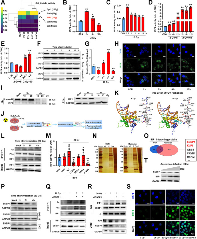
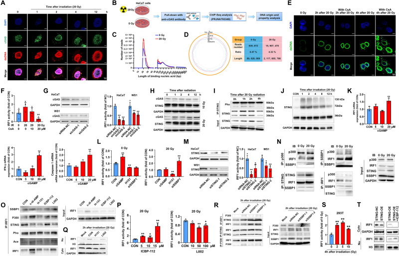
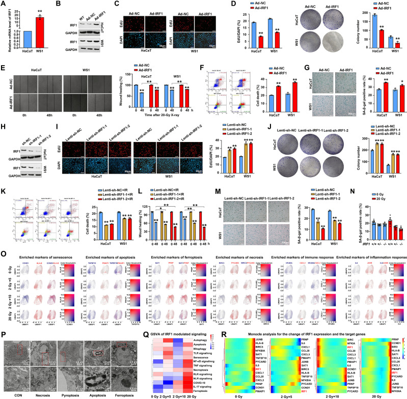
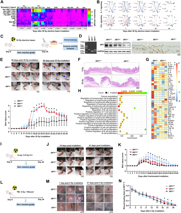
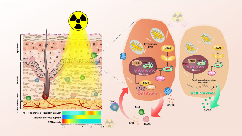
The key role of structural cells in immune modulation has been revealed with the advent of single-cell multiomics, but the underlying mechanism remains poorly understood. Here, we revealed that the transcriptional activation of interferon regulatory factor 1 (IRF1) in response to ionizing radiation, cytotoxic chemicals and SARS-CoV-2 viral infection determines the fate of structural cells and regulates communication between structural and immune cells. Radiation-induced leakage of mtDNA initiates the nuclear translocation of IRF1, enabling it to regulate the transcription of inflammation- and cell death-related genes. Novel posttranslational modification (PTM) sites in the nuclear localization sequence (NLS) of IRF1 were identified. Functional analysis revealed that mutation of the acetylation site and the phosphorylation sites in the NLS blocked the transcriptional activation of IRF1 and reduced cell death in response to ionizing radiation. Mechanistically, reciprocal regulation between the single-stranded DNA sensors SSBP1 and IRF1, which restrains radiation-induced and STING/p300-mediated PTMs of IRF1, was revealed. In addition, genetic deletion or pharmacological inhibition of IRF1 tempered radiation-induced inflammatory cell death, and radiation mitigators also suppressed SARS-CoV-2 NSP-10-mediated activation of IRF1. Thus, we revealed a novel cytoplasm-oriented mechanism of IRF1 activation in structural cells that promotes inflammation and highlighted the potential effectiveness of IRF1 inhibitors against immune disorders.

Keywords: Interferon regulatory factor 1 (IRF1); Ionizing radiation; Nuclear translocation; Posttranslational modification (PTM); Transcription regulation.


Conflict of interest statement

West China Second University Hospital has two patents pending related to the two effective IRF1-responsive inhibitors reported in this work, entitled “A small molecular compound against IRF1-responsive for treating inflammatory skin injury” and “A novel agent against genotoxic-induced tissue injuries”, respectively.

Figures

Fig. 1
Fig. 1
IRF1 expression dynamics in skin structural cells after irradiation. A Comparative analysis of IRF1 binding enrichment in irradiated tissues and cells. Immunohistochemistry (IHC) analysis of IRF1 expression in chronic radiogenic skin injury tissues obtained from patients exposed to radiation through treatment or accident (B) and in acute radiogenic skin injury models established in rodents and primates (C). D Schematic showing the workflow for investigating the IRF1 expression profiles with single-cell RNA-Seq (scRNA-Seq) of rat skin tissues at different times after irradiation. E Violin plot showing the distribution of IRF1 in different cell types and changes over time from 7 to 60 days after irradiation in keratinocytes and fibroblasts (F) of rat skin tissues. G Expression of IRF1 in different keratinocyte clusters visualized by t-SNE across scRNA-Seq datasets from irradiated rat skin tissues: IFE interfollicular epidermis, IFE-B interfollicular epidermis basal, IFE-D interfollicular epidermis differentiated, OB outer bulge, IB inner bulge, uHF upper hair follicle, SG sebaceous gland. H Dot plot depicting integrated keratinocyte clusters according to skin cell type-specific marker expression. I Changes in the proportions of keratinocyte clusters in rat skin tissues 7–60 days after irradiation. J Changes in IRF1 expression in patients exposed to iridium-192 at 250 days postirradiation and changes in IRF1 expression in different cell types in irradiated skin tissues compared with normal skin tissues. K Changes in IRF1 expression in the scRNA-Seq dataset of HaCaT cells exposed to fractional radiation (2 Gy ×5 and 2 Gy ×10) or a single high dose of radiation (20 Gy)
Fig. 2
Fig. 2
SSBP1 and PTMs determine the radiation-induced nuclear translocation and transient activation of IRF1. A CSI of module activity in keratinocyte cells 7–60 days after irradiation analyzed by SCENIC across scRNA-Seq datasets from irradiated rat skin tissues. Analysis of the effects of radiation, a single 20 Gy (B and C) or fractional 2 Gy doses (D and E) on IRF1 transcriptional activity in HaCaT and WS1 cells through a dual-luciferase assay. F Western blots showing IRF1 expression in HaCaT cells 1–12 h after 10 or 20 Gy irradiation in the presence or absence of 10 μM MG-132 (24 h). G qRT–PCR analysis of IRF1 mRNA expression in response to radiation. H, I Detection of radiation-induced nuclear translocation of IRF1 determined by immunofluorescence analysis and the separation of nuclear and cytoplasmic fractions followed by western blotting. J Schematic showing the proteomic analysis performed to investigate the interactome and PTMs of IRF1 in irradiated HaCaT cells, as determined by liquid chromatography with tandem mass spectrometry (LC‒MS/MS). K Molecular simulation of PTMs in the nuclear localization sequence domain of IRF1. L Verification of radiation-induced acetylation and phosphorylation of IRF1 through immunoprecipitation (IP) analysis. M Influence of mutations at different posttranslational modification sites on IRF1 transcriptional activation in primary skin cells from IRF1−/− mice, as detected through a dual-luciferase assay. N Silver staining of proteins pulled down with an anti-IRF1 antibody and a Venn diagram showing common and (O) specific interactomes detected by LC–MS/MS and representative proteins. P western blot analysis of SSBP1 expression in HaCaT cells 1–4 h after 20 Gy irradiation in the presence of CHX or MG132. Q Co-IP analysis of PTM marks in irradiated cells transfected with siRNAs against SSBP. Influence of SSBP1 knockdown on the radiation-induced nuclear translocation of IRF1 determined by separating the nucleus and cytoplasm followed by western blotting (R) and performing immunofluorescence analysis (S). T Expression of SSBP1 and IRF1 in adenovirus-infected HaCaT cells after 24 h (0–100 MOI). At least three replicate experiments were performed for each study. *P < 0.05 and **P < 0.01, compared with the control group
Fig. 3
Fig. 3
cGAS sensing cytosolic mtDNA initiates STING and p300-mediated PTM of IRF1. A Colocalization analysis showing cGAS and dsDNA in the cytoplasm of irradiated HaCaT cells. B Schematic showing the chromatin immunoprecipitation sequencing workflow used to investigate the origins and properties of the cGAS-binding nucleic acids. C Distribution profile showing the length of the bound nucleic acids. D Changes in the number, ratio and length of mtDNA sensed by cGAS in response to irradiation. E, F Effects of the mPTP inhibitor cyclosporin (CsA) (20 μM, 2 h) on IRF1 activity and cytosolic dsDNA formation 2 h after irradiation. G, M Dual-luciferase reporter assay showing changes in IRF1 activity caused by downregulation of cGAS and STING activity 2 h after 20 Gy irradiation. HJ Detection of cGAS and STING expression by western blotting, STING phosphorylation (Pho) by immunoprecipitation, and STING dimer formation by nonreducing PAGE at different times after irradiation. K Effects of the STING agonist 2’3’-cGAMP (20 μM, 24 h) on the mRNA expression of IRF1, IFN-γ and caspase 1. L Effects of the STING agonist 2’3’-cGAMP on IRF1 activity in nonirradiated cells and the effects of the STING antagonist H151 and the siRNA against SSBP1 on IRF1 activity in irradiated cells. N Co-IP analysis of the interaction between IRF1, P300, STING and SSBP1. O IP analysis of the change in the PTM of IRF1 and the interaction between IRF1/P300 or SSBP1 in irradiated cells pretreated with an agonist or antagonist of STING and p300. Effects of the p300 antagonists I-CBP112 and L002 on IRF1 transcriptional activity (P) and nuclear translocation (Q) in irradiated cells. R Co-IP analysis of the interaction between IRF1/STING and IRF1/p300 in irradiated cells transfected with siRNAs against SSBP1. S Changes in IRF1 activity in HEK-293T cells lacking TMEM173 4 h after 2–10 Gy irradiation. T Effect of exogenous STING and the p300 agonist ICBP-112 on the nuclear translocation of IRF1 in HEK-293T cells 4 h after 5 Gy irradiation, as determined by separating the nuclear and cytoplasm followed by western blotting. At least three replicate experiments were performed for each study. *P < 0.05 and **P < 0.01, compared with the control group
Fig. 4
Fig. 4
IRF1 triggers multiple kinds of cell death in irradiated skin cells. A Basel level of IRF1 mRNA expression in skin (HaCaT and WS1) cells. B, H Establishment of IRF1-overexpressing or IRF1-knockdown skin cells. C, I Effects of IRF1 on cell proliferation as determined by EdU staining in IRF1-overexpressing and IRF1-knockdown cells. D, J Effects of IRF1 on cell proliferation, as determined by a colony formation assay with IRF1-overexpressing and IRF1-knockdown cells. E, L Effects of IRF1 on the migratory ability of IRF1-overexpressing and IRF1-knockdown cells, as determined by wound healing analysis. F, K Effects of IRF1 on cell death, as determined by Annexin/propidium iodide (PI) staining, in IRF1-overexpressing and IRF1-knockdown cells. G, M and N Effects of IRF1 on the senescence of IRF1-overexpressing and IRF1-knockdown cells and primary skin cells from mice with different IRF1 genotypes, as determined by β-gal staining. O T-distributed stochastic neighbor embedding (t-SNE) plots showing different types of cell death identified by the expression of select biomarkers in irradiated HaCaT cells. P Typical morphological changes in apoptosis and ferroptosis caused by irradiation, as detected through transmission electron microscopy (TEM). Q Gene set variation analysis (GSVA) of IRF1 transcription-regulated genes involved in cell death-related signaling pathways. R Visualization of the IRF1-triggered progression of cell death as determined through pseudoanalysis of changes in biomarker expression. At least three replicate experiments were performed for each study. *P < 0.05 and **P < 0.01, compared with the control group
Fig. 5
Fig. 5
Activation of caspase 1 contributes to IRF1-induced cell death. A Transcriptome profiles of skin tissues from IRF1 wild-type (WT) and IRF1-mutant mice after 35 Gy electron beam irradiation. B Landscape of the function of IRF1 target genes in irradiated skin tissues based on Gene Ontology (GO) molecular function analysis. C qRT‒PCR analysis of alterations in the mRNA expression of caspase family members. D, E qRT‒PCR analysis of the mRNA expression of IRF1, caspase 1, GSDMD and IL-1 in irradiated cells transfected with Lenti-IRF1 or Lenti-shIRF1. F Investigation of the change in the enzymatic activity of caspase 1 and caspase 3 in irradiated skin tissues from WT IRF1 and IRF1-mutant mice using a specific assay kit. G, H western blotting showing changes in the cleavage of caspase 1, GSDMD and IL-1 in combined injury tissues and primary skin cells, irradiated or not, of mice with different IRF1 genotypes. I Western blots showing the expression of cell death-related biomarkers in irradiated HaCaT cells transfected with the indicated lentiviruses. J Translocation and degradation of Lamin B1 in irradiated HaCaT cells, as detected by an immunofluorescence assay. K Colocalization analysis of AIM2 and double-stranded DNA (dsDNA) in HaCaT cells at different times after 20 Gy irradiation. L western blotting showing the change in the ratio of cleaved/total caspase 1 in HaCaT cells 1–12 h after 20 Gy irradiation. M Schematic showing the workflow for investigating the origins and distribution profiles of AIM2-captured binding nucleic acids on the basis of their length in irradiated HaCaT cells, as determined by chromatin immunoprecipitation with sequencing (ChIP-Seq) and the corresponding results (N, O and P). Q qRT–PCR analysis of AIM2 mRNA expression in irradiated skin tissues from IRF1 wild-type and mutant mice and western blots showing AIM2 protein expression in cells transfected with Lenti-shIRF1 (R). S western blotting showing changes in the expression of AIM2 and ASC in primary skin cells, irradiated or not, from mice with different IRF1 genotypes. T, U Colocalization analysis of AIM2 and ASC in HaCaT cells at different times after 20 Gy irradiation. At least three replicate experiments were performed for each study. *P < 0.05 and **P < 0.01, compared with the control group
Fig. 6
Fig. 6
IRF1 tames radiation-induced inflammatory skin injury. A Strength of potential cell–cell interactions between structural cells and immune cells inferred from the gene expression of known receptor–ligand pairs in irradiated rat skin tissues. Ker keratinocyte, Fib fibroblast, End endothelium cell, NK natural killer cell, Mac macrophage, Mo monocyte, DC dendritic cell, Neut neutrophil. B Identification of signaling pathways among different cell populations. Different cell populations are color-coded, where the line color represents cells with outgoing events and the width represents the communication strength. C Schematic showing the workflow used to establish a radiogenic skin injury model by applying a single dose of radiation. D The expression of IRF1 in mice with different IRF1 genotypes was determined by western blotting and immunohistochemistry (IHC). E Pictures showing typical radiogenic skin injury 20 and 40 days after irradiation and the scoring curves of the whole course of radiogenic injury in mice with different IRF1 genotypes. F Different morphological changes were detected by hematoxylin and eosin (H&E) staining of irradiated skin tissues from wild-type (WT) IRF1 and IRF1 mutant mice. G Heatmap showing the differential expression of genes related to the inflammatory response in the irradiated skin tissues of the WT IRF1 and IRF1-mutant mice, as determined by a RayBiotech immunoassay (Guangzhou, China). H Gene Ontology (GO) molecular function analysis of altered inflammatory factor and chemokine levels in irradiated skin tissues. I Schematic showing the workflow used to establish a radiogenic skin injury model by fractional radiation (5.5 Gy ×4). J, K Pictures of typical radiogenic skin injury after irradiation and scoring curves of the whole course of radiogenic injury in mice with different IRF1 genotypes. L Schematic showing the workflow used to establish a combined radiogenic skin injury model by punch following total body irradiation (TBI) (4Gy). M, N Images of typical combined skin injury after irradiation and wound healing curves of the combined skin injury in mice with different IRF1 genotypes. *P < 0.05 and **P < 0.01, compared with the control group
Fig. 7
Fig. 7
Small molecules targeting the DBD of IRF1 attenuate radiation-induced inflammatory injury. A Schematic showing the workflow for identifying specific molecules targeting the DNA-binding domain (DBD) of IRF1. B Chemical formula and molecular stimulation of the two most effective inhibitors. C Effect of selected inhibitors on IRF1 transcriptional activity based on the dual-luciferase assay. D HaCaT cells were treated with 20 μM I-2 or I-19 for 12 h and then exposed to 20 Gy irradiation. At 2 h after irradiation, the recruitment of IRF1 to the caspase-1 promoter was determined by using a ChIP assay. E LDH analysis of 20 Gy-irradiated cells pretreated with different concentrations of select inhibitors. F Influence of IRF1 inhibitors on the colony formation ability of irradiated cells treated with the two specific inhibitors and the corresponding number of colonies and colony area. G Wound healing analysis of the migratory ability of irradiated cells treated with the two specific inhibitors. HK Influence of IRF1 inhibitors on the MMP, lipid peroxidation, ROS production and death of irradiated cells treated with the two specific inhibitors, as determined by JC-1, BIDOPY, dichlorodihydrofluorescein-diacetate and Annexin/propidium iodide (PI) staining, respectively. L Influence of IRF1 inhibitors on the senescence of irradiated primary skin cells treated with the two specific inhibitors, as detected by SA-β-Gal staining. M Influence of IRF1 inhibitors on the cell death of irradiated primary skin cells induced by the STING agonist G10 after treatment with the two specific inhibitors, as detected by Annexin/PI staining. NP Pictures of typical radiogenic skin and claw injuries after irradiation and scoring curves of the whole course of radiogenic injury in mice pretreated with specific inhibitors or dimethyl sulfoxide (DMSO). Q Heatmap showing the differential expression of inflammatory response genes in irradiated skin tissues pretreated with specific inhibitors or DMSO, as determined by an immunoassay at RayBiotech (Guangzhou, China). R Western blotting showing the change in the expression of protein markers related to the death of irradiated cells in the presence of specific inhibitors or DMSO. S Immunofluorescence analysis of the change in the cleavage of caspase 1 and GSDMD in irradiated cells in the presence of specific inhibitors or DMSO. *P < 0.05 and **P < 0.01, compared with the control group
Fig. 8
Fig. 8
Schematic representation of IRF1 triggering of radiation-induced inflammation. In response to ionizing radiation, IRF1 activity is triggered by the mtDNA-cGAS/STING signaling pathway, which exacerbates inflammatory injury. SSBP1 is a chaperone that inhibits IRF1 modification, translocation and activation. Administration of small molecules targeting the DNA binding domain (DBD) of IRF1 attenuates radiation-induced and IRF1-mediated cell death

References

    1. Peters A, Nawrot TS, Baccarelli AA. Hallmarks of environmental insults. Cell. 2021;184:1455–68. 10.1016/j.cell.2021.01.043 - DOI - PMC - PubMed
    1. Krausgruber T, Fortelny N, Fife-Gernedl V, Senekowitsch M, Schuster LC, Lercher A, et al. Structural cells are key regulators of organ-specific immune responses. Nature. 2020;583:296–302. 10.1038/s41586-020-2424-4 - DOI - PMC - PubMed
    1. Karki R, Sharma BR, Tuladhar S, Williams EP, Zalduondo L, Samir P, et al. Synergism of TNF-α and IFN-γ Triggers Inflammatory Cell Death, Tissue Damage, and Mortality in SARS-CoV-2 Infection and Cytokine Shock Syndromes. Cell. 2021;184:149–.e17. 10.1016/j.cell.2020.11.025 - DOI - PMC - PubMed
    1. Chabanon RM, Rouanne M, Lord CJ, Soria JC, Pasero P, Postel-Vinay S. Targeting the DNA damage response in immuno-oncology: developments and opportunities. Nat Rev Cancer. 2021;21:701–17. 10.1038/s41568-021-00386-6 - DOI - PubMed
    1. Mukherjee S, Abdisalaam S, Bhattacharya S, Srinivasan K, Sinha D, Asaithamby A. Mechanistic link between DNA damage sensing, repairing and signaling factors and immune signaling. Adv Protein Chem Struct Biol. 2019;115:297–324. 10.1016/bs.apcsb.2018.11.004 - DOI - PMC - PubMed

Substances

LinkOut - more resources